
Cephalic index for 5 marks according to narayanarerddy essential
cephalic index skull types dolichocephalic mesaticephalic brachycephalic forensic
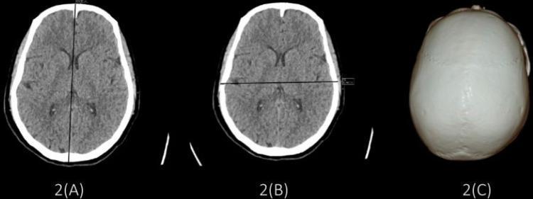
This composite diagnostic image illustrates the methodology for calculating the cephalic index using Computed Tomography (CT). Panels 2(A) and 2(B) show non-contrast axial CT scans of the head at the level of the lateral ventricles. Figure 2(A) demonstrates the measurement of the Maximum Cranial Length (MCL) via a mid-sagittal linear cursor extending from the glabella to the opisthocranion. Figure 2(B) illustrates the Maximum Cranial Breadth (MCB) measured as the maximum biparietal diameter perpendicular to the MCL. Visible neuroanatomical structures include the cerebral cortex, ventricles, and falx cerebri within the calvarium. Panel 2(C) presents a superior-view 3D volume-rendered reconstruction of the skull, highlighting its mesocephalic (ovoid) morphology and the sagittal suture. This image set is primarily used in radiology, forensic medicine, and physical anthropology to classify skull shapes (brachycephalic, dolichocephalic, or mesocephalic) based on the ratio of cranial breadth to length.
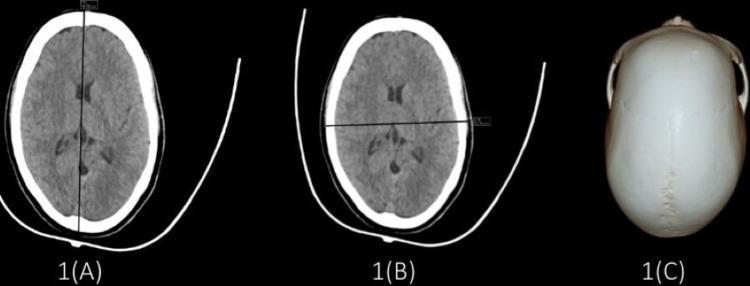
This composite educational image demonstrates the morphological characteristics of a dolichocephalic skull using radiological and anatomical modalities. Panels 1A and 1B show non-contrast axial computed tomography (CT) scans of the head at the level of the lateral ventricles. Panel 1A highlights the maximum cranial length (18.64 cm) along the anteroposterior axis, while Panel 1B shows the maximum cranial breadth (13.21 cm) along the transverse axis. The brain parenchyma and ventricular systems appear normal, with the hyperdense (white) calvarium outlining the elongated cranial vault. Panel 1C provides a superior-view clinical photograph of a dry skull specimen, clearly illustrating a cephalic index of 70.86, characteristic of dolichocephaly (long-headedness). Key anatomical landmarks visible in 1C include the sagittal suture and the overall narrow, elongated contour of the cranium. These visuals serve as a reference for cephalometry and anthropometric classification in forensic medicine, radiology, and neurosurgery.
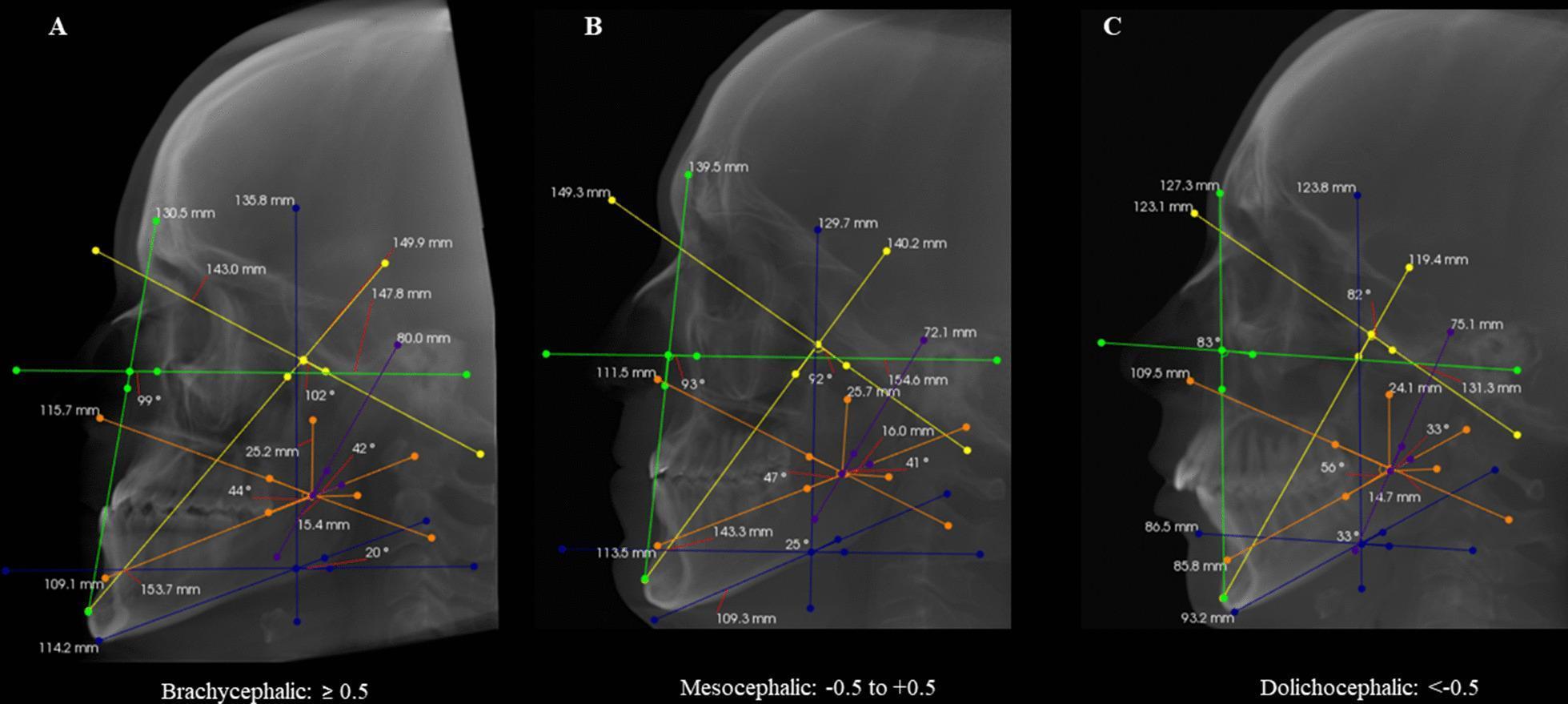
This diagnostic image set consists of three sagittal cone-beam computed tomography (CBCT) reconstructions illustrating cephalometric analysis for different facial types: Brachycephalic (A), Mesocephalic (B), and Dolichocephalic (C). Each scan displays standardized color-coded linear and angular measurements used to calculate the Vert index. Key measurements include: the facial axis (yellow line), facial depth (green line), mandibular plane (blue line), lower facial height (orange line), and mandibular arch (purple line). The images demonstrate the skeletal variation across patterns: the Brachycephalic type shows a shorter, wider facial structure with a flatter mandibular plane and steeper facial axis; the Dolichocephalic type exhibits a vertically elongated, narrower facial profile with a steeper mandibular plane and more acute facial axis. The Mesocephalic reconstruction represents a balanced intermediate skeletal relationship. This comparison is used in orthodontics and maxillofacial surgery to classify facial growth patterns and guide treatment planning for skeletal malocclusions.
Cephalic Index (5 Marks) - As Per Narayan Reddy's "Essentials of Forensic Medicine and Toxicology"
Note: K.S. Narayan Reddy's textbook is not directly available in this library, but the content below is drawn from standard forensic medicine references (Parikh's, PC Dikshit) which align with Narayan Reddy's teaching - all major Indian forensic medicine texts present this topic identically.
Definition
Formula
- Breadth = Greatest diameter across the skull above the mastoid process (measured at the widest point)
- Length = Distance between the glabella (anteriorly) and the external occipital protuberance (posteriorly)
- Measurements are done using sliding calipers (not a measuring tape). In the absence of sliding calipers, an osteometric board can be used.
Classification of Skulls Based on Cephalic Index
| Type of Skull | Cephalic Index | Alternate Name | Race |
|---|---|---|---|
| Dolichocephalic | 70 - 74.9 (or 70-75) | Long-headed | Aryans, Negroes (Blacks), Aborigines |
| Mesaticephalic | 75 - 79.9 (or 75-80) | Medium-headed | Europeans, Chinese |
| Brachycephalic | 80 and above (80-85) | Short-headed / Round-headed | Mongolians |
Visual Illustration
Other Skeletal Indices Used Along with Cephalic Index
-
Brachial Index (Radio-humeral index) = (Length of radius / Length of humerus) x 100
-
Crural Index (Tibio-femoral index) = (Length of tibia / Length of femur) x 100
-
Humero-femoral Index = (Length of humerus / Length of femur) x 100
Associated Indices for Race Determination
| Index | Blacks | Europeans | Mongols |
|---|---|---|---|
| Cephalic Index (CI) | 70-74.9 | 75-79.9 | 80 and above |
| Height Index (HI) | 72 | 71 | 75 |
| Nasal Index (NI) | 55 | 46 | 50 |
- Height Index (HI) = (Height of skull from tip of mastoid process to bregma / Length of skull) x 100
- Nasal Index (NI) = (Width of nasal aperture / Height of nasal aperture) x 100
Key Points for Exam
- CI is also called the breadth index.
- The surest method of identification is fingerprints - CI only helps determine race, not individual identity.
- Cephalic index can change with environment (Boas, 1912) - American-born children had different CI compared to their European-born parents, showing it is influenced by both genetics and environment.
- A CI of 80-85 indicates brachycephalic skull - seen in Mongolians.
- Measurements must always be done with sliding calipers, not a measuring tape.
Objectives and formalities of medicolegal autopsy for 5 marks according to k narayanareddy textbook fmt
Objectives and Formalities of Medicolegal Autopsy
As per K. Narayan Reddy's "Essentials of Forensic Medicine and Toxicology" (36th Ed., 2026)
Definition
OBJECTIVES OF MEDICOLEGAL AUTOPSY
1. Establishing Identity
2. Determining Cause, Mode, and Time Since Death
- Cause of death: Whether the cause is natural or unnatural; if unnatural, what exactly killed the person.
- Time since death: Based on postmortem changes - changes in the eye, muscles, postmortem staining, decomposition, mineral changes in skeletal remains, carbon dating, etc.
- Mode (mechanism) of death: Whether the cause of death led to the brain, heart, or lungs stopping first - the logical consequence of the cause on organ failure.
3. Manner of Death
- Natural
- Accidental
- Suicidal
- Homicidal
4. Collection and Documentation of Trace Evidence
5. Special Cases - Newborns
6. In Fragmented / Skeletal Remains
FORMALITIES (RULES) FOR MEDICOLEGAL AUTOPSY
1. Labelling and Identification
- The body must be labelled as soon as it arrives in the mortuary.
- The body must be identified by the police constable who accompanies it, and by relatives of the deceased.
- Names of those who identify the body must be recorded.
- In unidentified bodies: marks of identification, photographs, and fingerprints must be taken.
2. Location of Autopsy
- Must be conducted in a mortuary - never in a private room.
- Exceptions (autopsy may be done at the site when):
- Body is in an advanced state of putrefaction and transportation is difficult
- Materials of evidential value may be lost in transport
- Immediate examination without removal to hospital is essential for any special reason
3. Authorization
- Must be conducted only when there is an official order authorizing autopsy by the police or Magistrate.
4. Timing of Autopsy
- Should be performed as soon as possible after receiving requisition, without undue delay.
- Should ideally be conducted in daylight, because colour changes (jaundice, bruises, postmortem staining) cannot be appreciated in artificial light.
- If the body is received late in the evening: a preliminary external examination is done that day; the actual postmortem is conducted the next morning as early as possible.
- Decomposed bodies, homicide, and suspected foul play are NOT to be subjected to postmortem during nighttime.
5. Pre-Autopsy Review
- The medical officer should first read the inquest report carefully to note the apparent cause of death.
- All available details (case sheets, accident register) must be reviewed so attention may be directed to significant points.
- Appropriate ancillary investigations must be planned (toxicology, microbiology, virology, radiology, etc.).
- Importance of case sheet review: Helps identify nature of sutured wounds, surgical incisions through injuries, therapeutic wounds (tracheostomy, venesection), resuscitation injuries (artefacts from CPR), firearm wound entry/exit after surgical intervention, and negative autopsy causes (diabetes, asthma, epilepsy, etc.).
6. Conduct of the Autopsy
- Both positive and negative findings must be recorded.
- Even if the body is decomposed, autopsy should still be performed - certain important lesions may still be found.
- All findings, photographs, radiographs, sketches, measurements, and weights must be preserved in written records.
- Trace evidential material must be properly documented and preserved.
Key Point for Exams
"What the mind does not know, the eyes cannot see, and the hands cannot do" - this applies to the autopsy surgeon who plays an important role in giving justice to the departed soul. (Narayan Reddy, 36th Ed.)