
Pathology important questions with answers
What is your level/exam? · Which area of pathology do you want covered?
Pathology - Important Questions & Answers (MBBS)
UNIT 1: CELL INJURY, DEATH & ADAPTATIONS
- Robbins & Kumar Basic Pathology
- Adapt - achieve a new steady state, preserving viability (e.g., hypertrophy, hyperplasia, atrophy, metaplasia)
- Undergo reversible injury - if adaptive capability is exceeded but stimulus is mild/brief
- Die (irreversible injury) - if stress is severe or persistent
- Robbins & Kumar Basic Pathology
- Hypoxia and ischemia - most common; deprive cells of oxygen (needed for ATP generation)
- Physical agents - trauma, heat/cold, radiation, electrical injury
- Chemical agents and drugs - toxins, poisons, drugs (e.g., carbon tetrachloride)
- Infectious agents - bacteria, viruses, fungi, parasites
- Immunologic reactions - autoimmune diseases, hypersensitivity reactions
- Genetic derangements - mutations causing enzyme defects, structural protein abnormalities
- Nutritional imbalances - protein-calorie malnutrition, vitamin deficiencies, excess (obesity)
- Robbins & Kumar Basic Pathology
- Mitochondria - impaired oxidative phosphorylation → reduced ATP → membrane pump failure → cellular swelling
- Cellular membranes - damage to plasma and organelle membranes → loss of ion gradients, leakage of enzymes
- Nucleus (DNA) - strand breaks from free radicals or toxins → if not repaired, cell undergoes apoptosis
- Endoplasmic reticulum (ER) - accumulation of misfolded proteins triggers the unfolded protein response (UPR); severe ER stress triggers apoptosis
- Robbins & Kumar Basic Pathology
| Feature | Reversible Injury | Irreversible Injury |
|---|---|---|
| Mitochondria | Swelling, loss of phospholipids | Vacuolation, large amorphous densities |
| Plasma membrane | Blebbing, blunting of microvilli | Rupture |
| Nucleus | Chromatin clumping | Pyknosis, karyorrhexis, karyolysis |
| Lysosomes | Intact | Rupture, enzymatic digestion |
| Cell fate | Returns to normal | Necrosis or apoptosis |
- Robbins & Kumar Basic Pathology
| Feature | Necrosis | Apoptosis |
|---|---|---|
| Cause | Ischemia, toxins, infections (pathologic) | Physiologic or pathologic |
| Cell size | Enlarged (swelling) | Reduced (shrinkage) |
| Nucleus | Pyknosis → karyorrhexis → karyolysis | Fragmentation into nucleosome-sized pieces |
| Plasma membrane | Disrupted | Intact, blebbing |
| Cell contents | Leaked - causes inflammation | Packaged in apoptotic bodies, no inflammation |
| Adjacent inflammation | Yes (prominent) | No |
| Examples | MI, ischemic stroke | Embryogenesis, immune cell deletion, hormone-deprived tissue |
- Robbins & Kumar Basic Pathology, Robbins Pathologic Basis of Disease
| Pattern | Mechanism | Example |
|---|---|---|
| Coagulative | Protein denaturation preserves cell outlines | Myocardial infarction, kidney infarction |
| Liquefactive | Enzymatic digestion liquefies tissue | Brain infarction, abscess |
| Caseous | Combination: cheese-like, structureless | Tuberculosis (TB) |
| Fat necrosis | Lipase digestion of fat | Acute pancreatitis, breast trauma |
| Fibrinoid | Immune complexes + fibrin deposition in vessel walls | Vasculitis, malignant hypertension |
| Gangrenous | Usually coagulative; "wet" gangrene has superimposed liquefaction | Diabetic foot, bowel infarction |
- Robbins & Kumar Basic Pathology
- Hypertrophy - increase in cell size (not number). Occurs in non-dividing cells (cardiac muscle, skeletal muscle). E.g., cardiac hypertrophy in hypertension.
- Hyperplasia - increase in cell number. Occurs in dividing cells. E.g., endometrial hyperplasia with estrogen excess.
- Atrophy - decrease in cell size/number. E.g., muscle wasting from disuse, denervation.
- Metaplasia - replacement of one differentiated cell type by another, usually more resistant type. E.g., squamous metaplasia of bronchial epithelium in smokers (columnar → squamous). Reversible but predisposes to malignancy.
- Dysplasia - disordered growth; considered pre-malignant. E.g., cervical dysplasia (CIN).
- Robbins & Kumar Basic Pathology
- Steatosis (fatty change) - triglycerides in liver cells; caused by alcohol, obesity, diabetes, toxins
- Cholesterol accumulation - atherosclerosis, xanthomas (foam cells)
- Protein accumulation - Russell bodies in plasma cells (immunoglobulins)
- Hyaline change - intracellular pink, glassy material (e.g., alcoholic hyaline/Mallory bodies in hepatocytes)
- Glycogen accumulation - glycogen storage diseases (e.g., von Gierke disease - glucose-6-phosphatase deficiency)
- Pigments - lipofuscin ("wear and tear" pigment, brown), melanin, hemosiderin (iron), carbon (anthracosis)
- Pathologic calcification - dystrophic (in dead tissue, normal serum calcium) vs. metastatic (in normal tissue, elevated serum calcium)
- Robbins & Kumar Basic Pathology
UNIT 2: INFLAMMATION & REPAIR
- Rubor (redness) - increased blood flow
- Calor (heat) - increased blood flow
- Tumor (swelling) - increased vascular permeability
- Dolor (pain) - prostaglandins and bradykinin
- Functio laesa (loss of function) - Virchow's addition
- Robbins & Kumar Basic Pathology
| Feature | Acute | Chronic |
|---|---|---|
| Onset | Fast (minutes to days) | Slow (weeks to months) |
| Duration | Short | Prolonged |
| Key cells | Neutrophils | Lymphocytes, macrophages, plasma cells |
| Vascular changes | Prominent | Less prominent |
| Tissue injury | Mild, often reversible | Prominent, fibrosis |
| Causes | Infections, trauma | Persistent infections (TB, fungal), autoimmune, foreign material |
| Outcome | Resolution, abscess, or progression | Scarring/fibrosis |
- Histamine - first mediator released; from mast cells and basophils; causes vasodilation and increased permeability
- Prostaglandins - from arachidonic acid via COX pathway; cause vasodilation, pain, fever
- Leukotrienes - from arachidonic acid via lipoxygenase; LTB4 is chemotactic; LTC4/LTD4 increase permeability and cause bronchoconstriction
- Cytokines - TNF, IL-1 (from macrophages): systemic effects (fever, acute phase proteins); IL-8: chemotaxis
- PAF (Platelet Activating Factor) - bronchoconstriction, chemotaxis
- Nitric Oxide (NO) - vasodilation, kills microbes
-
Complement (C3a, C5a) - C5a is most important: chemotaxis, opsonization, MAC formation
-
Kinin system - bradykinin: pain, vasodilation, increased permeability
-
Coagulation system - fibrin, thrombin
-
Robbins & Kumar Basic Pathology
- Epithelioid cells (activated macrophages)
- Langhans giant cells (horseshoe-shaped nuclei) or foreign-body giant cells
- Peripheral lymphocytes
- Central caseous necrosis (in TB)
- S - Sarcoidosis (non-caseating)
- A - Allergic alveolitis
- T - Tuberculosis (caseating - most classic)
- C - Crohn's disease
- H - Histoplasmosis / other fungi
- B - Berylliosis, foreign body reaction, leprosy (syphilis)
- Hemostasis - platelet aggregation, fibrin clot
- Inflammation - neutrophils then macrophages clean debris
- Proliferation - angiogenesis, fibroblast proliferation, collagen synthesis
- Remodeling - collagen cross-linking, scar maturation
UNIT 3: HEMODYNAMICS, THROMBOSIS & SHOCK
- Endothelial injury - most important in arterial thrombosis; exposes subendothelial collagen → platelet activation. Causes: atherosclerosis, hypertension, vasculitis.
- Abnormal blood flow (turbulence/stasis) - prevents dilution of activated clotting factors, reduces laminar flow (keeps platelets away from endothelium). Turbulence causes arterial/cardiac thrombi; stasis causes venous thrombi.
- Hypercoagulability - primary (genetic: Factor V Leiden, prothrombin mutation, antithrombin III deficiency) or secondary (acquired: prolonged bed rest, cancer, pregnancy, OCP use).
- Thromboembolism - most common (95%+); DVT → pulmonary embolism (PE) most common
- Fat embolism - fractures of long bones, liposuction; fat globules obstruct pulmonary/cerebral vessels
- Air embolism - IV injections, surgery; >100 mL can be fatal
- Amniotic fluid embolism - obstetric complication; can cause DIC and respiratory failure
- Tumor embolism - cancer cells spread via hematogenous route
- Paradoxical embolism - embolus crosses from venous to arterial system through a patent foramen ovale
- White (anemic) infarct - solid organs with single blood supply (heart, kidney, spleen). Wedge-shaped.
- Red (hemorrhagic) infarct - loose tissue (lung, intestine) or organs with dual blood supply; also in venous occlusions.
- Nature of vascular supply (single vs. dual; end artery vs. collateral)
- Rate of occlusion (slow → collaterals can develop)
- Tissue vulnerability to hypoxia (neurons: 3-5 min; myocardium: 20-30 min; fibroblasts: hours)
- Oxygen-carrying capacity of blood (anemia worsens outcome)
| Type | Mechanism | Examples |
|---|---|---|
| Cardiogenic | Pump failure → reduced cardiac output | MI, cardiac tamponade, arrhythmia |
| Hypovolemic | Loss of blood/fluid volume | Hemorrhage, burns, severe diarrhea |
| Distributive (Septic) | Systemic vasodilation → maldistribution of flow | Gram-negative sepsis (endotoxin → NO release) |
| Neurogenic | Loss of vascular tone (autonomic dysfunction) | Spinal cord injury, general anesthesia |
| Anaphylactic | IgE-mediated mast cell degranulation → massive vasodilation | Bee sting, drug allergy |
UNIT 4: NEOPLASIA
| Feature | Benign | Malignant |
|---|---|---|
| Differentiation | Well differentiated | Poorly differentiated to anaplastic |
| Growth rate | Slow | Rapid |
| Invasion | No invasion | Locally invasive |
| Metastasis | Never | Yes (hallmark of malignancy) |
| Border | Encapsulated, well-defined | Irregular, infiltrating |
| Necrosis/hemorrhage | Rare | Common |
| Mitoses | Rare, normal | Frequent, atypical |
| Nuclear features | Normal N:C ratio | High N:C ratio, hyperchromatism, prominent nucleoli |
- RAS - most commonly mutated oncogene in human cancers; point mutation → constitutively active GTPase
- MYC - transcription factor; amplified in Burkitt lymphoma (t8;14 translocation)
- HER2/neu (ERBB2) - amplified in ~25% breast cancers; target of trastuzumab
- BCR-ABL - Philadelphia chromosome (t9;22); tyrosine kinase; CML; target of imatinib
- RB (retinoblastoma gene) - first tumor suppressor described; mutated in retinoblastoma, osteosarcoma
- TP53 - "guardian of the genome"; most commonly mutated tumor suppressor; mutated in >50% of all human cancers; Li-Fraumeni syndrome (germline p53 mutation)
- APC - mutated in familial adenomatous polyposis (FAP); loss → accumulation of β-catenin
- BRCA1/BRCA2 - DNA repair genes; mutations → breast and ovarian cancer
-
Lymphatic spread - most common for carcinomas (epithelial tumors). Sentinel lymph node is the first draining node. E.g., breast cancer → axillary nodes; colon cancer → mesenteric nodes.
-
Hematogenous spread - characteristic of sarcomas (connective tissue tumors) but also carcinomas. Travels via veins (portal → liver; systemic veins → lungs; vertebral venous plexus → spine - "Batson plexus").
- Liver - most common site of metastasis overall; GI cancers via portal vein
- Lungs - most common for cancers draining into systemic veins
- Bone - breast, prostate, lung, thyroid, kidney ("BPro-LTK")
- Brain - lung, breast, melanoma, kidney, colon
-
Seeding of body cavities (transcoelomic) - implantation on peritoneal/pleural surfaces. E.g., ovarian cancer → peritoneal seeding ("Krukenberg tumor" = ovarian metastasis from GI cancer via transcoelomic or hematogenous spread).
- DNA damage sensor - activated by DNA strand breaks, hypoxia, oncogene activation
- Cell cycle arrest - induces p21 (CDK inhibitor) → G1/S checkpoint arrest → allows DNA repair
- Apoptosis - if DNA damage is unrepairable, p53 activates BAX → apoptosis
- Senescence - permanent cell cycle arrest
- Inhibits angiogenesis - promotes TSP-1 expression
- Cells with damaged DNA continue to replicate → accumulation of mutations → cancer
- Li-Fraumeni syndrome: germline TP53 mutation → multiple cancers at young age
- Grade I: Well differentiated
- Grade II: Moderately differentiated
- Grade III: Poorly differentiated
- Grade IV: Anaplastic/undifferentiated
- T = Tumor size/local extent (T1-T4)
- N = Regional lymph node involvement (N0-N3)
- M = Distant metastasis (M0 or M1)
UNIT 5: IMMUNITY & HYPERSENSITIVITY
| Type | Name | Mechanism | Mediators | Examples |
|---|---|---|---|---|
| Type I | Immediate / Anaphylactic | IgE on mast cells → allergen crosslinks IgE → degranulation | Histamine, leukotrienes, prostaglandins | Anaphylaxis, asthma, urticaria, hay fever |
| Type II | Cytotoxic / Antibody-dependent | IgG/IgM bind cell surface antigens → complement activation or ADCC | Complement, NK cells | Hemolytic disease of newborn, Goodpasture's, autoimmune hemolytic anemia, myasthenia gravis |
| Type III | Immune complex | Antigen-antibody complexes deposited in tissues → complement → neutrophil recruitment | Complement, neutrophils | SLE, serum sickness, post-streptococcal GN, polyarteritis nodosa |
| Type IV | Delayed-type (DTH) / Cell-mediated | CD4+ T cells (Th1) or CD8+ CTLs react to antigen 24-72 hrs later | IFN-γ, TNF, cytotoxic T cells | Contact dermatitis, TB skin test (Mantoux), graft rejection, type 1 DM |
- Primary (AL) amyloidosis - light chains from plasma cell dyscrasia (multiple myeloma). Most common type. Affects heart, kidney, tongue, nerves.
- Secondary (AA) amyloidosis - serum amyloid A (SAA), an acute phase reactant, in chronic inflammatory diseases (TB, rheumatoid arthritis, IBD, osteomyelitis). Affects kidney (most common organ), liver, spleen.
- Hereditary (familial) amyloidosis - mutant transthyretin (TTR). E.g., familial Mediterranean fever → AA type.
- Dialysis-related - β2-microglobulin accumulates in long-term dialysis patients (carpal tunnel syndrome).
- Localized - e.g., Alzheimer disease (Aβ amyloid in brain plaques), type 2 DM (islet amyloid polypeptide in pancreatic islets).
UNIT 6: SELECTED ORGAN-SPECIFIC PATHOLOGY
| Time | Gross | Microscopic |
|---|---|---|
| 0-4 hrs | Normal | Normal (no changes) |
| 4-12 hrs | Pallor begins | Wavy fibers, coagulation necrosis begins |
| 12-24 hrs | Pallor | Coagulative necrosis, loss of nuclei/striations, neutrophil infiltration begins |
| 1-3 days | Yellow pallor with hyperemic border | Dense neutrophilic infiltrate |
| 3-7 days | Hyperemic border, central yellow-white | Macrophage infiltration, removal of dead cells |
| 1-3 weeks | Fibrovascular granulation tissue | Granulation tissue replaces necrotic muscle |
| >6 weeks | White fibrous scar | Dense collagen scar |
| Feature | HAV | HBV | HCV | HDV | HEV |
|---|---|---|---|---|---|
| Transmission | Fecal-oral | Parenteral, sexual, perinatal | Parenteral (IV drug use #1) | Parenteral (needs HBV) | Fecal-oral |
| Chronicity | None | 5-10% | 80% | Yes (with HBV) | None |
| Cirrhosis | No | Yes | Yes (most common cause in West) | Yes | No |
| HCC association | No | Yes (HBsAg carrier) | Yes | Yes | No |
| Vaccine | Yes | Yes | No | HBV vaccine prevents HDV | None widely available |
- First exposure, usually in children
- Subpleural focus (Ghon focus) + hilar lymph node involvement = Ghon complex
- Usually asymptomatic; heals with calcification
- Rarely: progressive primary TB or miliary TB in immunocompromised
- Reactivation of latent primary infection (usually >90% of adult cases)
- Usually upper lobe of lung (high O2 tension)
- Hallmark: caseating granuloma with central cheese-like necrosis
- Complications: cavity formation, bronchiectasis, massive hemoptysis, miliary TB, CNS TB, Pott's disease (vertebral TB), TB lymphadenitis
- Endothelial injury (from hypertension, hyperlipidemia, smoking, homocysteine) → endothelial dysfunction
- Increased permeability → LDL enters intima → oxidized LDL (ox-LDL)
- Monocyte recruitment → monocytes adhere to endothelium (via VCAM-1, ICAM-1) → enter intima → macrophages → engulf ox-LDL → foam cells
- Fatty streak - earliest visible lesion (reversible); foam cells + T lymphocytes
- Platelet aggregation → smooth muscle migration from media to intima → proliferation + ECM production
- Fibrous plaque - necrotic core (foam cells, cellular debris, cholesterol crystals) + fibrous cap (SMC + collagen) → advanced atheroma
- Plaque rupture/erosion → thrombosis → acute MI or stroke
- High-risk HPV types (16, 18, 31, 33) - responsible for >95% of cervical cancers
- HPV E6 protein → binds and degrades p53
- HPV E7 protein → binds and inactivates pRb (retinoblastoma protein)
- This releases E2F transcription factor → uncontrolled cell cycle progression
UNIT 7: HEMATOLOGY BASICS
- Microcytic hypochromic - iron deficiency anemia (most common), thalassemia, sideroblastic anemia, anemia of chronic disease
- Macrocytic (normochromic) - megaloblastic (B12/folate deficiency), liver disease, hypothyroidism, drugs (hydroxyurea)
- Normocytic normochromic - hemolytic anemia, aplastic anemia, anemia of chronic disease (early), blood loss (acute)
- Blood loss (acute vs. chronic)
- Increased destruction (hemolytic) - intrinsic (sickle cell, G6PD deficiency, hereditary spherocytosis) or extrinsic (autoimmune, microangiopathic)
- Decreased production - iron deficiency, B12/folate deficiency, aplastic anemia, renal failure
- Vaso-occlusive crises - pain crises (bones, chest, abdomen)
- Acute chest syndrome - fever + pulmonary infiltrates; most common cause of death in adults
- Stroke - especially in children
- Splenic sequestration/autosplenectomy - functional asplenia → susceptibility to encapsulated organisms (Streptococcus pneumoniae, Haemophilus influenzae, Neisseria meningitidis)
- Aplastic crisis - triggered by Parvovirus B19 infection
- Avascular necrosis - especially femoral head
- Renal papillary necrosis
QUICK RECALL TABLE: KEY STAINS IN PATHOLOGY
| Stain | What it identifies |
|---|---|
| H&E | Routine histology |
| PAS (Periodic Acid-Schiff) | Glycogen, fungi (Cryptococcus, Histoplasma), basement membranes |
| Congo red | Amyloid (apple-green birefringence under polarized light) |
| Ziehl-Neelsen (ZN/AFB) | Acid-fast bacilli (M. tuberculosis, M. leprae) |
| Gram stain | Bacteria (gram +ve = purple; gram -ve = pink) |
| Sudan IV / Oil Red O | Lipids/fat (frozen sections) |
| Prussian Blue (Perl's) | Hemosiderin (iron) |
| Silver stain (Gomori) | Fungi, Pneumocystis, reticulin fibers |
| India ink | Cryptococcus capsule |
| Giemsa | Malaria, Leishmania, chromosomes |
| Masson's Trichrome | Collagen (blue), muscle (red) |
QUICK RECALL: IMPORTANT TUMOR MARKERS
| Tumor Marker | Cancer |
|---|---|
| PSA | Prostate |
| AFP (alpha-fetoprotein) | Hepatocellular carcinoma, germ cell tumors (yolk sac) |
| β-hCG | Choriocarcinoma, hydatidiform mole |
| CEA | Colorectal cancer (also pancreas, lung, breast) |
| CA-125 | Ovarian cancer |
| CA 19-9 | Pancreatic cancer |
| CA 15-3 | Breast cancer |
| S-100 | Melanoma, neural tumors |
| Calcitonin | Medullary thyroid carcinoma |
| Chromogranin A | Neuroendocrine tumors |
Most imp diagram of which should be drawn in exams
Clean medical education diagram titled "Cell Injury & Death Pathways" for MBBS exam. Shows a central healthy cell at top. Three branches: LEFT branch = "Reversible Injury" showing cellular swelling, fatty change, vacuolization with arrows showing return to normal. CENTER branch = "Necrosis" with subtypes listed: Coagulative (MI, kidney), Liquefactive (brain abscess), Caseous (TB), Fat necrosis (pancreatitis), Fibrinoid (vasculitis). RIGHT branch = "Apoptosis" with features: cell shrinkage, chromatin condensation, apoptotic bodies, no inflammation. Bottom row shows comparison table of Necrosis vs Apoptosis with features: cell size, nucleus, membrane, inflammation, causes. White background, clear black text labels, color-coded boxes (blue for reversible, red for necrosis, green for apoptosis), arrows connecting concepts. Medical textbook style.
Medical education diagram titled "Virchow's Triad & Thrombosis" for MBBS exam. Central triangle with three labeled sides: TOP SIDE = "Endothelial Injury" (examples: atherosclerosis, hypertension, smoking), LEFT SIDE = "Abnormal Blood Flow / Stasis / Turbulence" (examples: DVT, atrial fibrillation, varicose veins), RIGHT SIDE = "Hypercoagulability" (primary: Factor V Leiden, prothrombin mutation; secondary: cancer, pregnancy, OCP). Inside triangle: "THROMBUS FORMATION". Below triangle: arrows showing outcomes - "Arterial thrombus → MI, Stroke" and "Venous thrombus → DVT → Pulmonary Embolism". Clean white background, bold labels, color-coded sides (red, blue, purple), medical textbook style suitable for hand-drawing.
Medical education diagram titled "Pathogenesis of Atherosclerosis" for MBBS exam. Step-by-step flowchart with numbered steps going downward: Step 1: Normal artery wall (intima, media, adventitia labeled). Step 2: Endothelial injury (causes listed: hypertension, smoking, hyperlipidemia, diabetes) → endothelial dysfunction. Step 3: LDL enters intima → oxidized LDL (oxLDL). Step 4: Monocytes adhere (VCAM-1, ICAM-1) → become macrophages → engulf oxLDL → FOAM CELLS → FATTY STREAK. Step 5: Platelet aggregation + smooth muscle cell migration from media. Step 6: Fibrous plaque = lipid core + fibrous cap (collagen + SMC). Step 7: Plaque rupture → thrombosis → MI or stroke. Cross-section artery drawings showing progression at each step. White background, clear arrows, red and yellow colors for plaque, medical textbook style.
Medical education diagram titled "Types of Hypersensitivity Reactions (Gell & Coombs)" for MBBS exam. Four horizontal rows, one per type. TYPE I (Immediate/Anaphylactic): allergen → IgE on mast cell → degranulation → histamine, leukotrienes → anaphylaxis, asthma. TYPE II (Cytotoxic): IgG/IgM bind cell surface antigen → complement activation / ADCC → cell death → examples: hemolytic disease of newborn, Goodpasture syndrome, myasthenia gravis. TYPE III (Immune Complex): Ag-Ab complex deposits → complement → neutrophils → tissue damage → SLE, serum sickness, post-strep GN. TYPE IV (Delayed/Cell-mediated): antigen → T cell sensitization → 48-72 hrs → CD4+ Th1 / CD8+ CTL → contact dermatitis, TB skin test, graft rejection. Each type in different color box (yellow, blue, green, orange). White background. Clean medical illustration style, exam-ready.
429 Too Many Requests. {'message': '{\n "error": {\n "code": 429,\n "message": "Resource exhausted. Please try again later. Please refer to https://cloud.google.com/vertex-ai/generative-ai/docs/error-code-429 for more details.",\n "status": "RESOURCE_EXHAUSTED"\n }\n}\n', 'status': 'Too Many Requests'}
We encountered an error while processing your request. Please try again or reach out to us for help.
Important questions with answers and with label of pathology
📚 PATHOLOGY - IMPORTANT Q&A WITH TOPIC LABELS
🏷️ TOPIC: INTRODUCTION TO PATHOLOGY
- Etiology = The "WHY" - the origin/cause of a disease, including underlying causes and modifying factors (e.g., genetic susceptibility + environmental triggers)
- Pathogenesis = The "HOW" - the step-by-step development of disease from the initial etiologic trigger to the cellular/molecular changes that give rise to structural and functional abnormalities
Exam tip: Etiology = cause; Pathogenesis = mechanism of development.
🏷️ TOPIC: CELL INJURY & DEATH
| # | Category | Examples |
|---|---|---|
| 1 | Hypoxia and Ischemia | Arterial obstruction (most common), cardiorespiratory failure, anemia |
| 2 | Physical agents | Trauma, heat, cold, radiation, electric shock |
| 3 | Chemical agents & drugs | Toxins, CCl₄, heavy metals, drugs |
| 4 | Infectious agents | Bacteria, viruses, fungi, parasites |
| 5 | Immunologic reactions | Autoimmune diseases, hypersensitivity |
| 6 | Genetic derangements | Enzyme defects, structural protein abnormalities (e.g., sickle cell) |
| 7 | Nutritional imbalances | Protein-calorie malnutrition, vitamin deficiencies, excess nutrition (obesity) |
CELL INJURY
│
├── 1. MITOCHONDRIA
│ → ATP depletion → membrane pump failure → swelling
│ → Irreversible: vacuolation + large amorphous densities
│
├── 2. CELLULAR MEMBRANES
│ → Damage to plasma, lysosomal, mitochondrial membranes
│ → Leakage of intracellular enzymes (↑ serum CK, LDH, AST)
│
├── 3. NUCLEUS (DNA)
│ → Strand breaks from free radicals / toxins
│ → If irreparable → apoptosis
│
└── 4. ENDOPLASMIC RETICULUM (ER)
→ Misfolded protein accumulation → Unfolded Protein Response (UPR)
→ Severe ER stress → apoptosis
| Feature | Reversible | Irreversible |
|---|---|---|
| Cause | Mild/brief injury | Severe/prolonged injury |
| Mitochondria | Swelling, loss of phospholipids | Vacuolization, large amorphous densities |
| Plasma membrane | Blebbing, blunting of microvilli | Rupture |
| Nucleus | Chromatin clumping | Pyknosis → karyorrhexis → karyolysis |
| Lysosomes | Intact | Ruptured |
| Outcome | Restored to normal | Necrosis or apoptosis |
- Cellular swelling (most common, earliest sign)
- Fatty change (steatosis)
- Cytoplasmic vacuolization
- Increased eosinophilia of cytoplasm
| Feature | NECROSIS | APOPTOSIS |
|---|---|---|
| Definition | Accidental, uncontrolled cell death | Programmed, regulated cell death |
| Stimulus | Ischemia, toxins, severe injury (pathologic) | Physiologic or pathologic |
| Cell size | Enlarged (swelling) | Reduced (shrinkage) |
| Nucleus | Pyknosis → karyorrhexis → karyolysis | Fragmentation into nucleosome-sized pieces |
| Plasma membrane | Disrupted, destroyed | Intact with altered lipid orientation (blebbing) |
| Cell contents | Leaked into extracellular space | Packaged into apoptotic bodies |
| Inflammation | PRESENT - major | ABSENT |
| Phagocytosis | By neutrophils & macrophages | By adjacent cells & macrophages |
| Energy (ATP) | Not required | Required (active process) |
| Examples | MI, ischemic stroke, TB, abscess | Embryogenesis, thymus T-cell deletion, viral hepatitis (councilman bodies) |
Key differentiator for exam: Necrosis = inflammation present; Apoptosis = no inflammation.
| Pattern | Mechanism | Appearance | Classic Example |
|---|---|---|---|
| Coagulative | Protein denaturation preserves cell outlines (ghost outline) | Pale, firm | MI (heart), renal infarct, splenic infarct |
| Liquefactive | Enzymatic digestion liquefies tissue | Liquid/pus-filled cavity | Brain infarct, abscess |
| Caseous | Cheese-like; structureless, amorphous eosinophilic material | Soft, white, cheese-like | Tuberculosis (classic) |
| Fat necrosis | Lipase digestion of adipose tissue; saponification | Chalky-white deposits (calcium soaps) | Acute pancreatitis, breast trauma |
| Fibrinoid | Deposition of fibrin-like material in vessel walls | Pink (eosinophilic) smudge in vessel wall | Vasculitis, malignant hypertension |
| Gangrenous | Coagulative + liquefactive | Dry (coagulative) or Wet (+ bacteria) | Diabetic foot, bowel infarction |
Exam mnemonic for patterns: "Can Lick Cats For Fun Generally" Coagulative, Liquefactive, Caseous, fat, Fibrinoid, Gangrenous
🏷️ TOPIC: CELLULAR ADAPTATIONS
CELLULAR ADAPTATIONS TO STRESS
│
┌────┴─────────────────────────────────────────────────┐
↓ ↓ ↓ ↓ ↓
HYPERTROPHY HYPERPLASIA ATROPHY METAPLASIA DYSPLASIA
(↑ cell size)(↑ cell no.) (↓ cell size) (cell type (disordered
change) growth)
| Adaptation | Definition | Stimulus | Example |
|---|---|---|---|
| Hypertrophy | ↑ cell size (no. unchanged) | Increased workload / hormones | Cardiac hypertrophy in HTN; skeletal muscle hypertrophy in bodybuilders |
| Hyperplasia | ↑ cell number | Hormonal stimulation / growth factors | Endometrial hyperplasia (↑ estrogen); benign prostatic hyperplasia |
| Atrophy | ↓ cell size + number | ↓ nutrients, disuse, denervation, ↓ blood supply | Muscle wasting; brain atrophy in dementia |
| Metaplasia | One adult cell type → another (both differentiated) | Chronic irritation | Columnar → Squamous in bronchus (smokers); Squamous → Columnar in lower esophagus (Barrett's) |
| Dysplasia | Disordered growth - size, shape, organization abnormal | Chronic irritation / pre-malignancy | Cervical dysplasia (CIN 1-3) |
Key exam point: Metaplasia is reversible; dysplasia is pre-malignant. Barrett's esophagus = intestinal metaplasia of lower esophagus (squamous → columnar with goblet cells) - predisposes to adenocarcinoma.
🏷️ TOPIC: INTRACELLULAR ACCUMULATIONS & CALCIFICATION
| Feature | Dystrophic | Metastatic |
|---|---|---|
| Location | Dead/necrotic tissue | Normal/viable tissue |
| Serum Ca²⁺ | NORMAL | ELEVATED (hypercalcemia) |
| Mechanism | Cell injury → membrane damage → local Ca²⁺ influx | Hypercalcemia → Ca²⁺ deposits in tissues |
| Causes | TB (caseous necrosis), atherosclerosis, dead parasites, old thrombi | Hyperparathyroidism, extensive bone destruction (metastases), Vit D toxicity, sarcoidosis, milk-alkali syndrome |
| Sites | At sites of necrosis | Kidneys, lungs, blood vessels, stomach (interstitial) |
| Pigment | Nature | Color | Disease |
|---|---|---|---|
| Lipofuscin | "Wear & tear" pigment; peroxidized lipid | Yellow-brown | Old age, atrophy; accumulates in heart & liver |
| Melanin | Endogenous; from tyrosine | Brown-black | Melanocytes; melanoma |
| Hemosiderin | Iron storage; derived from ferritin | Golden-yellow | Hemolysis, hemochromatosis - Prussian blue stain positive |
| Bilirubin | Hemoglobin breakdown | Yellow-green | Jaundice (hemolytic, obstructive, hepatocellular) |
| Carbon (anthracosis) | Exogenous | Black | Miners, smokers; lungs + hilar nodes |
🏷️ TOPIC: INFLAMMATION
- Vascular dilation (vasodilation) - redness + warmth (mediated by histamine)
- Increased vascular permeability - edema/exudate formation (mediated by histamine, bradykinin, leukotrienes)
- Leukocyte emigration - neutrophils first (24 hrs), then monocytes/macrophages (48-72 hrs)
Injury
↓
Transient vasoconstriction (seconds)
↓
Vasodilation → ↑ Blood flow → Redness + Warmth
↓
↑ Vascular permeability → Protein-rich exudate leaks → Swelling
↓
Stasis of blood → Margination of leukocytes
↓
Leukocyte rolling → Adhesion → Transmigration (diapedesis) → Chemotaxis
| Feature | Exudate | Transudate |
|---|---|---|
| Protein content | HIGH (>3 g/dL) | LOW (<3 g/dL) |
| Specific gravity | >1.020 | <1.015 |
| Cells | Many (neutrophils, etc.) | Few |
| Cause | Inflammation (↑ permeability) | Hydrostatic/osmotic imbalance (heart failure, cirrhosis, nephrotic syndrome) |
| LDH | High | Low |
| Appearance | Turbid, cloudy | Clear |
| Mediator | Source | Actions |
|---|---|---|
| Histamine | Mast cells, basophils, platelets | Vasodilation, ↑ permeability (earliest mediator) |
| Serotonin | Platelets | Vasoconstriction, ↑ permeability |
| Prostaglandins (PGs) | Arachidonic acid via COX | Vasodilation (PGI₂, PGE₂), pain, fever |
| Leukotrienes | Arachidonic acid via LOX | LTB₄: chemotaxis; LTC₄/LTD₄: ↑ permeability, bronchoconstriction |
| PAF | Leukocytes, endothelium | Platelet activation, chemotaxis, bronchoconstriction |
| Cytokines | Macrophages, lymphocytes | TNF + IL-1: fever, acute phase proteins; IL-8: neutrophil chemotaxis |
| Nitric oxide (NO) | Endothelium, macrophages | Vasodilation, kills microbes |
| ROS | Leukocytes | Kill bacteria, tissue damage |
| Mediator | Functions |
|---|---|
| Complement (C3a, C5a) | C5a: chemotaxis + opsonization; C3a: anaphylatoxin; MAC (C5b-9): kills microbes |
| Bradykinin (kinin system) | Vasodilation, ↑ permeability, PAIN |
| Coagulation factors | Thrombin, fibrin; reinforce inflammatory reaction |
Most important chemotactic agent: C5a, LTB₄, IL-8, bacterial products (f-Met-Leu-Phe)
ACUTE INFLAMMATION
│
┌────┴────────────────────┬──────────────────┬──────────────┐
↓ ↓ ↓ ↓
RESOLUTION SCARRING/FIBROSIS ABSCESS CHRONIC
(complete (if tissue FORMATION INFLAMMATION
restoration) destroyed) (pus-filled
cavity)
- Resolution - complete restoration if minimal tissue destruction (e.g., lobar pneumonia)
- Abscess formation - pus-filled cavity walled off by fibrous tissue
- Fibrosis/Scarring - extensive tissue destruction → replaced by connective tissue
- Chronic inflammation - if stimulus persists (e.g., TB, autoimmune)
- Infiltration by mononuclear cells (lymphocytes, macrophages, plasma cells) - NOT neutrophils
- Tissue destruction by activated macrophages
- Angiogenesis and fibrosis
- Macrophages are the central cell type - activated by IFN-γ from T cells
- Persistent infections (TB, fungal, viral, helminths)
- Autoimmune diseases (rheumatoid arthritis, SLE)
- Prolonged exposure to toxic substances (silica → silicosis)
- Some foreign bodies
Peripheral lymphocytes
↓ ↓
┌─────────────────────────┐
│ Epithelioid cells │
│ (activated macrophages)│ ← center
│ ± Caseous necrosis │
└─────────────────────────┘
Giant cells (Langhans type - horseshoe nuclei)
or Foreign-body type (random nuclei)
| Type | Necrosis | Causes |
|---|---|---|
| Caseating granuloma | YES (caseous) | Tuberculosis (classic), Histoplasmosis, Coccidioidomycosis |
| Non-caseating granuloma | NO | Sarcoidosis (most classic), Crohn's disease, Berylliosis, foreign body reaction, leprosy (tuberculoid type) |
Exam point: TB = caseating; Sarcoidosis = non-caseating
🏷️ TOPIC: TISSUE REPAIR & WOUND HEALING
| Feature | Primary Intention (1°) | Secondary Intention (2°) |
|---|---|---|
| Wound | Clean, incised, edges approximated | Open wound with large defect |
| Tissue loss | Minimal | Substantial |
| Inflammation | Minimal | Extensive |
| Granulation tissue | Little | Abundant |
| Contraction | Minimal | Significant (myofibroblasts) |
| Scar | Narrow, thin | Wide, prominent |
| Example | Surgical incision | Abscess, ulcer, large burn |
- Hemostasis (immediate) - platelet plug + fibrin clot
- Inflammation (days 1-3) - neutrophils → macrophages clean debris
- Proliferation (days 3-10) - angiogenesis + fibroblasts + collagen synthesis → granulation tissue
- Remodeling (weeks-months) - collagen cross-linking + scar contraction → mature scar
- Local: Infection, poor blood supply, foreign body, necrotic tissue
- Systemic: Malnutrition (Vitamin C deficiency impairs collagen synthesis), diabetes mellitus, corticosteroids (inhibit inflammation + collagen synthesis), immunosuppression
🏷️ TOPIC: HEMODYNAMIC DISORDERS
VIRCHOW'S TRIAD
┌───────────────────┐
│ THROMBOSIS │
└────────┬──────────┘
┌─────────┼──────────┐
↓ ↓ ↓
Endothelial Stasis / Hypercoag-
Injury Turbulence ulability
| Component | Significance | Examples |
|---|---|---|
| 1. Endothelial Injury | Most important for arterial thrombosis; exposes subendothelial collagen → platelet activation | Atherosclerosis, hypertension, vasculitis, trauma |
| 2. Abnormal Blood Flow (Stasis / Turbulence) | Prevents dilution of clotting factors; allows platelet-endothelium contact. Stasis → venous; turbulence → arterial thrombi | DVT (stasis), atrial fibrillation (stasis), aneurysm (turbulence) |
| 3. Hypercoagulability | Primary (inherited): Factor V Leiden (most common), prothrombin gene mutation, antithrombin III deficiency. Secondary (acquired): cancer, pregnancy, OCP, prolonged bed rest, DIC | Recurrent DVT/PE |
| Feature | Arterial Thrombus | Venous Thrombus |
|---|---|---|
| Main cause | Endothelial injury | Stasis (Virchow) |
| Composition | Platelet-rich ("white thrombus") | RBC-rich ("red thrombus" / "lines of Zahn") |
| Occlusion | Usually complete | Usually partial |
| Common sites | Coronary arteries, carotid, cerebral | Deep veins of legs (DVT), pelvic veins |
| Consequence | MI, stroke | Pulmonary embolism |
THROMBUS
│
├──► RESOLUTION (fibrinolysis) - thrombus dissolved by plasmin → best outcome
├──► PROPAGATION - grows larger
├──► EMBOLIZATION - detaches → travels to distant sites → PE, stroke
├──► ORGANIZATION & RECANALIZATION - fibroblasts/endothelium invade → new small channels form
└──► CALCIFICATION - phlebolith (in veins)
| Type | Description | Clinical |
|---|---|---|
| Thromboembolism | Most common (95%+); DVT → PE | Sudden dyspnea, chest pain, hemoptysis |
| Fat embolism | Long bone fractures, liposuction | Triad: dyspnea + neurologic symptoms + petechial rash (Fat Embolism Syndrome) |
| Air embolism | IV lines, surgery, decompression | >100 mL air fatal; "mill-wheel" cardiac murmur; "bends" in divers |
| Amniotic fluid embolism | Obstetric complication | Sudden dyspnea + DIC |
| Paradoxical embolism | Venous embolus crosses to arterial through patent foramen ovale | Systemic infarction from venous source |
| Tumor embolism | Cancer cells in bloodstream | Hematogenous metastasis |
| Feature | White (Anemic) Infarct | Red (Hemorrhagic) Infarct |
|---|---|---|
| Tissue | Solid organs with single blood supply | Loose tissue OR dual blood supply OR venous occlusion |
| Appearance | Pale, white-yellow, wedge-shaped | Red (blood-soaked) |
| Examples | Heart (MI), Kidney, Spleen | Lung, intestine, testis (torsion), ovary, brain (with reperfusion) |
| Mechanism | Blood cannot re-enter compressed parenchyma | Blood re-enters from collaterals or dual supply |
🏷️ TOPIC: NEOPLASIA
| Feature | BENIGN | MALIGNANT |
|---|---|---|
| Differentiation | Well differentiated | Poorly differentiated → anaplastic |
| Growth rate | Slow | Rapid |
| Mitoses | Rare, normal | Frequent, atypical |
| Invasion | No | Yes (local) |
| Metastasis | NEVER | Hallmark of malignancy |
| Capsule | Usually encapsulated | No capsule, infiltrating border |
| Necrosis/hemorrhage | Rare | Common |
| Nucleus | Normal N:C ratio | High N:C ratio, hyperchromatism, prominent nucleoli |
| Recurrence after surgery | Rare | Common |
| Cell of origin | Benign | Malignant |
|---|---|---|
| Epithelial - squamous | Squamous papilloma | Squamous cell carcinoma |
| Epithelial - glandular | Adenoma | Adenocarcinoma |
| Epithelial - transitional | Transitional cell papilloma | Transitional cell carcinoma (urothelial) |
| Fibroblast | Fibroma | Fibrosarcoma |
| Smooth muscle | Leiomyoma | Leiomyosarcoma |
| Skeletal muscle | Rhabdomyoma | Rhabdomyosarcoma |
| Bone | Osteoma | Osteosarcoma |
| Cartilage | Chondroma | Chondrosarcoma |
| Fat | Lipoma | Liposarcoma |
| Blood vessels | Hemangioma | Angiosarcoma |
| Lymphocytes | - | Lymphoma |
| Melanocytes | Nevus (mole) | Melanoma |
| Plasma cells | - | Multiple myeloma |
Exceptions (benign-sounding but malignant): Melanoma, hepatoma, seminoma, lymphoma, mesothelioma, glioma - all MALIGNANT.
| Oncogene | Mechanism | Cancer |
|---|---|---|
| RAS | Point mutation → constitutively active; most common oncogene mutation in human cancer | Pancreatic, colorectal, lung cancers |
| MYC | Transcription factor; amplification | Burkitt lymphoma (t8;14 translocation) |
| HER2/neu | ↑ Growth factor receptor; amplification | Breast cancer (~25%); target of trastuzumab |
| BCR-ABL | t(9;22) Philadelphia chromosome; tyrosine kinase | CML; target of imatinib |
| EGFR | Tyrosine kinase receptor; overexpression/mutation | Lung adenocarcinoma; erlotinib target |
| Gene | Function | Cancer |
|---|---|---|
| RB (retinoblastoma gene) | Cell cycle G1→S checkpoint control | Retinoblastoma, osteosarcoma |
| TP53 | "Guardian of the genome"; DNA damage → cell cycle arrest → repair or apoptosis | >50% of ALL human cancers; Li-Fraumeni syndrome |
| APC | Regulates β-catenin (WNT pathway) | Familial adenomatous polyposis (FAP); colorectal cancer |
| BRCA1/2 | DNA double-strand break repair | Breast cancer, ovarian cancer |
| CDKN2A (p16) | CDK4 inhibitor | Melanoma, pancreatic cancer |
| VHL | HIF regulation; prevents angiogenesis | Renal cell carcinoma |
DNA damage / hypoxia / oncogene activation
↓
p53 activated
(phosphorylated)
↓
┌─────────┴──────────────┐
↓ ↓
Cell cycle arrest Activate DNA
(induces p21 → repair genes
G1/S block) ↓
↓ DNA repaired?
(Time for repair) ↓
↑ YES NO ↓
└──── Resume ── p53 → BAX → Apoptosis
cycle OR → Senescence
- DNA damaged cells bypass G1 checkpoint
- Mutations accumulate
- → Malignant transformation
- Li-Fraumeni syndrome = germline TP53 mutation → multiple cancers (sarcomas, breast, brain, leukemia) at young age
- Follows lymphatics to regional nodes → sentinel node → distant nodes
- Examples: Breast → axillary nodes; colon → mesenteric nodes; gastric cancer → left supraclavicular (Virchow's node = Troisier's sign)
- Via veins (thinner walls than arteries)
- Portal vein → Liver (most common metastatic organ in GI cancers)
- Systemic veins → Lungs
- Vertebral plexus (Batson) → Spine
- Common targets: Liver, Lung, Bone, Brain, Adrenal glands
- Implantation on peritoneal/pleural/pericardial surfaces
- Classic: Ovarian carcinoma → peritoneal seeding
- Krukenberg tumor = bilateral ovarian metastases (usually from gastric adenocarcinoma; signet ring cells)
| GRADING | STAGING | |
|---|---|---|
| Definition | Histologic degree of differentiation | Extent of tumor spread |
| Assessment | Microscopic (pathologist) | Clinical + radiological + pathological |
| System | Grade I (well diff.) to Grade IV (anaplastic) | TNM: T=Tumor, N=Nodes, M=Metastasis |
| Clinical importance | Guides therapy selection | More important - determines prognosis + treatment |
| Examples | Gleason score (prostate), Fuhrman grade (renal) | Stage I-IV in most cancers |
Staging > Grading for prognosis because it reflects how far the cancer has spread.
🏷️ TOPIC: IMMUNOPATHOLOGY & HYPERSENSITIVITY
| Type | Name | Antibody/Cell | Mechanism | Examples |
|---|---|---|---|---|
| Type I | Immediate / Anaphylactic | IgE | IgE on mast cells → allergen crosslinks → degranulation → histamine, leukotrienes | Anaphylaxis, bronchial asthma, urticaria, hay fever, food allergy |
| Type II | Antibody-mediated Cytotoxic | IgG/IgM | Antibody binds cell surface Ag → complement-mediated lysis or ADCC | Hemolytic disease of newborn (Rh incompatibility), autoimmune hemolytic anemia, Goodpasture's disease, Graves' disease, myasthenia gravis |
| Type III | Immune Complex | IgG | Ag-Ab complexes deposit in tissues → complement activation → neutrophil recruitment → tissue damage | SLE, serum sickness, post-streptococcal GN, polyarteritis nodosa |
| Type IV | Delayed-type (DTH) / Cell-mediated | CD4+ T cells / CD8+ CTLs | T cell sensitization (24-72 hrs) → cytokine release (IFN-γ) → tissue damage | Tuberculin (Mantoux) skin test, contact dermatitis, graft rejection, type 1 DM |
Mnemonics: Types I-III = antibody-mediated; Type IV = T cell-mediated (no antibody) Time: Type I = immediate (mins); Type IV = delayed (48-72 hrs)
🏷️ TOPIC: AMYLOIDOSIS
| Type | Protein | Precursor | Causes | Organs affected |
|---|---|---|---|---|
| Primary (AL) | AL (amyloid light chain) | Immunoglobulin light chains | Multiple myeloma, MGUS | Heart, kidney, GI, tongue, nerves |
| Secondary (AA) | AA (amyloid-associated) | Serum amyloid A (SAA) acute phase protein | Chronic inflammation: TB, RA, IBD, osteomyelitis | Kidney (most common), liver, spleen |
| Familial/Hereditary | ATTR (transthyretin) | Mutant transthyretin | Familial Mediterranean fever (AA type); familial amyloid polyneuropathy | Peripheral nerves, heart |
| Dialysis-associated | Aβ₂M | β₂-microglobulin | Long-term hemodialysis | Carpal tunnel, joints |
| Alzheimer disease | Aβ | Amyloid precursor protein (APP) | Aging, Down syndrome | Brain (senile plaques) |
| Type 2 DM | AIAPP | Islet amyloid polypeptide | Type 2 diabetes | Pancreatic islets |
- Kidney → nephrotic syndrome (most common functional abnormality)
- Heart → restrictive cardiomyopathy ("nutmeg" or "rubber" consistency)
- Spleen → "sago spleen" (AL-periarteriolar) or "lardaceous spleen" (AA-sinusoidal)
🏷️ QUICK REFERENCE TABLES
Important Special Stains
| Stain | Identifies | Color |
|---|---|---|
| H&E | Routine (nuclei blue, cytoplasm pink) | Blue/pink |
| PAS | Glycogen, fungi, BM | Magenta |
| Congo Red | Amyloid | Apple-green (polarized) |
| Ziehl-Neelsen (ZN) | Acid-fast bacilli (TB, leprae) | Red on blue |
| Prussian Blue (Perl's) | Hemosiderin/iron | Blue |
| Sudan IV / Oil Red O | Lipid/fat | Orange-red |
| Silver (Gomori/GMS) | Fungi, Pneumocystis, reticulin | Black |
| India Ink | Cryptococcus capsule | Halo (negative stain) |
| Masson's Trichrome | Collagen | Blue |
| Giemsa | Malaria, Leishmania, chromosomes | Purple/blue |
Important Tumor Markers
| Marker | Tumor |
|---|---|
| PSA | Prostate carcinoma |
| AFP | Hepatocellular carcinoma, Yolk sac tumor |
| β-hCG | Choriocarcinoma, Hydatidiform mole |
| CEA | Colorectal cancer (also pancreas, lung, breast) |
| CA-125 | Ovarian carcinoma |
| CA 19-9 | Pancreatic carcinoma |
| Calcitonin | Medullary thyroid carcinoma |
| S-100 | Melanoma, Neural tumors, Langerhans cell histiocytosis |
| Chromogranin A | Neuroendocrine tumors, carcinoid |
| Inhibin | Granulosa cell tumor (ovary) |
| PLAP | Seminoma |
High-Yield One-Liners
| Fact | Answer |
|---|---|
| Earliest morphologic change in cell injury | Cellular swelling |
| Most common type of necrosis | Coagulative |
| Hallmark of irreversible injury | Karyorrhexis + mitochondrial amorphous densities |
| First cell in acute inflammation | Neutrophil (PMN) |
| Predominant cell in chronic inflammation | Macrophage |
| Most important chemotactic agent | C5a |
| Central cell in granuloma | Epithelioid macrophage |
| Most common oncogene mutation in human cancer | RAS (point mutation) |
| Most commonly mutated tumor suppressor | TP53 (>50% of all cancers) |
| Two-hit hypothesis related to | RB (retinoblastoma gene) |
| Hallmark of malignancy | Metastasis |
| Most common site of hematogenous metastasis | Liver |
| Congo red + apple-green birefringence | Amyloid |
| Caseous necrosis = | TB (most classic) |
| Non-caseating granuloma = | Sarcoidosis (most classic) |
| HPV types in cervical cancer | 16, 18 (high risk) |
| Most common tumor marker used in practice | PSA (prostate) |
Important diagram of pathology exam oriented and histopathology slide with labelling
🔬 IMPORTANT PATHOLOGY DIAGRAMS & HISTOPATHOLOGY SLIDES
1. 🏷️ TOPIC: CELL DEATH - APOPTOSIS (Diagram from Robbins)
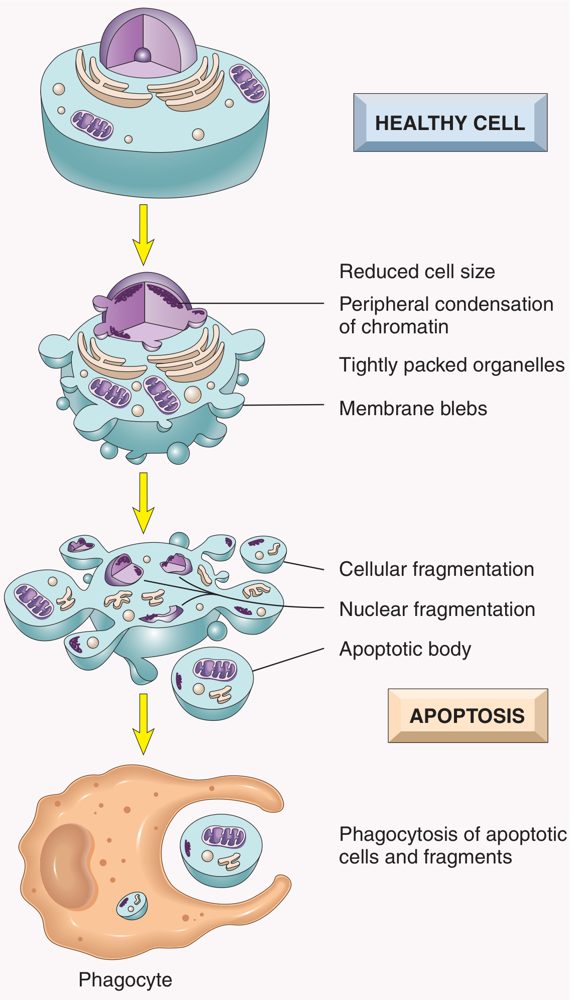
| Structure seen | What it means |
|---|---|
| Reduced cell size | Cell shrinks (opposite of necrosis = swells) |
| Peripheral condensation of chromatin | Nucleus becomes dark, crescent-shaped at periphery |
| Membrane blebs | Intact plasma membrane forms bubble-like protrusions |
| Cellular fragmentation | Cell breaks into fragments (not lysis) |
| Nuclear fragmentation | Nucleus breaks into pieces |
| Apoptotic bodies | Membrane-bound packages of cell contents |
| Phagocyte engulfs apoptotic body | No inflammation - silent death |
2. 🏷️ TOPIC: CELL INJURY - Reversible vs. Irreversible (H&E: Kidney tubules)
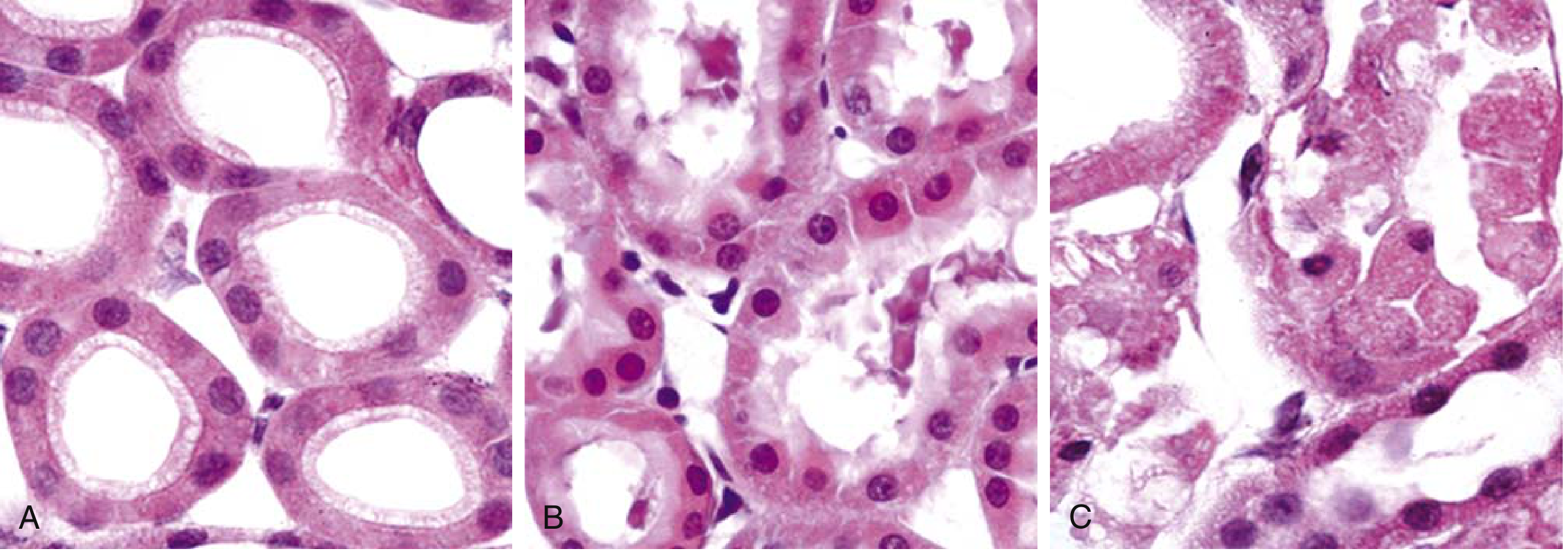
| Panel | What you see | Diagnosis |
|---|---|---|
| A (Normal) | Regular tubular cells, round nuclei, well-defined borders | Normal kidney tubule epithelium |
| B (Reversible) | Surface blebs on luminal surface, ↑ eosinophilia (pink), occasional cell swelling | Reversible ischemic injury |
| C (Irreversible/Necrosis) | Loss of nuclei (anucleate cells), fragmented cells, pink "ghost" outlines, leakage of contents | Coagulative necrosis (irreversible) |
3. 🏷️ TOPIC: COAGULATIVE NECROSIS - Kidney Infarct (H&E)
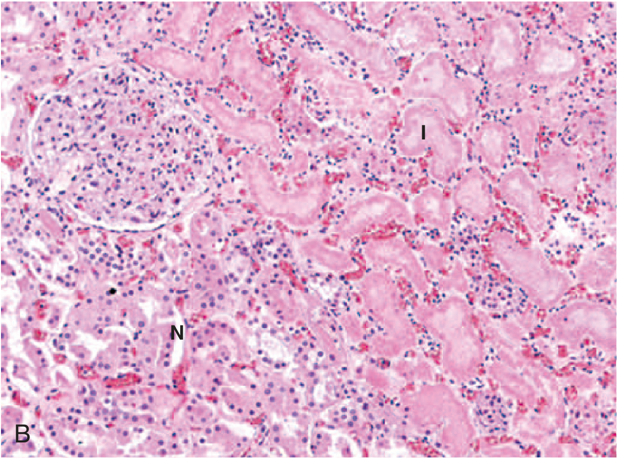
- N = Normal renal tubules (nuclei visible, structured)
- I = Infarcted/necrotic area - tubular outlines preserved but nuclei absent = ghost cells
- Also visible: glomerulus on left side (normal)
4. 🏷️ TOPIC: FIBRINOID NECROSIS - Vasculitis (H&E)
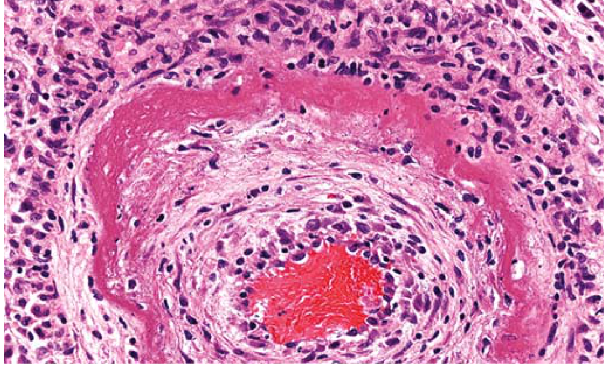
- Bright pink (eosinophilic) smudgy material in the vessel wall = fibrinoid deposits (immune complexes + plasma proteins)
- Vessel lumen (red blood cells visible centrally)
- Dense inflammatory infiltrate (dark nuclei) surrounding the vessel wall
- Destruction of normal vessel architecture
5. 🏷️ TOPIC: GRANULOMATOUS INFLAMMATION - Tuberculosis (H&E)
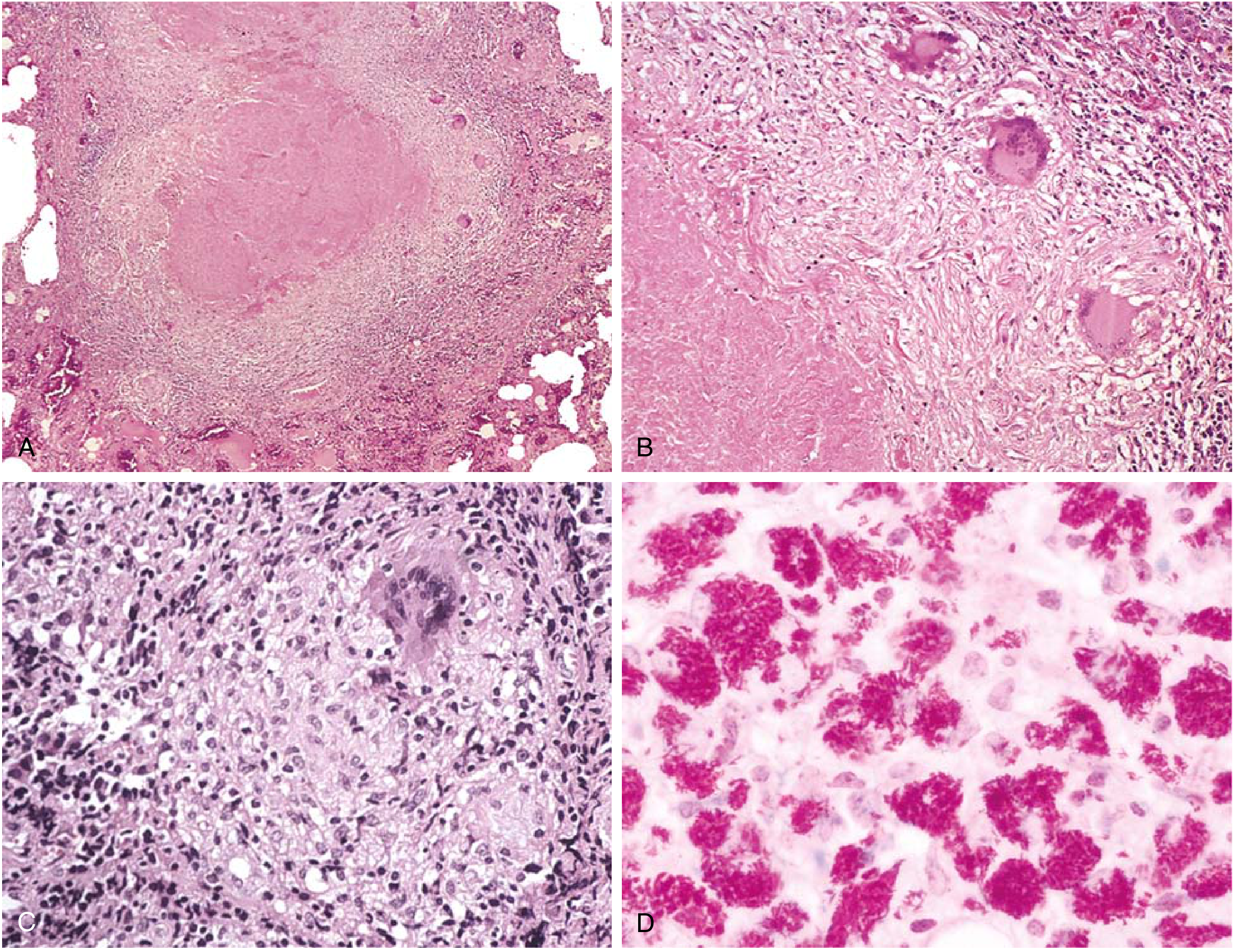
| Panel | Features | What to write |
|---|---|---|
| A (Low power) | Central pale pink area = caseous necrosis, surrounded by cellular zone | Typical TB tubercle at low power |
| B (Medium power) | Central caseous necrosis (granular, pink, amorphous), peripheral epithelioid macrophages, Langhans giant cells (round cells with multiple nuclei arranged in horseshoe/peripheral pattern) | Classic caseating granuloma |
| C (Non-caseating) | Granuloma WITHOUT central necrosis; epithelioid cells + giant cells + lymphocytes | Non-caseating granuloma (Sarcoidosis-like) |
| D (AFB/Acid-fast stain) | Bright red bacilli inside macrophages on blue background | Immunocompromised TB - macrophages packed with AFB |
6. 🏷️ TOPIC: ATHEROSCLEROSIS - Gross Lesions (Aorta)
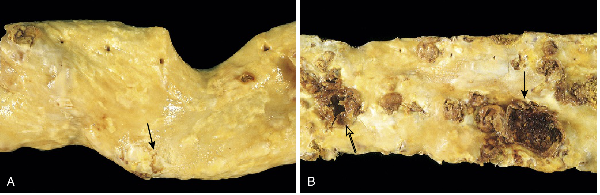
| Image A | Image B |
|---|---|
| Aorta opened longitudinally | Aorta with advanced disease |
| Raised yellowish fibrous plaque (arrow) = early atheroma | Multiple ulcerated plaques |
| Minimal luminal narrowing | Calcification (brown hard deposits) |
| Intimal surface relatively smooth elsewhere | Thrombus over ruptured plaque (dark areas) |
Normal vessel
↓
Endothelial injury (HTN, smoking, hyperlipidemia)
↓
LDL enters intima → oxidized LDL
↓
Monocytes adhere (VCAM-1) → Macrophages → engulf oxLDL
↓
FOAM CELLS → FATTY STREAK (reversible)
↓
Platelet aggregation + SMC migration from media
↓
FIBROUS PLAQUE (necrotic core + fibrous cap)
↓
Plaque rupture → THROMBOSIS → MI / Stroke
7. 🏷️ TOPIC: FATTY STREAK - Atherosclerosis Microscopy (H&E)
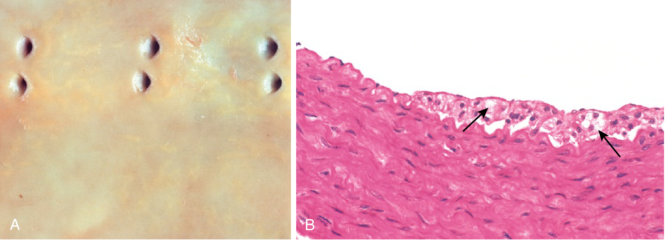
- Panel A (Gross): Yellowish linear deposits near ostia of branch vessels = fatty streaks. Earliest visible atherosclerotic lesion.
- Panel B (Microscopy): Foam cells in intima (arrows) = macrophages that have engulfed oxidized LDL. Cells appear pale with vacuolated cytoplasm (lipid washed out in processing).
8. 🏷️ TOPIC: AMYLOIDOSIS - Congo Red Staining (Polarized Light)
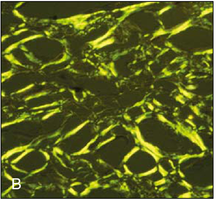
- Yellow-green (apple-green) birefringent material = AMYLOID FIBRILS
- Deposits in tissue architecture (vessel walls, glomeruli, or interstitium depending on organ)
- This is only seen under POLARIZED LIGHT after Congo red staining
- Under ordinary light: pink/red deposits; under polarized: apple-green
🗂️ SUMMARY: What to Draw & What to Write - Exam Cheat Sheet
| Slide/Diagram | Topic | Key Labels | Key Exam Line |
|---|---|---|---|
| Apoptosis diagram | Cell death | Cell shrinkage, chromatin condensation, apoptotic bodies, phagocyte | "No inflammation" - distinguishes from necrosis |
| Kidney tubules H&E (3 panels) | Reversible vs. irreversible injury | Normal → blebs → ghost cells | Ghost cells + no nuclei = coagulative necrosis |
| Coagulative necrosis kidney | Infarction | N = normal, I = infarcted; ghost tubules | Preserved architecture, lost nuclei |
| Fibrinoid necrosis artery | Vasculitis / immune complex | Bright pink amorphous vessel wall, inflammatory infiltrate | Only seen microscopically |
| TB granuloma H&E | Chronic granulomatous inflammation | Caseous necrosis, epithelioid cells, Langhans giant cells, lymphocytes | ZN stain for AFB |
| Aortic plaques (gross) | Atherosclerosis | Fatty streak → fibrous plaque → ulcerated plaque | Plaque rupture → thrombosis → MI |
| Fatty streak (micro) | Atherosclerosis (earliest) | Foam cells in intima | Vacuolated macrophages = foam cells |
| Congo red polarized | Amyloidosis | Apple-green birefringence | Pathognomonic for amyloid |
All histopathology images are from Robbins & Kumar Basic Pathology (10th ed.), accessed via the medical textbook library.
Sustemi Systemic pathology important questions with answers.
🫀🫁🧠 SYSTEMIC PATHOLOGY — IMPORTANT Q&A (MBBS)
🏷️ SYSTEM 1: CARDIOVASCULAR PATHOLOGY (CVS)
↓ Cardiac output (forward failure)
+
↑ Pulmonary venous back-pressure (backward failure)
↓
↑ Pulmonary capillary pressure → Pulmonary edema
- Frank-Starling mechanism — ventricular dilation → increased preload → increased contractility (compensated failure)
- Neurohumoral activation — norepinephrine (↑ HR, ↑ contractility), RAAS activation (salt/water retention, vasoconstriction), ANP release (counterbalances RAAS)
- Myocardial hypertrophy — new sarcomeres added; concentric (pressure overload — HTN) vs. eccentric (volume overload — regurgitation)
| Organ | Change |
|---|---|
| Lungs | Pulmonary edema, "cardiac asthma," pink frothy sputum, brown induration (hemosiderin-laden macrophages = "heart failure cells") |
| Kidneys | ↓ perfusion → prerenal azotemia; RAAS activation → fluid retention |
| Brain | Hypoxic encephalopathy |
| Organ | Change |
|---|---|
| Liver | "Nutmeg liver" (centrilobular congestion → cardiac cirrhosis) |
| Spleen | Congestive splenomegaly |
| Legs | Pitting edema (dependent) |
| Body cavities | Ascites, pleural effusion, anasarca |
- Angina pectoris — chest pain on exertion; no necrosis
- Stable angina (fixed plaque, predictable)
- Unstable angina (plaque disruption, unpredictable — "acute coronary syndrome")
- Prinzmetal/variant angina (coronary vasospasm)
- Acute Myocardial Infarction (MI) — ischemic necrosis of myocardium
- Chronic IHD / Ischemic cardiomyopathy — progressive heart failure from cumulative ischemia
- Sudden cardiac death — usually due to lethal arrhythmia from ischemia
- LAD (left anterior descending) → anterior wall + septum (40-50%)
- RCA (right coronary artery) → posterior wall + right ventricle
- LCX (left circumflex) → lateral wall
| Time | Gross | Microscopy |
|---|---|---|
| 0-4 hours | Normal (no change) | Normal / wavy fiber change |
| 4-12 hours | Pallor begins | Early coagulative necrosis, wavy fibers, ↑ eosinophilia |
| 12-24 hours | Pallor + yellow-tan | Coagulative necrosis - loss of nuclei, striations; neutrophil infiltration begins |
| 1-3 days | Yellowing with hyperemic (red) border | Dense neutrophilic infiltrate, nuclear pyknosis → karyolysis |
| 3-7 days | Soft yellow-white center, red border | Macrophage infiltration begins; dead cells cleared |
| 1-3 weeks | Vascular granulation tissue (pink-red, soft) | Granulation tissue - angiogenesis + fibroblasts + collagen |
| >6 weeks | White fibrous scar | Dense collagen scar (permanent) |
- Arrhythmia — most common cause of death in 1st hour (ventricular fibrillation)
- Cardiogenic shock — pump failure
- Mural thrombus → systemic embolism
- Cardiac rupture — days 3-7 (at softest point of healing)
- Ventricular aneurysm — paradoxical bulge on ECG
- Pericarditis — early (fibrinous, 1-3 days), late (Dressler syndrome — autoimmune, weeks later)
- Reinfarction
- Papillary muscle rupture → acute mitral regurgitation
RISK FACTORS: Hypertension, hyperlipidemia (LDL), smoking, diabetes
↓
1. ENDOTHELIAL INJURY / DYSFUNCTION
↓
2. LDL enters intima → oxidized LDL (ox-LDL)
↓
3. Monocytes adhere to endothelium (VCAM-1, ICAM-1)
→ Enter intima → Macrophages
→ Engulf ox-LDL via scavenger receptors
→ FOAM CELLS
↓
4. FATTY STREAK (reversible earliest lesion)
↓
5. SMC migration from media → intima
SMC proliferation + ECM (collagen) production
Platelet activation → growth factors (PDGF, FGF)
↓
6. FIBROUS PLAQUE = Lipid core + Fibrous cap
↓
7. VULNERABLE PLAQUE: thin fibrous cap,
large lipid core, many inflammatory cells
↓
8. PLAQUE RUPTURE → Thrombosis
↓
Acute MI / Stroke / Peripheral vascular disease
| Type | Chamber | Mechanism | Causes | Key Features |
|---|---|---|---|---|
| Dilated (DCM) | All chambers dilated; ↓ EF | Systolic dysfunction | Alcohol, viral myocarditis, idiopathic, genetic, pregnancy (PPCM) | "Flabby" heart; mural thrombi; S3 gallop |
| Hypertrophic (HCM) | Massive ventricular hypertrophy (especially septum) | Diastolic dysfunction; LVOTO | Autosomal dominant — sarcomere protein mutations (MYH7, MYBPC3) | Most common cause of sudden cardiac death in young athletes |
| Restrictive (RCM) | Normal size but stiff walls; ↓ filling | Diastolic dysfunction | Amyloidosis, sarcoidosis, hemochromatosis, fibrosis | Normal EF; ↑ filling pressures |
- Follows Group A β-hemolytic Streptococcal (GAS) pharyngitis → autoimmune reaction
- Antibodies against streptococcal M protein cross-react with cardiac proteins ("molecular mimicry")
- Pancarditis = myocarditis + pericarditis + endocarditis
- Aschoff bodies (pathognomonic) = foci of fibrinoid necrosis surrounded by activated macrophages (Aschoff cells / caterpillar cells) + lymphocytes
- Mitral valve most commonly affected (96%) → stenosis (most common valvular lesion in RHD)
- Mitral stenosis → left atrial dilation → pulmonary HTN → right heart failure
- "Fish-mouth" / "Button-hole" deformity of mitral valve on gross
- Verrucous endocarditis — small vegetations along line of closure
🏷️ SYSTEM 2: RESPIRATORY PATHOLOGY
| Type | % | Smoking | Location | Key Features |
|---|---|---|---|---|
| Adenocarcinoma | 50% (most common) | Moderate (also in non-smokers) | Peripheral | Most common in women, non-smokers, <45 yr; EGFR mutation (target: erlotinib/gefitinib); TTF-1+; Clara cell origin |
| Squamous cell carcinoma (SCC) | 20% | Strong | Central (hilar) | Arises from bronchial mucosa after squamous metaplasia → dysplasia → CIS; cavitation common; PTHrP → hypercalcemia |
| Small cell carcinoma (SCLC) | 15% | Strongest | Central | Most aggressive; neuroendocrine origin; ACTH/ADH secretion; Kulchitsky (oat) cells; never surgically resectable |
| Large cell carcinoma | 2% | Strong | Peripheral | Undifferentiated; exclusion diagnosis; poor prognosis |
- SCC → PTHrP → hypercalcemia (most common)
- SCLC → ACTH → Cushing syndrome; ADH → SIADH; anti-Hu antibodies → Lambert-Eaton syndrome
- Carcinoid → Serotonin → carcinoid syndrome
| Feature | Lobar Pneumonia | Bronchopneumonia |
|---|---|---|
| Distribution | Entire lobe (or segment) | Patchy; multiple lobules; bilateral |
| Age | Young adults | Extremes of age (infants, elderly) |
| Common organisms | Streptococcus pneumoniae (most common) | S. aureus, H. influenzae, Klebsiella, E. coli, mixed |
| Consolidation | Homogeneous lobe consolidation | Patchy foci of consolidation |
| Exudate | Intraalveolar fibrinous exudate | Purulent intra-alveolar exudate |
- Congestion (Day 1-2) — vascular engorgement, edema fluid in alveoli, few bacteria
- Red hepatization (Day 2-4) — alveoli packed with RBCs + neutrophils + fibrin; lung solid, red, liver-like
- Grey hepatization (Day 4-8) — RBCs lysed; alveoli filled with neutrophils + fibrin; grey, firm
- Resolution (Day 8+) — enzymatic digestion of exudate; macrophages clear debris; lung returns to normal
| Feature | Emphysema | Chronic Bronchitis |
|---|---|---|
| Definition | Permanent enlargement of airspaces distal to terminal bronchioles with destruction of walls | Productive cough for ≥3 months/year for ≥2 consecutive years |
| Pathology | Destruction of alveolar walls; loss of elastic recoil | Mucus gland hyperplasia in bronchi → ↑ Reid Index (gland/wall ratio >0.5) |
| Types | Centriacinar (smoking — upper lobe) / Panacinar (α1-antitrypsin deficiency — lower lobe) | - |
| "Pink puffer" vs "Blue bloater" | Pink puffer (maintain O₂ via ↑ respiratory effort, thin, barrel chest) | Blue bloater (cyanosis, edema, cor pulmonale, obese) |
| Pathogenesis | Protease-antiprotease imbalance: smoking → macrophages/neutrophils → elastase destroys alveolar walls | Smoking → mucus hypersecretion + ciliary dysfunction → infection → chronic inflammation |
| Embolus size | Effect |
|---|---|
| Massive (>60% pulmonary circulation) | Sudden death, right heart failure (acute cor pulmonale) |
| Medium | Pulmonary infarction (hemorrhagic — triangular, pleural-based) — only if collateral circulation compromised |
| Small/multiple | Pulmonary hypertension (chronic) |
🏷️ SYSTEM 3: GASTROINTESTINAL PATHOLOGY (GIT)
- Flagella — motility through viscous mucus
- Urease — converts urea → ammonia → neutralizes acid (protects bacteria, damages mucosa)
- Adhesins — binds surface foveolar cells
- Toxins (CagA, CagE) → IL-8 release → neutrophil chemotaxis → mucosal damage
H. pylori in antrum
↓
Stimulates G cells → ↑ Gastrin → ↑ Acid secretion
↓
Antral gastritis → Peptic ulcer (duodenum > gastric)
↓ (if spreads to body)
Parietal cell loss → Gastric atrophy + Intestinal metaplasia
↓
Risk of Gastric Adenocarcinoma
- Chronic inflammation → submucosal lymphoid aggregates (MALT)
- MALT lymphoma develops; may regress with H. pylori eradication
| Feature | Ulcerative Colitis (UC) | Crohn's Disease (CD) |
|---|---|---|
| Location | Colon and rectum ONLY; rectum always involved | Any part of GIT (mouth to anus); terminal ileum most common |
| Distribution | Continuous (no skip lesions) | Skip lesions (patchy, discontinuous) |
| Inflammation depth | Mucosal only (superficial) | Transmural (full thickness) |
| Gross | Pseudopolyps, ulcers, "lead-pipe" colon | Cobblestone mucosa, fat wrapping, fistulas, strictures |
| Microscopy | Crypt abscesses (cryptitis), goblet cell depletion | Non-caseating granulomas (50%), lymphoid aggregates |
| Complications | Toxic megacolon, colorectal cancer (↑ risk with long-standing UC), hemorrhage | Fistulas, abscesses, strictures/obstruction, malabsorption |
| Extraintestinal | Pyoderma gangrenosum, primary sclerosing cholangitis | Erythema nodosum, uveitis, ankylosing spondylitis |
| Cancer risk | HIGH (colorectal carcinoma) | Slightly increased |
- Alcoholic liver disease (most common in West) — fatty liver → alcoholic hepatitis → cirrhosis
- Viral hepatitis (HBV, HCV — most common in developing world)
- Non-alcoholic fatty liver disease (NAFLD/NASH)
- Biliary obstruction (primary biliary cirrhosis, sclerosing cholangitis)
- Autoimmune hepatitis
- Wilson's disease (copper accumulation), Hemochromatosis (iron accumulation)
CIRRHOSIS
│
┌────────────┼──────────────┐
↓ ↓ ↓
PORTAL HEPATO- HEPATO-
HYPERTENSION CELLULAR CELLULAR
│ FAILURE CARCINOMA
│
┌──┴────────────────────────────┐
↓ ↓ ↓ ↓
Splenomegaly Varices Ascites Caput medusae
(cytopenias) (esophageal → (SBP risk) (skin veins)
hemorrhage)
- Esophageal varices → life-threatening hemorrhage (most dangerous complication)
- Ascites → spontaneous bacterial peritonitis (SBP)
- Splenomegaly → hypersplenism → pancytopenia
- Hepatic encephalopathy → ↑ ammonia → CNS dysfunction
- Hepatorenal syndrome
Normal colon
↓ APC loss (FAP gene)
Hyperproliferative epithelium
↓ KRAS mutation
Early adenoma
↓ DCC/SMAD loss
Intermediate adenoma
↓ TP53 loss
Late adenoma / Carcinoma in situ
↓ Additional changes
INVASIVE CARCINOMA → METASTASIS (liver first via portal vein)
- Right colon — polypoid/exophytic, large, bleeds → iron-deficiency anemia
- Left colon — annular/napkin-ring → obstruction, constipation/diarrhea alternation
🏷️ SYSTEM 4: RENAL PATHOLOGY
| Feature | Nephrotic Syndrome | Nephritic Syndrome |
|---|---|---|
| Pathology | ↑ Glomerular permeability (podocyte/GBM damage) | Glomerular inflammation (proliferative) |
| Proteinuria | Massive (>3.5 g/day) - selective/non-selective | Mild-moderate (subnephrotic) |
| Hematuria | Absent or microscopic | Macroscopic hematuria (red cell casts) |
| Hypertension | Absent (or mild) | Present |
| Edema | Generalized (periorbital, pitting, ascites) | Mild |
| Hypoalbuminemia | Severe (<3 g/dL) | Mild |
| Hyperlipidemia | Yes (↑ hepatic synthesis) | No |
| Lipiduria | Yes (oval fat bodies, fatty casts) | No |
| GFR | Normal or slightly ↓ | ↓ (azotemia) |
| Disease | Key Features | Microscopy |
|---|---|---|
| Minimal Change Disease (MCD) | Most common in children (90%); idiopathic; excellent response to steroids | LM normal; EM: podocyte foot process effacement |
| Membranous Nephropathy | Most common in adults; idiopathic (anti-PLA2R Ab) or secondary (SLE, HBV, drugs) | LM: GBM thickening; EM: subepithelial deposits; "spike and dome" pattern |
| Focal Segmental Glomerulosclerosis (FSGS) | Adults, HIV, heroin nephropathy, obesity | Focal (<50% glomeruli) segmental sclerosis; podocyte foot process effacement |
| Diabetic nephropathy | Kimmelstiel-Wilson nodules; diffuse glomerulosclerosis | Nodular mesangial expansion |
| Disease | Mechanism | Key Feature |
|---|---|---|
| Post-streptococcal GN | Type III hypersensitivity; subepithelial deposits | Hump-shaped subepithelial deposits; "lumpy bumpy" IF |
| Rapidly Progressive GN (RPGN/Crescentic GN) | Crescents in Bowman's space (parietal cell + macrophage proliferation) | Anti-GBM (Goodpasture), pauci-immune (ANCA), immune complex |
| IgA Nephropathy (Berger's disease) | IgA immune complexes in mesangium | Most common GN worldwide; hematuria with URTI |
| Lupus Nephritis (WHO class) | SLE; immune complex deposition | "Wire-loop" lesions; full house IF pattern |
- Pre-renal AKI — ↓ renal perfusion (hypovolemia, shock, cardiac failure) — most common
- Intrinsic AKI — renal parenchymal damage
- Post-renal AKI — obstruction (stones, BPH, tumor)
- Ischemic ATI — shock, sepsis, surgery → tubular epithelial cell necrosis (especially PCT and medullary thick ascending limb)
- Nephrotoxic ATI — aminoglycosides, cisplatin, radiocontrast, myoglobin (crush injury/rhabdomyolysis), hemoglobin
- Initiation phase — onset; oliguria begins
- Maintenance phase — established oliguria (BUN/creatinine rise); lasts 1-2 weeks
- Recovery phase — tubular regeneration; polyuria (tubules can't concentrate yet); recovery usually complete
🏷️ SYSTEM 5: HEMATOLOGY & LYMPH NODE PATHOLOGY
LEUKEMIAS
│
┌────┴──────┐
↓ ↓
ACUTE CHRONIC
│ │
┌─┴──┐ ┌──┴──┐
↓ ↓ ↓ ↓
ALL AML CLL CML
| Feature | Acute Leukemia | Chronic Leukemia |
|---|---|---|
| Cell differentiation | Blasts >20% (immature) | Mature appearing cells |
| Onset | Abrupt | Insidious |
| Prognosis | Rapidly fatal if untreated | Slower progression |
| Bone marrow | Packed with blasts | Hypercellular with mature forms |
| Peripheral blood | Many blasts | Mature lymphocytes (CLL) or mature granulocytes (CML) |
| Feature | Hodgkin Lymphoma | Non-Hodgkin Lymphoma |
|---|---|---|
| Cell of origin | B cells (Reed-Sternberg cell) | B cells (85%) or T cells (15%) |
| Pathognomonic cell | Reed-Sternberg (RS) cell (owl-eye bilobed nuclei) | Depends on type |
| Spread | Contiguous node-to-node | Non-contiguous, widespread |
| Extranodal disease | Rare | Common |
| B symptoms | Fever, night sweats, weight loss | Less common |
| EBV association | Yes (especially mixed cellularity) | Burkitt lymphoma |
| Prognosis | Generally good (curable ~80-90%) | Variable |
- Nodular sclerosis — most common (60-70%); young women; nodular bands of fibrosis; lacunar RS cells; mediastinal mass
- Mixed cellularity — second most common; EBV association; classic RS cells
- Lymphocyte rich — best prognosis
- Lymphocyte depleted — worst prognosis; older; many RS cells
- Diffuse Large B-cell Lymphoma (DLBCL) — most common NHL in adults; aggressive; BCL6 mutations
- Follicular lymphoma — t(14;18) → BCL2 overexpression; indolent; CD10+; ↑ BCL2 = ↑ apoptosis resistance
- Burkitt lymphoma — t(8;14) → MYC overexpression; "starry sky" pattern; jaw in African children (endemic); EBV+
- Mantle cell lymphoma — t(11;14) → cyclin D1; aggressive
- Malignant plasma cells proliferate in bone marrow → replace normal marrow
- Produce monoclonal Ig (IgG most common; IgA second)
- Bence Jones protein = free light chains (κ or λ) excreted in urine
- C = hypercalCemia (bone destruction → ↑ Ca²⁺) → polyuria, constipation, confusion
- R = Renal failure (Bence Jones protein → cast nephropathy, light chain deposition)
- A = Anemia (marrow replacement)
- B = Bone pain / lesions ("punched-out" lytic lesions on X-ray; pathological fractures)
- Recurrent infections (↓ normal Ig)
- Rouleaux formation of RBCs on blood smear
- ↑ ESR, ↑ total protein, ↑ β2-microglobulin
- "Rain-drop skull" on X-ray (punched-out lytic lesions)
- Primary amyloidosis (AL type) in ~10%
- Serum protein electrophoresis → M-spike (monoclonal band)
- Bone marrow biopsy → >10% plasma cells
- Urine Bence Jones protein (heat test — precipitates at 56°C, dissolves at 100°C)
🏷️ SYSTEM 6: ENDOCRINE PATHOLOGY
| Feature | Type 1 DM (T1DM) | Type 2 DM (T2DM) |
|---|---|---|
| Onset | Childhood/adolescence (usually) | Adults (increasingly also children) |
| Body habitus | Normal weight or weight loss | Obese (80%) |
| Mechanism | Autoimmune destruction of β-cells → absolute insulin deficiency | Insulin resistance + relative β-cell failure |
| Insulin levels | Very low/absent | Elevated (early) → normal/low (late) |
| Autoantibodies | Anti-GAD65, anti-IA2, anti-Znt8 antibodies | None |
| HLA | HLA-DR3, DR4 (MHC class II) | No HLA linkage |
| Pathology | Insulitis (T-cell + macrophage infiltrate) → β-cell depletion + islet atrophy | Amyloid deposition in islets (IAPP); mild β-cell depletion |
| DKA risk | HIGH (no insulin → ketoacidosis) | Low; hyperosmolar non-ketotic coma more common |
| Treatment | Insulin required | Lifestyle → oral hypoglycemics → insulin |
DIABETES MELLITUS
│
┌────┴────────────────────────────────────────┐
↓ ↓
MICROVASCULAR MACROVASCULAR
│ │
├─ Diabetic nephropathy (Kimmelstiel- ├─ Coronary artery disease
│ Wilson nodules → ESRD) ├─ Stroke
├─ Diabetic retinopathy (leading cause └─ Peripheral vascular disease
│ of new blindness in adults)
└─ Diabetic neuropathy (glove-stocking
pattern; autonomic neuropathy)
Also: Diabetic foot (neuropathy + vascular disease + infection)
Increased susceptibility to infections
- Diffuse hyperplasia of thyroid (diffuse goiter)
- Tall follicular cells with papillary infolding
- Colloid appears scalloped/pale ("moth-eaten")
- Lymphocytic infiltration
- Hyperthyroidism — weight loss, heat intolerance, tachycardia, tremor, ↑ BMR
- Exophthalmos (proptosis) — lymphocytic infiltration + glycosaminoglycan deposition in orbital fat
- Pretibial myxedema — thickening of skin over tibia (localized myxedema)
🏷️ SYSTEM 7: CNS PATHOLOGY
| Type | Artery/Vessel | Cause | Features |
|---|---|---|---|
| Extradural (Epidural) | Middle meningeal artery | Temporal bone fracture | Lucid interval then rapid deterioration; lens/biconvex hematoma on CT; ARTERIAL bleed |
| Subdural | Bridging cortical veins | Head injury (elderly, infants — shaken baby) | Crescent-shaped hematoma; no lucid interval; VENOUS bleed; slow accumulation |
| Subarachnoid | Berry (saccular) aneurysm (most common) | Spontaneous or trauma | "Worst headache of life"; blood in CSF (xanthochromia); Kernig's/Brudzinski's signs |
| Intracerebral (Intraparenchymal) | Cerebral vessels | Hypertension (most common) | Basal ganglia/internal capsule most common site; devastating |
- At branching points of Circle of Willis
- Most common: junction of anterior communicating artery with ACA
- Associated with polycystic kidney disease, coarctation of aorta, Marfan's syndrome
-
Amyloid plaques (Senile/neuritic plaques):
- Extracellular deposits of Aβ (amyloid β-peptide) derived from APP (amyloid precursor protein)
- Core of amyloid surrounded by dystrophic (swollen) neurites and reactive astrocytes
- Congo red positive
-
Neurofibrillary tangles (NFTs):
- Intracellular bundles of hyperphosphorylated tau protein (normally stabilizes microtubules)
- Silver stain positive; "flame-shaped" in pyramidal neurons
- NFT burden correlates better with dementia severity than plaque count
- Cortical atrophy (especially temporal, parietal, frontal lobes)
- Widened sulci + narrowed gyri
- Dilated ventricles (hydrocephalus ex vacuo)
🏷️ QUICK REFERENCE: SYSTEMIC PATHOLOGY HIGH-YIELD ONE-LINERS
| Fact | Answer |
|---|---|
| Most common cause of MI | Left anterior descending (LAD) artery occlusion |
| Aschoff bodies are pathognomonic of | Rheumatic heart disease |
| Most common valvular lesion in RHD | Mitral stenosis |
| Heart failure cells (hemosiderin-laden macrophages) found in | Lungs (in left heart failure) |
| "Nutmeg liver" = | Right heart failure (centrilobular congestion) |
| Most common cause of sudden death in young athletes | Hypertrophic cardiomyopathy (HCM) |
| Most common primary lung tumor | Adenocarcinoma |
| Small cell carcinoma → ectopic ACTH → | Cushing syndrome |
| SCC of lung → PTHrP → | Hypercalcemia |
| Reid index >0.5 | Chronic bronchitis |
| α1-antitrypsin deficiency → | Panacinar emphysema (lower lobes) |
| Most common cause of hepatic cirrhosis (West) | Alcoholic liver disease |
| H. pylori is Group I carcinogen → | Gastric adenocarcinoma + MALT lymphoma |
| Pseudopolyps in IBD | Ulcerative colitis |
| Skip lesions + transmural + granulomas | Crohn's disease |
| Kimmelstiel-Wilson nodules | Diabetic nephropathy |
| Foot process effacement only on EM | Minimal change disease (MCD) |
| "Starry sky" pattern on histology | Burkitt lymphoma |
| t(9;22) Philadelphia chromosome | CML (BCR-ABL); also ALL (poor prognosis) |
| t(14;18) → BCL2 overexpression | Follicular lymphoma |
| t(8;14) → MYC | Burkitt lymphoma |
| Auer rods | AML (myeloblasts) |
| Reed-Sternberg cells (owl eyes) | Hodgkin lymphoma |
| Most common HL subtype | Nodular sclerosis |
| M-spike on electrophoresis + lytic bone lesions | Multiple myeloma |
| Bence Jones protein | Free light chains in multiple myeloma |
| Berry aneurysm → worst headache of life | Subarachnoid hemorrhage |
| Lucid interval + temporal fracture | Extradural/Epidural hematoma |
| Amyloid plaques + NFTs | Alzheimer's disease |
| Tau hyperphosphorylation | Neurofibrillary tangles (Alzheimer's) |
| ApoE-ε4 allele | Sporadic Alzheimer's risk |
| Insulitis (T-cell infiltrate) | Type 1 DM |
| Amyloid in islets of Langerhans | Type 2 DM |
| TSI (anti-TSH receptor Ab) | Graves' disease |
Make flashcards or mind maps of the high-yield one-liners from this guide
Which format do you want? · How do you want the output?
A clean, colorful medical mind map on white background titled "CVS PATHOLOGY — High-Yield One-Liners". Central node: "CARDIOVASCULAR" in red circle. Six branches radiating out, each with bold label and answer text: Branch 1 (blue): "MI → Most common artery" → "LAD (Left Anterior Descending)". Branch 2 (green): "Aschoff bodies" → "Rheumatic Heart Disease". Branch 3 (orange): "Most common valvular lesion in RHD" → "Mitral Stenosis". Branch 4 (purple): "Heart failure cells in lungs" → "Hemosiderin-laden macrophages". Branch 5 (red): "Nutmeg liver" → "Right Heart Failure". Branch 6 (teal): "Sudden death in young athletes" → "Hypertrophic Cardiomyopathy (HCM)". Each branch has a small colored circle node. Arrows connect question to answer. Medical education style, clean sans-serif font, no clutter.
A clean, colorful medical mind map on white background titled "RESPIRATORY & GIT PATHOLOGY — High-Yield One-Liners". Central node: "RESP + GIT" in dark green circle. Eight branches: Branch 1 (blue): "Most common primary lung tumor" → "Adenocarcinoma". Branch 2 (red): "Small cell carcinoma + ectopic ACTH" → "Cushing Syndrome". Branch 3 (orange): "SCC lung + PTHrP" → "Hypercalcemia". Branch 4 (purple): "Reid Index >0.5" → "Chronic Bronchitis". Branch 5 (teal): "α1-antitrypsin deficiency" → "Panacinar emphysema (lower lobes)". Branch 6 (green): "Most common cause of cirrhosis (West)" → "Alcoholic liver disease". Branch 7 (pink): "H. pylori → Group I carcinogen" → "Gastric adenocarcinoma + MALT lymphoma". Branch 8 (brown): "Skip lesions + Transmural + Granulomas" → "Crohn's Disease". Each branch has bold question text and answer. Clean medical education diagram, white background, colored connecting lines.
A clean, colorful medical mind map on white background titled "RENAL & HEMATOLOGY PATHOLOGY — High-Yield One-Liners". Central node: "RENAL + HAEM" in dark blue circle. Eight branches radiating: Branch 1 (blue): "Kimmelstiel-Wilson nodules" → "Diabetic Nephropathy". Branch 2 (green): "Foot process effacement on EM ONLY" → "Minimal Change Disease (MCD)". Branch 3 (red): "Starry sky pattern" → "Burkitt Lymphoma". Branch 4 (orange): "t(9;22) Philadelphia chromosome" → "CML / ALL (poor prognosis)". Branch 5 (purple): "t(14;18) → BCL2 overexpression" → "Follicular Lymphoma". Branch 6 (teal): "t(8;14) → MYC" → "Burkitt Lymphoma". Branch 7 (pink): "Auer rods in blasts" → "AML". Branch 8 (brown): "Reed-Sternberg cells (owl-eye nuclei)" → "Hodgkin Lymphoma". Clean white background, medical education style, bold labels, colored branches with arrows.
A clean, colorful medical mind map on white background titled "NEOPLASIA, ENDOCRINE & CNS PATHOLOGY — High-Yield One-Liners". Central node: "NEOPLASIA + ENDO + CNS" in dark purple circle. Eight branches: Branch 1 (red): "Most common HL subtype" → "Nodular Sclerosis". Branch 2 (blue): "M-spike + lytic bone lesions" → "Multiple Myeloma". Branch 3 (green): "Bence Jones protein" → "Free light chains in myeloma". Branch 4 (orange): "Berry aneurysm → worst headache of life" → "Subarachnoid Hemorrhage". Branch 5 (purple): "Lucid interval + temporal fracture" → "Epidural Hematoma". Branch 6 (teal): "Amyloid plaques + NFTs" → "Alzheimer's Disease". Branch 7 (pink): "Insulitis (T-cell infiltrate in islets)" → "Type 1 DM". Branch 8 (brown): "Amyloid in islets + TSI antibodies" → "Type 2 DM / Graves' Disease". Bold text, clean sans-serif font, colored branches with small circle nodes, medical education style.
CVS drugs classification
💊 CVS DRUGS — COMPLETE CLASSIFICATION
🏷️ GROUP 1: ANTIHYPERTENSIVE DRUGS
Classification by Mechanism:
ANTIHYPERTENSIVE DRUGS
│
┌─────┼──────────────────────────────────┐
↓ ↓ ↓ ↓ ↓
DIUR- RAAS SYMPATH- CALCIUM DIRECT
ETICS DRUGS OLYTICS CHANNEL VASODILATORS
BLOCKERS
A. DIURETICS
| Class | Drugs | Mechanism | Use |
|---|---|---|---|
| Thiazides | Hydrochlorothiazide (HCTZ), Chlorthalidone, Indapamide | Block NaCl co-transporter in distal tubule | First-line HTN; mild HF |
| Loop diuretics | Furosemide, Bumetanide, Torsemide, Ethacrynic acid | Block Na-K-2Cl in thick ascending loop of Henle | Acute pulmonary edema, severe HF, hypercalcemia |
| K-sparing | Spironolactone, Eplerenone (aldosterone antagonists); Amiloride, Triamterene (ENaC blockers) | Block aldosterone receptor or ENaC | Hyperaldosteronism, HF (spironolactone reduces mortality), prevent K loss |
| Osmotic | Mannitol | Osmotic effect in tubule | Cerebral edema, raised ICP (not for HTN) |
| Carbonic anhydrase inhibitors | Acetazolamide | Block CA → ↓ H⁺/Na⁺ exchange | Glaucoma, altitude sickness (not primary HTN) |
Mnemonic for loop diuretics: "BEEF" — Bumetanide, Ethacrynic acid, furosEmide, Furosemide (Lasix)
B. RAAS (Renin-Angiotensin-Aldosterone System) Drugs
| Class | Drugs | Mechanism | Key Features |
|---|---|---|---|
| ACE Inhibitors (ACEi) | Captopril, Enalapril, Lisinopril, Ramipril, Perindopril | Block ACE → ↓ Angiotensin II + ↑ Bradykinin | 1st line in DM nephropathy, HF, post-MI; SE: dry cough (bradykinin), angioedema, hyperkalemia |
| ARBs (AT1 blockers) | Losartan, Valsartan, Irbesartan, Candesartan, Olmesartan | Block AT1 receptor directly | Same indications as ACEi; no cough (no bradykinin ↑); preferred if ACEi cough |
| Direct Renin Inhibitor | Aliskiren | Block renin enzyme directly | Rarely used; avoid in pregnancy; CI with ACEi/ARB |
| Aldosterone Antagonists | Spironolactone, Eplerenone | Block mineralocorticoid receptor | HF (mortality benefit), hyperaldosteronism |
ACEi mnemonic: "CELLAR PREP" — Captopril, Enalapril, Lisinopril, Lisinopril, Altace (Ramipril), Ramipril, Perindopril, Enalapril, Benazepril, Perindopril
C. SYMPATHOLYTICS
| Class | Drugs | Mechanism | Notes |
|---|---|---|---|
| β-blockers (non-selective) | Propranolol, Nadolol, Timolol, Carvedilol (α+β) | Block β1 + β2 receptors | CI: asthma, COPD, peripheral vascular disease |
| β-blockers (selective β1) | Metoprolol, Atenolol, Bisoprolol, Nebivolol | Block β1 (cardioselective) | Preferred in asthmatics (relatively safe); Post-MI; HF |
| α1-blockers | Prazosin, Doxazosin, Terazosin | Block α1 → vasodilation | Hypertension + BPH; SE: first-dose syncope |
| Central α2 agonists | Clonidine, Methyldopa | Stimulate central α2 → ↓ sympathetic outflow | Methyldopa = safe in pregnancy; Clonidine: rebound HTN on abrupt withdrawal |
| Ganglionic blockers | Hexamethonium, Mecamylamine | Block nicotinic Nn receptors | Rarely used today |
| Adrenergic neuron blockers | Reserpine, Guanethidine | Deplete NE from nerve terminals | Rarely used; severe depression (reserpine) |
D. CALCIUM CHANNEL BLOCKERS (CCBs)
| Sub-class | Drugs | Selectivity | Uses |
|---|---|---|---|
| Dihydropyridines (DHPs) | Amlodipine, Nifedipine, Felodipine, Nimodipine, Nicar-dipine | Vascular smooth muscle >> cardiac | HTN, angina (Amlodipine = 1st line); Nimodipine = cerebral vasospasm after SAH |
| Non-DHPs | Verapamil (phenylalkylamine) | Heart > vessels | SVT, rate control in AF, angina; ↓ HR + contractility |
| Non-DHPs | Diltiazem (benzothiazepine) | Heart + vessels (intermediate) | SVT, AF rate control, angina |
Memory trick:
- "Nifedipine = Nipple" = vascular (peripheral) → used in HTN
- Verapamil = "Very cardiac"
- Diltiazem = in between
E. DIRECT VASODILATORS
| Drug | Mechanism | Use | SE |
|---|---|---|---|
| Hydralazine | ↑ cGMP → relaxes arterioles | Hypertensive emergency in pregnancy | Reflex tachycardia, lupus-like syndrome (SLE) |
| Minoxidil | Opens K⁺ channels → hyperpolarizes → vasodilation | Severe refractory HTN | Hypertrichosis (hair growth — topical use), fluid retention, tachycardia |
| Sodium nitroprusside | Releases NO → ↑ cGMP | Hypertensive crisis (IV) | Cyanide toxicity (contains CN⁻) |
| Diazoxide | Opens K⁺ channels | Hypertensive emergency; hypoglycemia | Hyperglycemia (blocks insulin release) |
F. DRUGS FOR HYPERTENSIVE EMERGENCIES
| Drug | Route | Use |
|---|---|---|
| Sodium nitroprusside | IV infusion | All hypertensive crises |
| Labetalol | IV | Hypertensive emergencies, aortic dissection |
| Nicardipine | IV | Hypertensive emergency + neuro patients |
| Hydralazine | IV/IM | Hypertension in pregnancy (eclampsia) |
| Methyldopa | Oral | HTN in pregnancy (chronic) |
🏷️ GROUP 2: ANTIARRHYTHMIC DRUGS
Singh-Vaughan Williams Classification (Most Important for Exams)
CLASS I → Na⁺ Channel Blockers
CLASS II → β-Blockers
CLASS III → K⁺ Channel Blockers (prolong APD)
CLASS IV → Ca²⁺ Channel Blockers
+ MISC: Adenosine, Digoxin, Magnesium
CLASS I — Sodium Channel Blockers
| Sub-class | Drugs | Action Potential | Kinetics | Uses |
|---|---|---|---|---|
| Class IA | Quinidine, Procainamide, Disopyramide | Prolongs APD & QT | Intermediate | AF, VT, WPW; Quinidine → "cinchonism" (tinnitus, headache); Disopyramide → anticholinergic SE |
| Class IB | Lidocaine, Mexiletine, Phenytoin | Shortens APD | Fast | Post-MI VT/VF (IV lidocaine); oral mexiletine for chronic VT |
| Class IC | Flecainide, Propafenone, Encainide | No change in APD | Slow | SVT, AF; proarrhythmic (CAST trial) — CI post-MI |
Mnemonic Class IA: "Q-PD" — Quinidine, Procainamide, Disopyramide Mnemonic Class IB: "Likely MexPheny" — Lidocaine, Mexiletine, Phenytoin Mnemonic Class IC: "FPE" — Flecainide, Propafenone, Encainide
CLASS II — β-Blockers
| Drugs | Use |
|---|---|
| Propranolol, Metoprolol, Atenolol, Esmolol (IV, ultra-short acting) | SVT, rate control in AF/atrial flutter, post-MI VT prevention, AVNRT |
CLASS III — Potassium Channel Blockers (Prolong APD/QT)
| Drug | Key Features | Use |
|---|---|---|
| Amiodarone | Most powerful; all 4 classes; iodine-containing; longest t½ (40-55 days) | VT, VF, AF — drug of choice for most arrhythmias; SE: pulmonary fibrosis, thyroid (hypo/hyper), photosensitivity, corneal microdeposits, liver toxicity, grey-blue skin |
| Sotalol | β-blocker + Class III (prolongs QT) | AF, VT |
| Dronedarone | Amiodarone analog (no iodine, less toxic) | AF; CI in severe HF |
| Dofetilide | Pure IKr blocker; prolongs QT | AF cardioversion/maintenance |
| Ibutilide | IV only; prolongs QT | Acute AF/flutter cardioversion |
CLASS IV — Calcium Channel Blockers (Non-DHP)
| Drug | Use |
|---|---|
| Verapamil | AVNRT, SVT, rate control in AF — drug of choice for SVT |
| Diltiazem | SVT, AF rate control |
MISCELLANEOUS ANTIARRHYTHMICS
| Drug | Mechanism | Use | Notes |
|---|---|---|---|
| Adenosine | Activates A1 receptors → ↑ K⁺ → hyperpolarizes SA/AV node | Drug of choice for acute SVT/AVNRT (terminates) | IV bolus, t½ = 10 sec; SE: transient flushing, chest tightness, AV block |
| Digoxin | Na/K ATPase inhibitor → ↑ intracellular Ca²⁺ → positive inotropy; also vagomimetic → ↓ HR | Rate control in AF + HF | Narrow therapeutic index; toxicity: visual disturbances (yellow-green), nausea, arrhythmias |
| Magnesium sulphate | Blocks NMDA + Ca²⁺ channels | Torsades de Pointes (drug of choice), digitalis toxicity, eclampsia | IV infusion |
| Atropine | Muscarinic antagonist | Bradycardia, AV block | IV for acute bradycardia |
🏷️ GROUP 3: ANTIANGINAL DRUGS
Three Drug Groups:
ANTIANGINAL DRUGS
│
┌────┼────────────────────────┐
↓ ↓ ↓
NITRATES β-BLOCKERS CALCIUM CHANNEL
BLOCKERS
| Class | Drugs | Mechanism | Use |
|---|---|---|---|
| Organic Nitrates | GTN (nitroglycerin), Isosorbide dinitrate (ISDN), Isosorbide mononitrate (ISMN) | Donate NO → ↑ cGMP → venodilation (↓ preload) >> arterodilation (↓ afterload) | Acute angina (GTN sublingual), prophylaxis (ISMN long-acting) |
| β-blockers | Metoprolol, Atenolol, Propranolol | ↓ HR, ↓ contractility → ↓ O₂ demand | Stable/unstable angina, post-MI |
| CCBs | Amlodipine (stable angina), Verapamil/Diltiazem (vasospastic angina) | Vasodilation, ↓ HR (non-DHP) | Stable angina, Prinzmetal/variant angina |
| Ranolazine | Blocks late Na⁺ current → ↓ intracellular Ca²⁺ → ↓ wall tension | Chronic angina (adjunct) | Does NOT lower BP or HR |
| Ivabradine | Blocks If (funny current) in SA node → ↓ HR without affecting contractility | Stable angina + HFrEF | Useful when β-blockers CI |
Nitrate tolerance: Develops with continuous use; prevented by nitrate-free interval (8-12 hrs). GTN sublingual = onset 2 min, duration 30 min.
🏷️ GROUP 4: DRUGS USED IN HEART FAILURE
HEART FAILURE DRUGS
│
┌────┼────────────────────────────┐
↓ ↓ ↓ ↓
RAAS DIUR- β-BLOCKERS POSITIVE
DRUGS ETICS INOTROPES +
OTHERS
| Drug | Class | Mechanism | Mortality benefit? |
|---|---|---|---|
| ACE Inhibitors (Enalapril, Lisinopril) | RAAS | ↓ Ang II, ↓ aldosterone, ↑ bradykinin → vasodilation + ↓ remodeling | YES |
| ARBs (Valsartan, Losartan) | RAAS | Block AT1 | YES (when ACEi not tolerated) |
| Sacubitril/Valsartan (Entresto) | ARNi (ARB + Neprilysin inhibitor) | ↑ BNP (vasodilatory) + ↓ AT1 | YES (superior to ACEi) |
| Spironolactone / Eplerenone | Aldosterone antagonist | Block aldosterone → ↓ fibrosis + K-sparing | YES |
| Carvedilol, Metoprolol, Bisoprolol | β-blockers | ↓ sympathetic activation → ↓ remodeling | YES |
| Furosemide | Loop diuretic | Relieve congestion (↓ preload) | Symptom relief only |
| Digoxin | Cardiac glycoside | Na/K ATPase inhibitor → ↑ Ca²⁺ → ↑ contractility; also ↑ vagal tone → ↓ HR | Reduces hospitalizations; NO mortality benefit |
| Ivabradine | If channel blocker | ↓ HR | YES (in HFrEF with HR >70) |
| SGLT2 inhibitors (Dapagliflozin, Empagliflozin) | Glucose/sodium co-transporter | Osmotic diuresis + ↓ cardiac preload/afterload | YES (latest - major benefit) |
| Hydralazine + Nitrates | Vasodilators | ↓ Preload + afterload | YES (in Black patients intolerant of ACEi) |
| Dobutamine | β1 agonist | ↑ contractility | Acute decompensated HF (short-term IV) |
| Milrinone | PDE3 inhibitor | ↑ cAMP → ↑ contractility + vasodilation | Acute HF, bridge to transplant |
🏷️ GROUP 5: ANTITHROMBOTIC DRUGS
ANTITHROMBOTIC DRUGS
│
┌─────┼─────────────┐
↓ ↓ ↓
ANTI- ANTI- THROMBO-
PLATE- COAG- LYTICS
LETS ULANTS
A. ANTIPLATELET DRUGS
| Drug | Mechanism | Use |
|---|---|---|
| Aspirin | Irreversibly blocks COX-1 → ↓ TXA2 | ACS, post-MI, stroke prevention; low dose 75-100 mg |
| Clopidogrel | Irreversibly blocks P2Y12 ADP receptor | ACS, PCI (stent), stroke; prodrug (CYP2C19 activation) |
| Ticagrelor | Reversibly blocks P2Y12 receptor | ACS (more potent than clopidogrel); direct acting |
| Prasugrel | Irreversibly blocks P2Y12 | ACS + PCI; CI in prior TIA/stroke |
| Abciximab, Eptifibatide, Tirofiban | GP IIb/IIIa receptor blockers | ACS, PCI (IV use) |
| Dipyridamole | PDE inhibitor → ↑ cAMP; also adenosine uptake inhibitor | Stroke prevention (with aspirin = Aggrenox) |
B. ANTICOAGULANTS
| Drug | Mechanism | Route | Key Points |
|---|---|---|---|
| Unfractionated Heparin (UFH) | Activates antithrombin III → inhibits IIa (thrombin) + Xa | IV/SC | Monitor aPTT; antidote = Protamine sulphate; HIT (thrombocytopenia) |
| LMWH (Enoxaparin, Dalteparin) | Activates AT-III → mainly inhibits Xa | SC | More predictable; no monitoring needed; preferred in pregnancy; antidote = Protamine (partial) |
| Fondaparinux | Synthetic pentasaccharide; selectively inhibits Xa via AT-III | SC | No HIT risk; no antidote |
| Warfarin | Inhibits Vitamin K epoxide reductase → ↓ clotting factors II, VII, IX, X + protein C, S | Oral | Monitor PT/INR; antidote = Vitamin K (slow) or FFP (fast); many drug interactions |
| Dabigatran | Direct thrombin (IIa) inhibitor | Oral | DOAC; antidote = Idarucizumab; AF, DVT/PE |
| Rivaroxaban, Apixaban, Edoxaban | Direct factor Xa inhibitors | Oral | DOACs; antidote = Andexanet alfa; AF, DVT/PE, ACS |
Warfarin vs. Heparin:
- Heparin = parenteral, immediate onset, used in ACUTE settings
- Warfarin = oral, delayed onset (3-5 days), used for CHRONIC anticoagulation
- Bridge therapy = use both during transition
C. THROMBOLYTICS (FIBRINOLYTICS)
| Drug | Mechanism | Use | SE |
|---|---|---|---|
| Streptokinase | Activates plasminogen → plasmin; antigenic (1st generation) | STEMI, DVT, PE | Allergic reactions, bleeding |
| Alteplase (tPA) | Recombinant tPA; fibrin-specific (2nd generation) | STEMI, ischemic stroke (within 4.5 hr), massive PE | Intracranial hemorrhage |
| Tenecteplase, Reteplase | Modified tPA; longer t½ (3rd generation) | STEMI (single IV bolus) | Bleeding |
Antidote to thrombolytics: Aminocaproic acid (inhibits fibrinolysis)
🏷️ GROUP 6: LIPID-LOWERING DRUGS (ANTIHYPERLIPIDEMICS)
| Class | Drugs | Mechanism | Effect | Use |
|---|---|---|---|---|
| Statins (HMG-CoA reductase inhibitors) | Atorvastatin, Rosuvastatin, Simvastatin, Pravastatin, Lovastatin | Block HMG-CoA reductase → ↓ cholesterol synthesis → ↑ LDL receptor expression | ↓↓ LDL (primary), ↓ TG, ↑ HDL | 1st line for hypercholesterolemia, atherosclerosis prevention; SE: myopathy/rhabdomyolysis, ↑ LFTs |
| PCSK9 inhibitors | Evolocumab, Alirocumab | Block PCSK9 → ↑ LDL receptor recycling → ↓ LDL | ↓↓↓ LDL | Familial hypercholesterolemia, statin-intolerant patients |
| Fibrates | Fenofibrate, Gemfibrozil, Bezafibrate | Activate PPARα → ↑ lipoprotein lipase → ↑ TG clearance | ↓↓ TG, ↑ HDL, ↓ LDL (modest) | Hypertriglyceridemia; SE: myopathy (↑ risk with statins) |
| Niacin (Nicotinic acid) | Inhibits lipolysis in adipose → ↓ VLDL → ↓ LDL + ↑ HDL | ↑↑ HDL, ↓ TG, ↓ LDL | Now rarely used; SE: flushing (↓ by aspirin pretreatment), hyperglycemia, gout | |
| Ezetimibe | Blocks NPC1L1 transporter → ↓ intestinal cholesterol absorption | ↓ LDL (10-20%) | Used with statins; SE minimal | |
| Bile acid sequestrants | Cholestyramine, Colestipol, Colesevelam | Bind bile acids in gut → ↑ bile acid synthesis → ↑ LDL receptor | ↓ LDL | ↑ TG (contraindicated in hypertriglyceridemia); SE: constipation, GI |
| Omega-3 fatty acids | Icosapentaenoic acid (EPA) | ↓ VLDL synthesis | ↓ TG | Severe hypertriglyceridemia |
🏷️ QUICK SUMMARY TABLE — CVS DRUGS AT A GLANCE
| Indication | Drug of Choice / First-Line |
|---|---|
| Hypertension (uncomplicated) | Thiazide / ACEi / ARB / Amlodipine |
| HTN + Diabetes / CKD | ACEi or ARB |
| HTN + Heart failure | ACEi + β-blocker + Aldosterone antagonist |
| HTN + Angina | β-blocker or CCB |
| HTN in pregnancy | Methyldopa (chronic), Hydralazine/Labetalol (acute) |
| Hypertensive crisis | Sodium nitroprusside IV |
| Acute MI (STEMI) | Aspirin + Clopidogrel + Thrombolysis / PCI |
| Stable angina | GTN (acute) + β-blocker (prophylaxis) |
| Prinzmetal/Vasospastic angina | CCB (Amlodipine); β-blockers contraindicated |
| Acute SVT | Adenosine IV (first-line) |
| AF rate control | β-blockers / Diltiazem / Verapamil / Digoxin |
| AF rhythm control | Amiodarone / Flecainide |
| VT/VF | Amiodarone (first-line antiarrhythmic) |
| Torsades de Pointes | IV Magnesium sulphate |
| Heart failure (HFrEF) | ACEi/ARBi + β-blocker + Aldosterone antagonist + SGLT2i |
| DVT/PE treatment (acute) | LMWH → DOAC (rivaroxaban/apixaban) |
| Long-term anticoagulation (AF) | DOAC (Dabigatran/Rivaroxaban/Apixaban) or Warfarin |
| Ischemic stroke (thrombolysis) | Alteplase (tPA) within 4.5 hrs |
| High LDL / Dyslipidemia | Statin (1st line always) |
| High Triglycerides | Fibrate or Omega-3 |
| Familial hypercholesterolemia | High-dose statin + PCSK9 inhibitor |
🏷️ KEY DRUG ANTIDOTES — CVS (Must Know!)
| Drug | Antidote |
|---|---|
| Heparin (UFH/LMWH) | Protamine sulphate |
| Warfarin | Vitamin K (slow, 12-24 hrs) / FFP (immediate) |
| Dabigatran | Idarucizumab |
| Rivaroxaban / Apixaban | Andexanet alfa |
| Fibrinolytics (Streptokinase, tPA) | Aminocaproic acid |
| Digoxin toxicity | Digoxin-specific antibody fragments (Digibind) |
| Nitroprusside (cyanide toxicity) | Sodium thiosulfate + Hydroxocobalamin |