
Tension pneumothorax in surgery
Reading File
Now I'll search the medical library and supplementary sources in parallel.
Finding Sources
Finding Sources
Searching Images
tension pneumothorax chest X-ray diagram tracheal deviation
Excellent results. Now I'll read the most relevant surgical and emergency sections in parallel:
Reading File
Reading File
Reading File
Good data. Let me fetch the CXR image and read the Bailey & Love section continuation plus the Roberts & Hedges section for needle decompression details:
Loading Image
Reading File
Reading File
I now have comprehensive data from multiple surgical and emergency medicine textbooks. Let me also fetch the pathophysiology diagram:
Loading Image
Searching PubMed
"tension pneumothorax" needle decompression management
I now have all the information needed for a thorough, well-illustrated answer.
Tension Pneumothorax in Surgery
Tension pneumothorax is the most life-threatening of all respiratory emergencies encountered in surgical practice. It is one of the six immediately life-threatening chest injuries in the "deadly dozen" described by ATLS.
Pathophysiology
A tension pneumothorax develops when a one-way valve mechanism allows air to enter the pleural space during inspiration but prevents its escape during expiration. Air progressively accumulates under increasing pressure, leading to:
- Complete ipsilateral lung collapse - abolishing oxygenation and ventilation on that side
- Mediastinal shift away from the affected side - compressing the contralateral lung
- Compression of the superior and inferior vena cava - severely reducing venous return (preload)
- Falling cardiac output - resulting in obstructive shock and cardiorespiratory arrest if untreated
The diagram below illustrates the one-way valve mechanism during inspiration (air enters) and expiration (valve closes, trapping air):
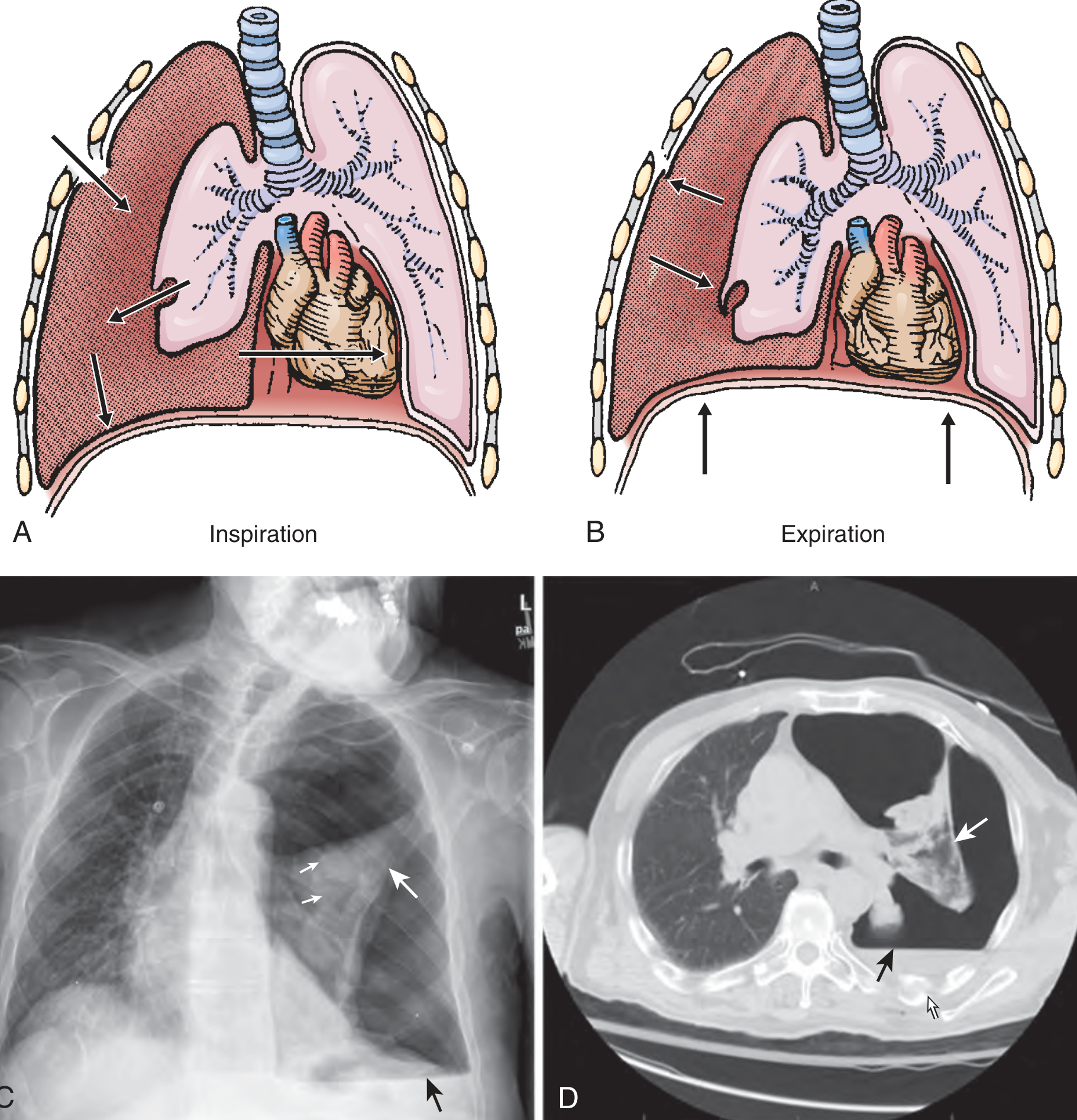
Roberts and Hedges' Clinical Procedures in Emergency Medicine, p. 264
Causes in the Surgical Context
| Category | Examples |
|---|---|
| Penetrating chest trauma | Stab wounds, gunshot wounds (most common cause) |
| Blunt trauma | Parenchymal tear that fails to seal spontaneously |
| Iatrogenic | Central venous access (subclavian, internal jugular), mechanical PPV, thoracocentesis |
| PPV-related | Any patient on positive-pressure ventilation with a pneumothorax is at very high risk of tension developing |
| Occlusive dressings | Covering an open pneumothorax without a flutter valve |
| Airway injuries | Fractured trachea or bronchus, ruptured esophagus |
| CPR | Rib fractures penetrating lung + PPV = a well-recognised intraoperative/resuscitation hazard |
Key surgical pearl: Any patient with penetrating thoracic injury about to be intubated and placed on mechanical ventilation should be considered for a prophylactic chest tube even without immediate CXR evidence of pneumothorax - Roberts and Hedges', p. 264.
Clinical Features
This is a clinical diagnosis - treatment must never be delayed for radiological confirmation.
Classic Signs
- Respiratory distress - air hunger, tachypnea, increasing hypoxia
- Absent/decreased breath sounds on the affected side
- Hyperresonance to percussion on the affected side
- Tracheal deviation away from the affected side (a late finding; may be masked by an ETT)
- Distended neck veins (JVD) - may be absent if the patient is also hypovolaemic
- Hypotension + tachycardia - haemodynamic collapse
In Ventilated Patients (Intraoperative)
The earliest sign on the ventilator is a sudden increase in peak airway pressure (resistance to ventilation). Blood pressure falls and CVP rises. This pattern during anaesthesia should trigger immediate suspicion.
Differentiation from Pericardial Tamponade
Both cause distended neck veins, hypotension, and a feeling of impending doom. The key differentiating features:
| Feature | Tension Pneumothorax | Cardiac Tamponade |
|---|---|---|
| Breath sounds | Absent/reduced unilaterally | Normal bilaterally |
| Percussion | Hyperresonant | Normal |
| Tracheal deviation | Yes (away from affected side) | No |
| Heart sounds | Normal | Muffled |
| eFAST | No pericardial fluid | Pericardial fluid present |
Bailey and Love's Short Practice of Surgery 28th Ed., p. 396
Radiology
Radiological confirmation is not required and actively not recommended if the patient is haemodynamically unstable. Nonetheless, the CXR appearance when the patient is stable enough to image:
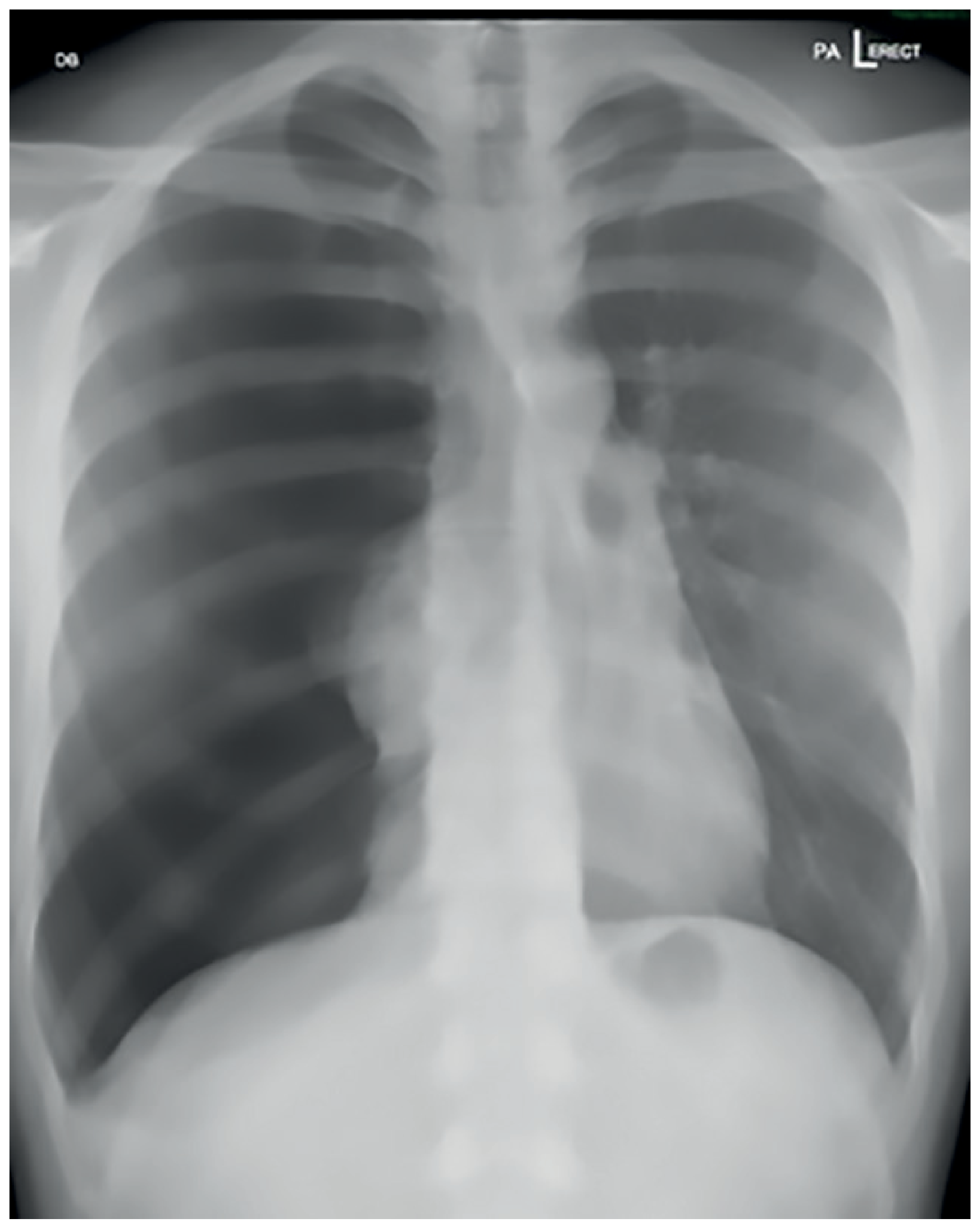
Bailey and Love's Short Practice of Surgery 28th Ed., Figure 29.3 - note complete left lung collapse and dramatic rightward mediastinal shift
Management
Step 1: Immediate Needle Decompression (Needle Thoracostomy)
Insert a large-bore needle/angiocatheter (14G or larger, ≥5 cm length) to convert tension pneumothorax to a simple pneumothorax:
Two sites are accepted:
- 2nd intercostal space, mid-clavicular line (2ICS MCL) - traditional ATLS approach; insert above the upper border of the 3rd rib to avoid the neurovascular bundle
- 4th/5th intercostal space, anterior axillary line (4-5ICS AAL) - now preferred in current teaching (Bailey & Love "safe triangle"); the chest wall is thinner here, making success more reliable especially in obese patients
A rush of air confirms correct placement and should produce immediate haemodynamic improvement.
Note: A 2025 meta-analysis (PMID 40383767) reviewed the optimal needle length and decompression site, providing updated consensus recommendations relative to current ATLS/ETC guidelines - the lateral site (AAL) has higher success rates due to thinner chest wall.
Step 2: Chest Tube (Tube Thoracostomy)
Needle decompression is a bridge, not a definitive treatment. Always follow with a formal chest drain through the 5th intercostal space, anterior axillary line ("safe triangle"). The chest tube:
- Provides definitive, sustained decompression
- Drains any associated haemothorax
- Allows lung re-expansion to seal the parenchymal defect
Step 3: Finger Thoracostomy
In extremis (cardiac arrest, no equipment available), a finger thoracostomy at the same safe-triangle location gives immediate pleural access while the chest tube is prepared. This is particularly valuable in the pre-hospital or damage-control setting.
Intraoperative / Perioperative Considerations
- Any patient under positive-pressure ventilation (GA or ICU) is at heightened risk - a small pneumothorax can rapidly tension under PPV
- An abrupt increase in peak inspiratory pressure with haemodynamic deterioration = tension pneumothorax until proven otherwise
- Patients undergoing laparoscopic surgery (CO2 pneumoperitoneum) who develop intraoperative haemodynamic instability should also prompt consideration - diaphragmatic defects can allow CO2 entry into the pleural space
- Post-subclavian/internal jugular central line insertion: always consider iatrogenic pneumothorax if patient deteriorates after line placement
Summary of Management Algorithm
Suspect tension pneumothorax
        │
        ▼
Clinical diagnosis: absent BS + hyperresonance + haemodynamic instability
        │
        ▼
IMMEDIATE needle decompression
(14G, ≥5cm - 2ICS MCL or 4-5ICS AAL)
        │
        ▼
Rush of air + haemodynamic improvement
        │
        ▼
Chest tube (5ICS AAL, "safe triangle")
        │
        ▼
CXR to confirm lung re-expansion
Sources: Current Surgical Therapy 14e, p. 1310 | Bailey and Love's 28th Ed., p. 395-396 | Rosen's Emergency Medicine, pp. 2557-2565 | Roberts and Hedges' Clinical Procedures, p. 264 | Meta-analysis on needle decompression, PMID 40383767
This is a shared conversation. Sign in to Orris to start your own chat.