
In detail about femoroacetabular impingement for Orthopaedic postgraduate exam don't miss any points
femoroacetabular impingement FAI orthopaedic exam complete notes classification diagnosis treatment
"femoroacetabular impingement"[MeSH Terms] AND treatment
Femoroacetabular Impingement (FAI) - Complete Orthopaedic PG Notes
1. DEFINITION
- First described and popularized by Ganz et al.
- Consensus on FAIS (FAI Syndrome) established in 2016 (Warwick Agreement)
- FAIS is a triad: symptoms + clinical signs + bony deformity (all three required)
2. CLASSIFICATION / TYPES
A. CAM Impingement (Femoral-sided)
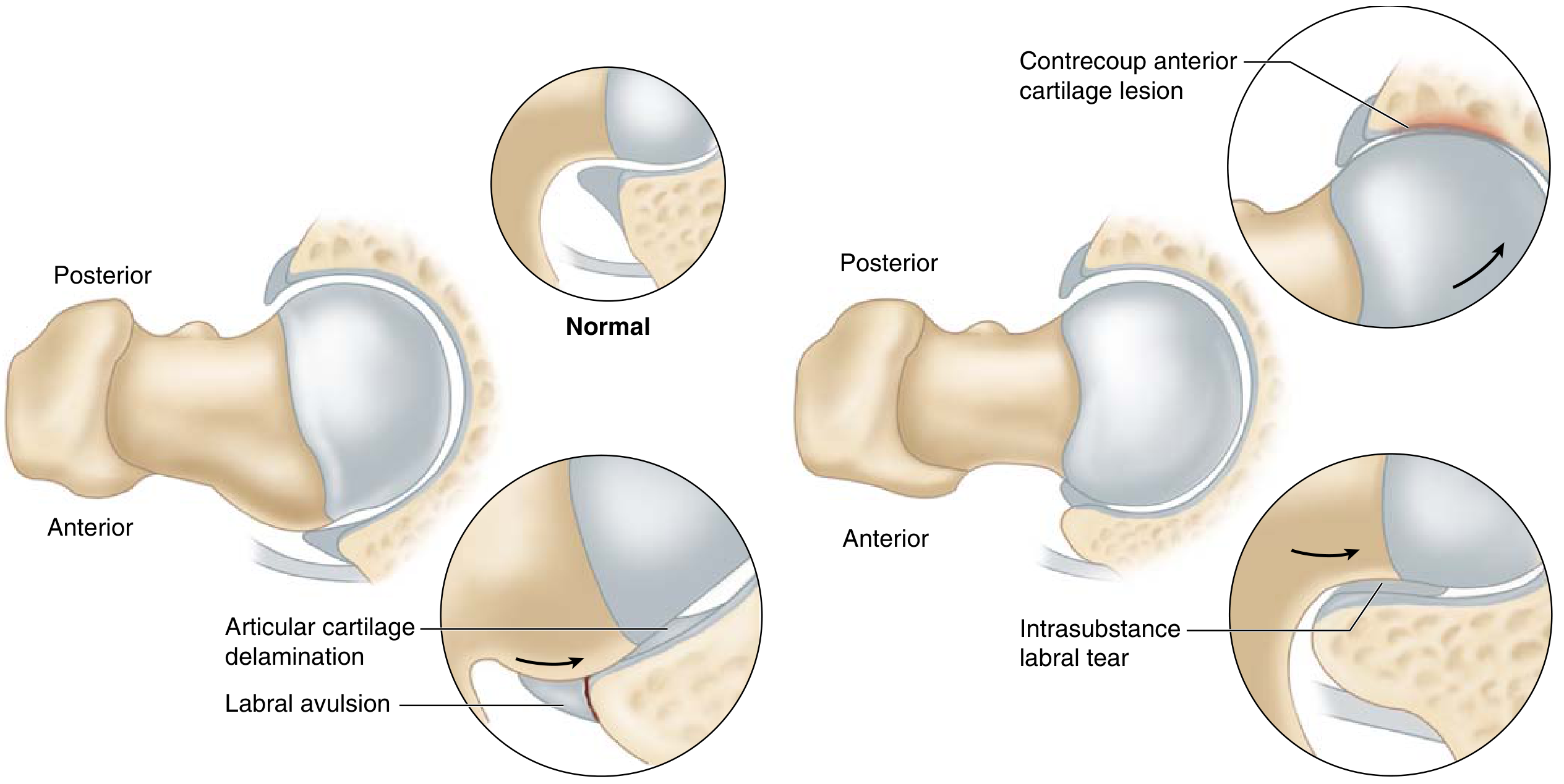
- Abnormal bony prominence at the anterosuperior femoral head-neck junction (reduced offset / non-spherical head)
- During flexion + internal rotation, the non-spherical portion rotates into the acetabulum
- Results in labral avulsion at the labro-chondral junction and articular cartilage delamination (carpet lesion) progressing from the acetabular rim inward
- The labrum is relatively spared in cam impingement; cartilage damage predominates
- More common in young athletic males
- Etiology: possibly a developmental abnormality of the lateral femoral physis related to high-intensity sports during adolescence
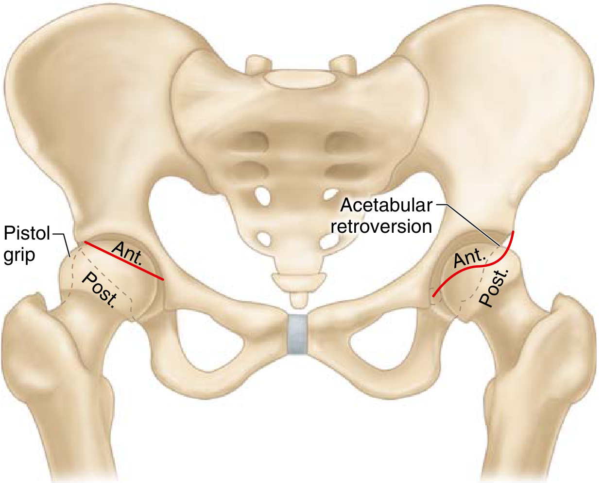
- Pistol-grip deformity = non-spherical femoral head on AP radiograph
B. PINCER Impingement (Acetabular-sided)
- Overcoverage of the femoral head by the acetabulum causes the femoral neck to abut the acetabular rim during motion
- Causes:
- Acetabular retroversion (crossover sign)
- Coxa profunda (acetabular fossa medial to ilioischial line)
- Protrusio acetabuli (femoral head crosses ilioischial line)
- Global overcoverage / deep acetabulum
- Results in intrasubstance labral tears (typically anterosuperior quadrant); labrum is primarily injured
- Contrecoup injury = posteroinferior acetabular cartilage damage from levering of femoral neck on rim
- More common in middle-aged women
- Worsens with time due to reactive bone growth / labral calcification increasing overcoverage
C. Combined CAM + PINCER (Most Common)
- Both morphologies coexist
- Damages both acetabular rim cartilage and labrum to varying degrees
- Worst clinical prognosis of all three types
D. Femoral Retrotorsion
- Contributes to anterior hip impingement as an additional mechanism
3. EPIDEMIOLOGY & RISK FACTORS
- Young to middle-aged adults (teens to 50s)
- Male sex (cam type); Female sex (pincer type)
- Athletes in high-intensity sports: hockey, soccer, football, tennis, ballet, cycling
- Caucasian ethnicity
- Familial FAI morphology
- High-impact activities during adolescent skeletal development
- Cam morphology prevalence in elite athletes approaches 60-70%
4. PATHOANATOMY
| Type | Primary lesion | Secondary lesion |
|---|---|---|
| Cam | Cartilage delamination (acetabular rim → central) | Labral avulsion |
| Pincer | Labral intrasubstance tear (anterosuperior) | Contrecoup posterior cartilage lesion |
| Combined | Both cartilage + labrum | Worse outcomes |
- Labral tear site: anterosuperior quadrant is most frequently affected in both types
- Carpet lesion: acetabular cartilage delamination from subchondral bone - specific to cam FAI
- FAI is a leading cause of secondary hip osteoarthritis in young adults
5. CLINICAL FEATURES
Symptoms
- Groin pain - the most common presentation; typically slow onset, persistent
- Pain worsens with: deep hip flexion, prolonged sitting, twisting, sporting activity
- C-sign: Patient cups the hand over the lateral hip in a "C" shape to describe pain location
- Hip stiffness and limited range of motion
Clinical Signs
- Limited internal rotation in flexion (most consistent finding)
- Patients have more passive external rotation than internal rotation
- Positive anterior impingement test (FADIR test): Flexion + ADduction + Internal Rotation reproduces groin pain
- Most sensitive test for FAI (~95% sensitive but low specificity)
- FABER test (Flexion + ABduction + External Rotation): assesses posterior impingement and labral tears; pain = positive
- Posterior impingement test: Extension + external rotation reproduces posterior hip pain
- Log roll test: patient supine, passive internal/external rotation - pain suggests intraarticular pathology
- Stinchfield test: resisted hip flexion at 30° reproduces pain
6. IMAGING
A. Plain Radiographs
- AP pelvis (gold standard initial view)
- 45° Dunn lateral view (preferred by most hip arthroscopists - best for measuring alpha angle; identifies subtle cam deformities not visible on AP)
- Cross-table lateral
- False-profile view: evaluates anterior coverage, subspine deformities, posterior joint space narrowing
| Sign | Type | Description |
|---|---|---|
| Alpha angle >55° | Cam | Angle measuring loss of femoral head sphericity on Dunn/lateral view |
| Pistol-grip deformity | Cam | Non-spherical femoral head on AP view |
| Head-neck offset ratio reduced | Cam | <0.17 indicates loss of offset |
| Crossover sign | Pincer | Anterior acetabular wall line crosses lateral to posterior wall = acetabular retroversion |
| Figure-of-8 sign | Pincer | Exaggerated crossover |
| Coxa profunda | Pincer | Acetabular fossa medial to ilioischial line (may be normal variant in women) |
| Protrusio acetabuli | Pincer | Femoral head crosses the ilioischial line |
| Lateral centre-edge angle >40° | Pincer | Overcoverage (normal: 25-40°) |
| Sclerosis/cysts at anterolateral head-neck junction | Both | Present in ~1/3 of symptomatic patients |
| Calcified labrum | Pincer | Worsens overcoverage |
B. CT Scan / 3D-CT
- Best for assessing bony morphology and version of femur and acetabulum
- 3D reconstruction guides surgical planning for amount of bony resection
- Delineates extent of cam and pincer deformity
- Measures femoral version and acetabular version accurately
C. MRI / MR Arthrography (MRA)
- MRA (intraarticular gadolinium) is the gold standard for labral pathology
- Sensitivity increases from ~60% (plain MRI) to >90% with contrast
- Reveals labral tears, cartilage delamination, paralabral cysts
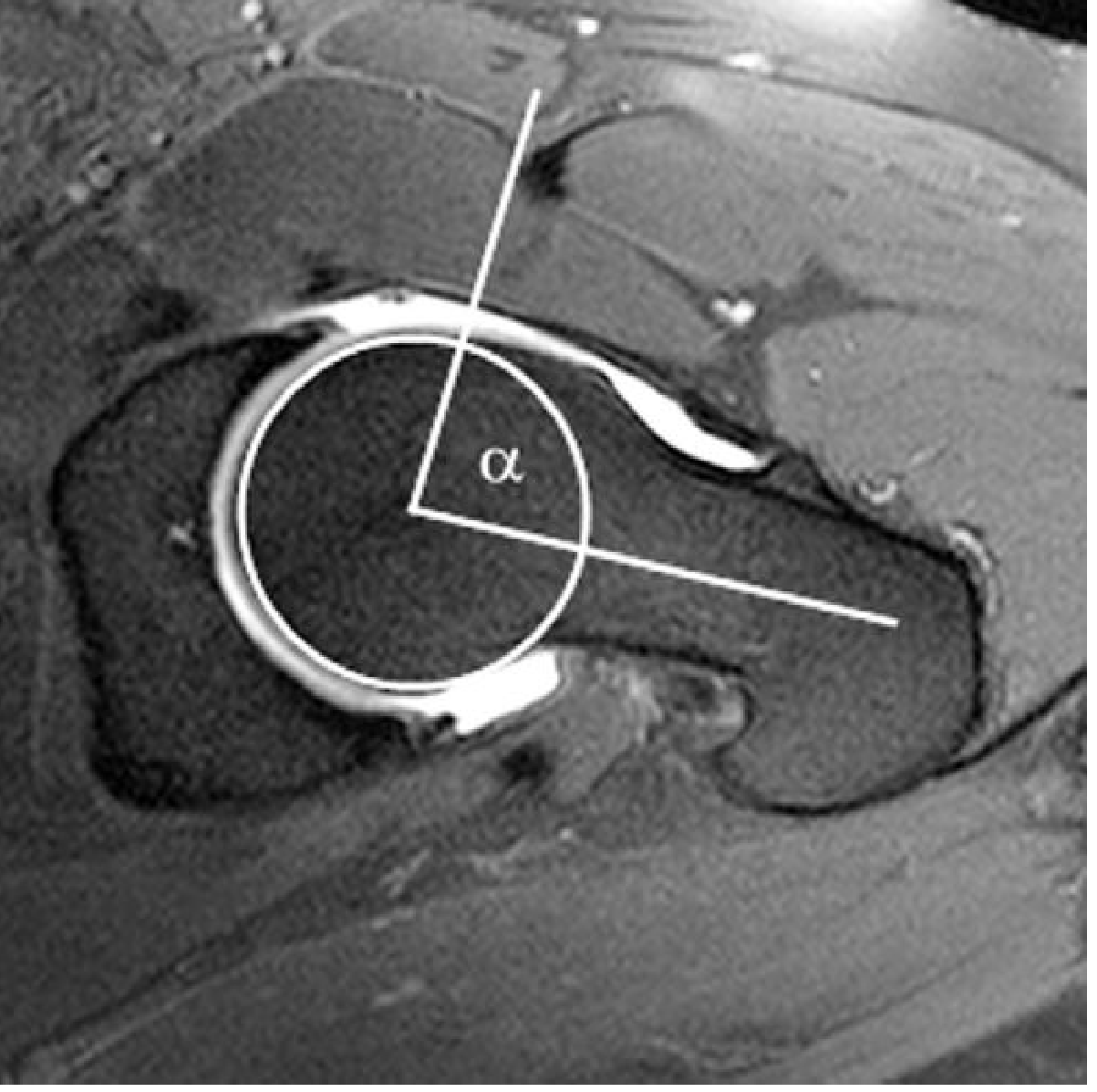
- Axial oblique sequence: alpha angle measured on MRI (see diagram below)
- Contrast tracking beneath articular cartilage suggests delamination
- Higher-resolution 3T MRI may eventually replace MRA
D. Intraarticular Injection
- Diagnostic/therapeutic: Fluoroscopy-guided intraarticular LA ± steroid
- If injection relieves pain, confirms intraarticular source of pain
7. KEY MEASUREMENTS (Exam High-Yield)
| Measurement | Normal | Abnormal (FAI) |
|---|---|---|
| Alpha angle | <50-55° | >55° = cam |
| Lateral centre-edge angle (LCEA) | 25-40° | >40° = pincer overcoverage |
| Anterior centre-edge angle (ACEA) | >25° | - |
| Tönnis angle (acetabular index) | <10° | >10° = dysplasia (low coverage) |
| Head-neck offset ratio | >0.17 | <0.17 = cam |
| Joint space | >2 mm | <2 mm = high THA conversion risk |
8. CLASSIFICATION OF ARTICULAR CARTILAGE DAMAGE
- Grade 0: No signs of OA
- Grade 1: Slight narrowing, sclerosis
- Grade 2: Small cysts, moderate narrowing, loss of sphericity
- Grade 3: Large cysts, severe narrowing, avascular necrosis signs
9. DIFFERENTIAL DIAGNOSIS
- Hip dysplasia (DDH) - LCEA <20°, acetabular index >10°
- Labral tear without FAI
- Avascular necrosis (AVN) of femoral head
- Iliopsoas tendinitis / internal snapping hip
- Trochanteric bursitis
- Hip OA
- Stress fracture of femoral neck
- Lumbar spine pathology
- Sports hernia / athletic pubalgia
- Piriformis syndrome
10. TREATMENT
A. Conservative (Non-Operative) - First Line
- Activity modification: avoid deep flexion, high-impact sports
- Physiotherapy: core strengthening, hip rotator strengthening, ROM exercises
- NSAIDs / analgesics
- Intraarticular corticosteroid injection (diagnostic + therapeutic)
- Conservative management for 3-6 months before surgery
B. Operative Treatment
- Failed conservative treatment (3-6 months)
- Symptomatic FAI with confirmed labral/chondral pathology on MRI
- Good/adequate joint space (Tönnis grade 0-1; joint space >2 mm)
- No severe osteoarthritis
- Tönnis grade 2+ OA (relative contraindication)
- Joint space <2 mm (predicts high THA conversion rate)
- Advanced age with significant OA → proceed to THA
B1. Hip Arthroscopy (Most Common Surgical Approach)
- Preferred method for most FAI cases
- Performed through 2-3 portals (anterolateral, anterior, mid-anterior)
- Traction applied to distract the joint
- Procedures performed:
- Cam resection (femoral osteochondroplasty): reshaping the femoral head-neck junction
- Pincer resection (rim trimming / acetabuloplasty): removing excess acetabular rim
- Labral repair: preferred over debridement (superior outcomes); labral refixation using suture anchors
- Labral reconstruction: if labrum is irreparable (using IT band or ligamentum teres graft)
- Chondral treatment: microfracture, fibrin glue, cartilage repair as needed
- Capsular plication or repair (especially in borderline dysplasia)
- Return to sport: median 6 months
- Outcomes: Excellent in properly selected patients; 80-90% patient satisfaction
B2. Surgical Hip Dislocation (Ganz Trochanteric Flip Osteotomy)
- Allows excellent 360° exposure of the femoral head and acetabulum
- Indicated for: severe deformities not amenable to arthroscopy, complex combined pathology
- Technique (Ganz):
- Trochanteric flip osteotomy
- Z-shaped capsulotomy (anterior Z-capsulotomy)
- Safe surgical dislocation preserving posterior blood supply (medial femoral circumflex artery)
- Preserves femoral head blood supply - very important
- Allows complete labral repair and chondral flap treatment
- Complications (<5%): trochanteric nonunion, heterotopic ossification
- Anterior Z-capsulotomy preserves posterior vessels to femoral neck, minimizing AVN risk
B3. Periacetabular Osteotomy (PAO) / Bernese PAO
- For acetabular overcoverage with significant retroversion
- Posterior column is preserved - maintains pelvic stability
- Allows correction of acetabular tilt and version
- Less commonly used for FAI but helpful for retroverted acetabula
B4. Reverse PAO (Anti-protrusio / Derotation)
- Reduces anterior overcoverage in retroverted acetabulum
- Less commonly performed
B5. Femoral Osteotomy
- For femoral retrotorsion or other femoral-sided deformities
- Derotation osteotomy of the proximal femur
B6. Total Hip Arthroplasty (THA)
- Reserved for patients with significant arthritic changes (Tönnis grade 2-3)
- Joint space <2 mm = high conversion to THA risk after arthroscopy
11. PROGNOSIS / PROGNOSTIC FACTORS
- Younger age
- Tönnis grade 0-1 OA
- Joint space >2 mm
- Isolated cam impingement (better than pincer)
- Labral repair > labral debridement (superior 10-year survivorship)
- Normal acetabular coverage (LCEA 25-40°)
- Tönnis grade ≥ 2 OA
- Joint space < 2 mm → risk of conversion to THA
- Borderline dysplasia (LCEA 18-25°) - capsular plication important
- Combined FAI morphology
- Advanced age
- Significant chondral damage at time of surgery
12. COMPLICATIONS OF HIP ARTHROSCOPY
- Traction neuropraxia (lateral femoral cutaneous nerve, pudendal nerve)
- Instrument breakage
- Avascular necrosis (rare)
- Heterotopic ossification
- Capsular instability (if over-release without repair)
- Incomplete resection → recurrence of symptoms
- Conversion to THA
13. SUBSPINE IMPINGEMENT (AIIS Impingement)
- Impingement between the anterior inferior iliac spine (AIIS) and the femoral head-neck junction
- A form of extra-articular impingement
- Common in patients with prior AIIS avulsion injuries or prominent AIIS
- Addressed arthroscopically with AIIS decompression
14. SPECIAL POINTS FOR PG EXAM
| High-Yield Fact | Detail |
|---|---|
| Alpha angle cut-off | >55° (cam); ideally measured on Dunn lateral / axial MRI |
| LCEA cut-off | >40° (pincer overcoverage); <20° = dysplasia |
| Most common type | Combined CAM + Pincer |
| Cam more common in | Young athletic males |
| Pincer more common in | Middle-aged women |
| Primary labral injury in cam | Labro-chondral junction avulsion + cartilage delamination |
| Primary labral injury in pincer | Intrasubstance labral tear (anterosuperior) |
| Contrecoup injury | Posteroinferior acetabular cartilage (in pincer) |
| Specific radiographic sign of cam | Pistol-grip deformity on AP; alpha angle on Dunn/lateral |
| Specific sign of pincer | Crossover sign (acetabular retroversion) |
| Gold standard for labral pathology | MR Arthrography (sensitivity >90%) |
| Intraoperative OA grading | Tönnis grade |
| Poor prognostic indicator | Tönnis grade ≥ 2; joint space <2 mm |
| Labral repair vs debridement | Repair = better long-term survivorship |
| Blood supply at risk in open dislocation | Medial femoral circumflex artery (protected by Z-capsulotomy) |
| Return to sport after arthroscopy | Median 6 months |
| Ganz procedure complication | Trochanteric nonunion, heterotopic ossification (<5%) |
| FAI leads to | Labral tears → early hip osteoarthritis |
| Impingement test | FADIR (Flexion, ADduction, Internal Rotation) = anterior impingement test |
15. SUMMARY OF RADIOGRAPHIC ANGLES
Alpha angle:
  - Measured on: 45° Dunn lateral / cross-table lateral / axial oblique MRI
  - Method: Circle of best fit around femoral head → angle between
    (1) femoral neck axis and
    (2) line from head center to point where head protrudes from the circle
  - Normal: <50-55° | Abnormal (cam): >55°

Lateral Centre-Edge Angle (LCEA / Wiberg angle):
  - Measured on: AP pelvis
  - Method: Angle between vertical line from femoral head center
    and line from femoral head center to lateral acetabular edge
  - Normal: 25-40° | Overcoverage (pincer): >40° | Dysplasia: <20°

Head-Neck Offset Ratio:
  - Normal: >0.17 | Reduced: <0.17 = cam morphology
In detail about intoeing for Orthopaedic postgraduate exam in simple words
intoeing pediatric orthopaedics causes metatarsus adductus internal tibial torsion femoral anteversion evaluation treatment
intoeing gait torsional profile children femoral anteversion tibial torsion metatarsus adductus
Intoeing - Complete Notes for Orthopaedic PG Exam (Simple Words)
1. What is Intoeing?
- Measured by Foot Progression Angle (FPA) = angle between the line of walking and the long axis of the foot.
- Negative angle = intoeing
- Positive angle = out-toeing
2. Three Main Causes (Remember by Age and Site)
| Site | Cause | Typical Age |
|---|---|---|
| Foot | Metatarsus adductus | Infancy (first year of life) |
| Tibia (shin bone) | Internal tibial torsion | Toddlers (1-3 years) |
| Femur (thigh bone) | Increased femoral anteversion | Children <10 years (most noticeable 3-6 years) |
3. Metatarsus Adductus (Foot-level problem)
- Lateral border of the foot looks convex/curved (instead of straight) - like a "bean-shaped" foot
- Seen from birth or in the first year of life
- 10-15% of cases are associated with hip dysplasia (DDH) - always check the hips
- 85% resolve on their own without treatment
- If foot can be passively straightened to neutral (heel bisector lines up with 2nd toe) → just do stretching exercises
- If foot is rigid (cannot be corrected passively) → serial casting (results are mixed)
- Surgery (lateral column shortening + medial column lengthening) only rarely needed, in children older than 5 years
4. Internal Tibial Torsion (Shin bone problem)
- Most common cause of intoeing in toddlers (1-3 years)
- Usually bilateral
- Often goes along with physiological bow legs (tibia vara) in infants
- Measured by thigh-foot angle (angle between the thigh and the foot with the child lying on their tummy, knee bent to 90°)
- Normal thigh-foot angle: 0 to +10 degrees (external)
- Internal tibial torsion: around -10 degrees (negative/internal)
- Resolves on its own with growth by around age 4 (as the tibia naturally rotates outward with growth)
- Braces/shoes do NOT help - no proven benefit
- Surgery (supramalleolar derotation osteotomy) very rarely needed - only in severe cases, done between age 7-10 years
5. Femoral Anteversion (Thigh bone problem) - Most Common Overall Cause
- Most common overall cause of intoeing perceived by parents in school-age children
- More common in girls
- Typically bilateral
- All babies are born with some degree of femoral anteversion; normally it decreases as the child grows because the femur derotates with growth
- Child often prefers to sit in "W" position (both knees bent, feet out to the sides)
- Runs with a "circumduction" or "egg-beater" type gait
- On examination (child lying prone/on tummy, knee bent to 90°):
- Excessive internal rotation of the hip (can go up to 80-90°)
- Reduced external rotation of the hip
- Normal total rotation arc, just shifted toward internal rotation
- Craig's test (trochanteric prominence test): measure internal rotation when the greater trochanter is most prominent (most lateral position) - this estimates the degree of anteversion
- Most cases improve spontaneously by age 8-10
- If it persists beyond 10-12 years AND causes real functional problems (tripping, difficulty with sports) → corrective derotation osteotomy of the femur may be considered
- Children who cannot externally rotate the extended hip at all should be watched carefully
- Happens when increased femoral anteversion + compensatory external tibial torsion occur together
- The foot progression angle looks normal (because the inward hip twist is cancelled out by the outward shin twist), BUT the child has:
- Knee pain
- Patella (kneecap) maltracking / instability
- Feeling of the knee "giving way"
- This is a trap in exams - normal-looking gait can still hide significant torsional abnormality, so always check hip and tibial rotation separately, not just the foot progression angle.
6. How to Examine - The Torsional Profile (Very Important for Exam)
- Foot progression angle - observed while child walks
- Hip rotation (internal and external) - hip extended, knee bent 90°, rotate the leg like a clock hand
- Assesses femoral anteversion/retroversion
- Thigh-foot angle - angle between thigh axis and foot axis
- Assesses tibial torsion
- Foot shape / heel bisector - looking at the sole of the foot
- Assesses metatarsus adductus
7. Red Flags - When Intoeing is NOT Simple/Physiological
- Pain, swelling, or limp
- Asymmetric findings (one leg different from the other)
- Progressive worsening instead of improvement with age
- Associated with:
- Developmental dysplasia of the hip (DDH)
- Clubfoot
- Skew foot
- Neuromuscular disease (e.g., cerebral palsy)
- Unilateral out-toeing in an adolescent → think of Slipped Capital Femoral Epiphysis (SCFE) until proven otherwise (this is a classic exam trap - out-toeing, not intoeing, but commonly tested alongside torsional problems)
8. General Treatment Principles
- Reassurance and observation is the mainstay for almost all physiological intoeing - most resolve by age 8 without any treatment
- No good evidence for special shoes, twister cables, bars, or night splints - these are outdated and NOT recommended
- Surgery is reserved for:
- Severe deformity persisting beyond age 8-10
- Causing a real functional problem (frequent tripping, cosmetic/psychological distress, sports limitation)
- Confirmed by clinical exam + imaging (CT or MRI for precise version measurement)
9. Quick Comparison Table (High-Yield for Exam)
| Feature | Metatarsus Adductus | Internal Tibial Torsion | Femoral Anteversion |
|---|---|---|---|
| Level | Foot | Tibia | Femur/Hip |
| Peak age | Infancy | 1-3 years | 3-10 years (most common overall) |
| Sex | No clear predilection | No predilection | More common in girls |
| Key sign | Convex lateral foot border | Negative thigh-foot angle | Increased internal rotation, decreased external rotation of hip |
| Special test | Heel bisector line | Thigh-foot angle | Craig's test |
| Natural resolution | ~85% by 2-4 yrs | By age 4 | By age 8-10 |
| Surgery age (if needed) | >5 years | 7-10 years | 10-12 years |
Summary in One Line
In detail about genu valgus deformity for Orthopaedic postgraduate exam in simple words
genu valgum knock knee children orthopaedics causes classification treatment osteotomy
genu valgum knock knee deformity children x-ray mechanical axis