
Necrotising pneumonia treatment
necrotizing pneumonia treatment management
necrotising pneumonia treatment guidelines 2024 2025 antibiotics surgery
https://pmc.ncbi.nlm.nih.gov/articles/PMC11597800
PMID: 42336503
necrotizing pneumonia CT scan cavitation lung abscess
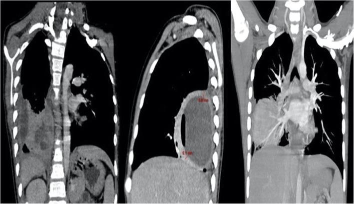
This diagnostic image consists of three thoracic CT scan views (coronal and sagittal) demonstrating cavitated necrotizing pneumonia in the right lower lobe. The primary finding is a large, well-defined lung abscess involving the medial, posterior, and lateral basal segments of the right lung. On the sagittal view, the abscess appears as a large circular lesion with a thick, enhancing wall and a low-density core, with a measurement indicated (approximately 75.7 mm). The coronal views reveal heterogeneous consolidation in the surrounding lung parenchyma and internal lucencies within the main cavity, suggesting multifocal cavitation. Additionally, significant mediastinal lymphadenopathy is visible as enlarged soft-tissue density nodes located in the paratracheal and hilar regions. The pathology shows mass effect on the right hemi-diaphragm. The image serves as an educational example of severe pulmonary tuberculosis complications and secondary bacterial infections (such as Streptococcus anginosus group), highlighting the imaging characteristics of large cavitary lesions and associated reactive or infectious adenopathy.
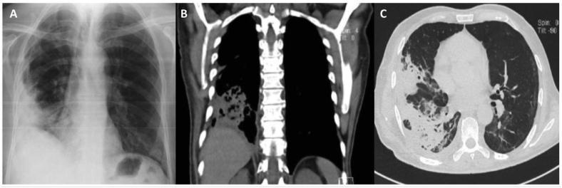
A composite of three clinical diagnostic images illustrating necrotizing pneumonia in the right lung. (A) Posteroanterior chest X-ray shows a large, heterogeneous opacity in the right basal region with ill-defined borders, indicating consolidation. (B) Coronal CT scan in mediastinal window demonstrates a hyperdense area in the right lower lobe consistent with lung consolidation, featuring internal low-attenuation areas suggestive of tissue breakdown or early cavitation. (C) Axial CT scan in lung window reveals a large, peripheral pulmonary opacity in the right lung with intimate pleural contact. The opacity exhibits internal air-filled lucencies and a distinct central cavitation, characteristic of necrotizing pneumonia and pulmonary abscess formation. This series is an educational example of progressive infectious lung disease, highlighting the transition from radiographic opacification to CT-confirmed cavitation and parenchymal necrosis. Key concepts include imaging signs of consolidation, cavitation, and pleural involvement in lower respiratory tract infections.
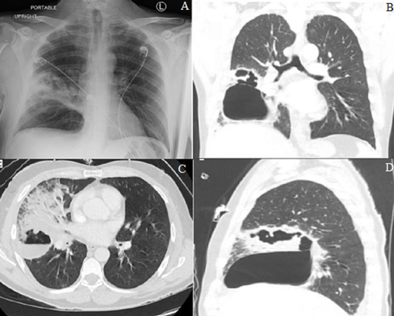
This composite figure presents a radiographic and tomographic evaluation of necrotizing pneumonia with abscess formation in the right lung. Panel A is a portable frontal chest X-ray showing a dense focal infiltrate in the right mid-lung zone involving the minor fissure, with internal lucencies suggestive of early cavitation. Panels B, C, and D are CT lung window images in coronal, axial, and sagittal planes, respectively. These images demonstrate extensive consolidation of the right middle lobe (RML) and right lower lobe (RLL). A large, thick-walled abscess cavity is clearly visible, containing a prominent air-fluid level—a hallmark of pulmonary abscess. The surrounding parenchyma shows heterogeneous attenuation and tissue destruction consistent with necrotizing pneumonia. This visual series illustrates the progression from simple lobar consolidation to complex cavitary disease and the importance of multiplanar CT imaging in characterizing the extent of parenchymal necrosis and abscess localization within specific pulmonary segments.
Necrotising Pneumonia: Treatment
Definition & Key Pathogens
- Staphylococcus aureus (including PVL-producing SA and MRSA) - most feared, associated with rapid cavitation
- Streptococcus pneumoniae (especially serotypes 3, 8, 19A)
- Klebsiella pneumoniae - classic in diabetics/alcoholics, tends toward upper lobe "bulging fissure"
- Anaerobes - common in aspiration pneumonia
- Group A Streptococcus (GAS)
- Pseudomonas aeruginosa - particularly in hospital-acquired / immunocompromised patients
- Mycoplasma pneumoniae in children (occasionally)
1. General Principles
- Admit all patients - consider ICU for haemodynamic instability or respiratory failure
- Obtain cultures (blood, sputum, BAL) before starting antibiotics
- Chest CT with contrast to define extent of necrosis and pleural involvement
- Monitor inflammatory markers (CRP, procalcitonin) - though not used to initiate antibiotics, they may guide duration
2. Antimicrobial Treatment
Empirical Approach
| Clinical Context | Empirical Regimen |
|---|---|
| CAP (community-acquired) | Beta-lactam (e.g. IV ampicillin-sulbactam, piperacillin-tazobactam) + respiratory fluoroquinolone (or azithromycin) |
| MRSA risk factors present | Add vancomycin or linezolid to the above |
| Hospital-acquired (HAP/VAP) | Anti-pseudomonal beta-lactam + MRSA coverage |
| Aspiration/lung abscess | Add anaerobic coverage (metronidazole, clindamycin, or piperacillin-tazobactam) |
PVL-Producing Staphylococcus aureus (PVL-SA) - Special Protocol
- IV linezolid 600 mg twice daily
- IV clindamycin 1.2 g four times daily
- IV rifampicin 600 mg twice daily
Antibiotic Duration
- Minimum 2-4 weeks in most patients (from paediatric literature, extrapolated to adults)
- Up to 6 weeks for cavitary/abscess disease (Goldman-Cecil Medicine)
- Biomarkers (CRP, procalcitonin) are increasingly used to guide de-escalation in individual cases, though not yet formally recommended
3. Management of Pleural Complications
| Stage | Description | Treatment |
|---|---|---|
| Stage I (simple exudate) | Free-flowing, non-purulent, glucose normal | Antibiotics alone; +/- thoracocentesis |
| Stage II (fibrinopurulent) | Loculations, fibrin deposits, no organised peel | Chest tube drainage; VATS if incomplete drainage |
| Stage II/III mixed | Areas of fibrinous organisation | Surgical consultation - VATS |
| Stage III (organising) | Frank organised pleural peel | VATS and/or decortication |
4. Role of Bronchoscopy
- Obtain BAL specimens for targeted microbiological diagnosis
- Assess for endobronchial obstruction
- Therapeutic lavage of necrotic debris
- Manage bronchopleural fistula (bronchoscopic occlusion techniques)
5. Surgery
- Failure of medical management (persistent sepsis, non-resolving infection)
- Significant or persistent haemoptysis (consider bronchial artery embolisation first)
- Extensive gangrene
- Tension pneumatocele or bronchopleural fistula not amenable to bronchoscopic intervention
6. Supportive and Adjunctive Measures
- Oxygen/ventilatory support - NIV or invasive ventilation for respiratory failure; ECMO in refractory cases (increases surgical risk but not a contraindication)
- Haemodynamic support - Sepsis bundles (IV fluids, vasopressors) per Surviving Sepsis Campaign
- Haemoptysis - Bronchial artery embolisation is first-line for significant haemoptysis before considering surgery
- Oral hygiene and aspiration precautions - Prevention of aspiration in at-risk patients
- Steroids - Not routinely recommended in adults; insufficient evidence in children too
7. Paediatric Considerations
Summary Algorithm
Suspected Necrotising Pneumonia
        │
        ▼
CT chest with contrast + Blood/sputum cultures + ICU assessment
        │
        ▼
Start IV empirical antibiotics (CAP or HAP regimen)
  → Add MRSA cover if risk factors
  → Add anaerobic cover if aspiration/abscess
  → PVL-SA suspected: add linezolid + clindamycin + rifampicin
        │
        ▼
Pleural effusion present?
  → Yes: Thoracocentesis/chest drain; stage and manage
  → No: Monitor
        │
        ▼
Failing to improve?
  → Bronchoscopy (BAL + therapeutic)
  → Consider IR-guided drainage of abscess
  → BAE for haemoptysis
  → Surgery if all else fails
        │
        ▼
Duration: Minimum 2-4 weeks IV → step down when stable