
Leucorrhoea
leucorrhoea vaginal discharge etiology management
vaginal discharge leukorrhea etiology treatment
vaginal discharge types comparison trichomoniasis bacterial vaginosis candida
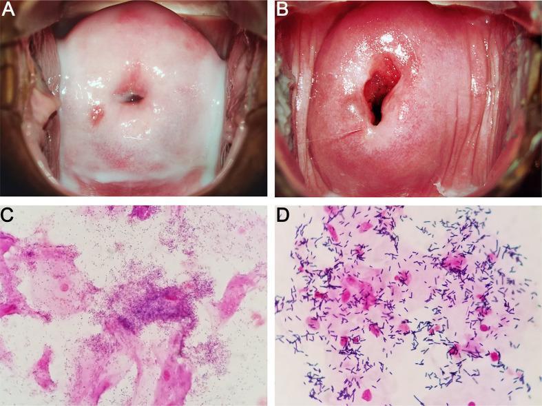
This four-panel image provides a side-by-side comparison of Bacterial Vaginosis (BV) and a normal vaginal microbiome through colposcopic and microscopic examinations. Panels A and B are colposcopic clinical photographs of the cervix and vaginal vault. Panel A (BV) displays characteristic thin, homogeneous, milk-white discharge coating the vaginal walls. In contrast, Panel B (Normal) shows a healthy cervix with clear, physiological secretions. Panels C and D show Gram-stained microscopic slides of vaginal smears. Panel C (BV) demonstrates the presence of 'clue cells'—vaginal epithelial cells heavily encrusted with coccobacilli (Gardnerella-like microbes) that obscure the cell margins. Panel D (Normal) shows a healthy microbiome dominated by large, purple-stained Gram-positive rods, consistent with Lactobacillus species, and clear epithelial cells without bacterial adherence. This comparison illustrates the clinical and laboratory findings used in Amsel criteria and Nugent scoring for the diagnosis of dysbiosis in the female reproductive tract.
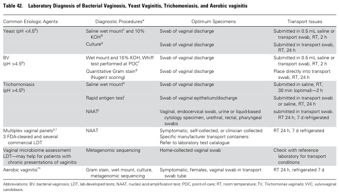
Table 42. Laboratory Diagnosis of Bacterial Vaginosis, Yeast Vaginitis, Trichomoniasis, and Aerobic vaginitis <table><thead><tr><th>Common Etiologic Agents</th><th>Diagnostic Procedures<sup>a</sup></th><th>Optimum Specimens</th><th>Transport Issues</th></tr></thead><tbody><tr><td>Yeast (pH <4.5<sup>b</sup>)</td><td>Saline wet mount<sup>c</sup> and 10% KOH<sup>d</sup></td><td>Swab of vaginal discharge</td><td>Submitted in 0.5 mL saline or transport swab, RT, 2 h</td></tr><tr><td></td><td>Culture<sup>e</sup></td><td>Swab of vaginal discharge</td><td>Submitted in transport swab, RT, 24 h</td></tr><tr><td>BV (pH >4.5<sup>b</sup>)</td><td>Wet mount and 10% KOH, Whiff test performed at POC<sup>f</sup></td><td>Swab of vaginal discharge</td><td>Submitted in 0.5 mL saline or transport swab, RT, 2 h</td></tr><tr><td></td><td>Quantitative Gram stain<sup>g</sup> (Nugent scoring)</td><td>Swab of vaginal discharge</td><td>Place directly into transport swab, RT, 24 h</td></tr><tr><td>Trichomoniasis (pH >4.5<sup>b</sup>)</td><td>Saline wet mount<sup>h</sup></td><td>Swab of vaginal discharge</td><td>Submitted in saline, RT, 30 min (optimal)—2 h</td></tr><tr><td></td><td>Rapid antigen test<sup>i</sup></td><td>Swab of vaginal epithelium/discharge</td><td>Submitted in transport swab or saline, RT, 24 h</td></tr><tr><td></td><td>NAAT<sup>j</sup></td><td>Vaginal, endocervical swab, urine or liquid-based cytology specimen, urethral, rectal, pharyngeal swabs</td><td>submitted in transport swab, RT 24 h, 7 d refrigerated</td></tr><tr><td>Multiplex vaginal panels<sup>k,l</sup> 3 FDA-cleared and several commercial LDT</td><td>NAAT</td><td>Symptomatic, self-collected, or clinician collected Specific manufacturer transport containers Refer to laboratory test catalogue</td><td>RT 24 h, 7 d refrigerated</td></tr><tr><td>Vaginal microbiome assessment LDT—may help for patients with chronic presentations of vaginitis</td><td>Metagenomic sequencing</td><td>Home-collected vaginal swab</td><td>Check with reference laboratory for transport conditions</td></tr><tr><td>Aerobic vaginitis<sup>m</sup></td><td>Gram stain, wet mount, culture, metagenomic sequencing</td><td>Symptomatic, females, vaginal swab in transport swab tube</td><td>RT 24 h, refrigerated 7 d</td></tr></tbody></table> Abbreviations: BV, bacterial vaginosis; LDT, lab-developed tests; NAAT, nucleic acid amplification test; POC, point-of-care; RT, room temperature; TV, Trichomonas vaginalis; VVC, vulvovaginal candidiasis.
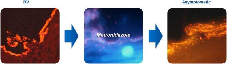
This comparison chart utilizes optical coherence tomography (OCT) or similar high-resolution diagnostic imaging to illustrate the progression of a polymicrobial bacterial vaginosis (BV) biofilm. The visual is divided into three sequential panels. The first panel, labeled 'BV', depicts a dense, continuous, and brightly fluorescent orange-red layer adhering to the vaginal epithelial surface, representing a mature pathogenic biofilm often composed of Gardnerella vaginalis and Atopobium vaginae. The middle panel shows the clinical intervention with 'Metronidazole', showing a blue-tinted transitional phase where the biofilm structure is being disrupted. The final panel, labeled 'Asymptomatic', demonstrates the state after treatment. In this state, the biofilm appears less dense, fragmented, and more diffuse, indicating successful thinning and disruption of the adhering microbial community. This educational visual emphasizes the persistence of subclinical biofilms even in asymptomatic states, which contributes to the high recurrence rates of BV. It is designed for medical professionals and students focusing on gynecology, microbiology, and infectious disease management.
Leucorrhoea
1. Physiological Leucorrhoea
- Neonates: Many newborn girls have physiological leukorrhea in the first 2-3 weeks of life as maternal hormone levels decline. The discharge is thin, slippery, and clear or white. - Tintinalli's Emergency Medicine
- Puberty and reproductive years: Estrogen stimulates cervical glands and vaginal epithelium to produce secretions
- Mid-cycle (ovulation): Increased, clear, stretchy mucus
- Pregnancy: Increased estrogen causes more vaginal secretions
- Physiological discharge has normal vaginal pH (<4.5), no odour, and no associated symptoms
2. Pathological Leucorrhoea - Causes
Vaginitis Causes:
| Organism | Discharge Character | pH | Key Finding |
|---|---|---|---|
| Candida albicans | Curdy/cottage cheese-like, white | 3.8-4.5 (normal) | Pseudohyphae on KOH |
| Trichomonas vaginalis | Profuse, yellow-green, frothy | >5 | Motile trichomonads on wet mount |
| Bacterial vaginosis (BV) | Thin, grey-white, homogeneous, malodorous | >4.5 | Clue cells; positive Whiff test |
| Aerobic vaginitis | Heavy purulent discharge, foul odour | >4.5 | Cocci or coarse rods |
| Atrophic vaginitis | Scant, thin; dryness/pain | >4.5 | RBCs, WBCs on wet mount |
| Allergic/irritant vaginitis | Variable | Any | Negative microscopy |
- Textbook of Family Medicine 9e (Table 25-7)
Cervicitis Causes (discharge originates from the cervix):
- Neisseria gonorrhoeae
- Chlamydia trachomatis
- Trichomonas vaginalis
- Herpes simplex virus
3. Clinical Features by Type
Bacterial Vaginosis (BV)
- Thin, homogeneous, white-grey discharge uniformly coating the vaginal mucosa
- Characteristic fishy (amine) odour, especially after sex
- Caused by loss of hydrogen-peroxide-producing Lactobacillus spp. and overgrowth of anaerobes (Gardnerella vaginalis, Mobiluncus, Prevotella, Mycoplasma hominis)
- Associated with increased risk of acquiring HIV, C. trachomatis, and N. gonorrhoeae
- Also associated with preterm delivery and spontaneous abortion
- Increased white homogeneous vaginal discharge
- Vaginal pH >4.5
- Positive "Whiff test" - fishy odour with 10% KOH
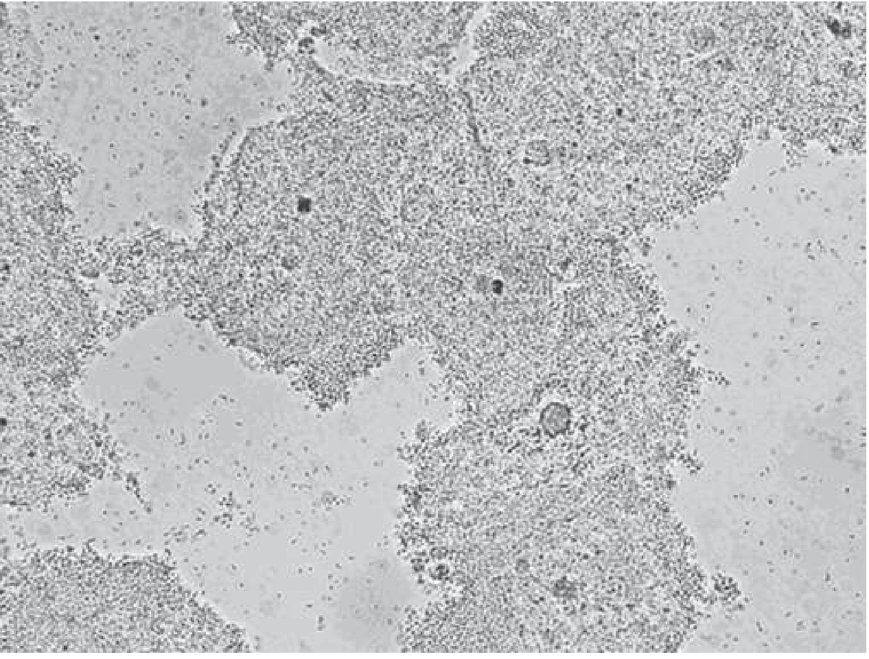
- Clue cells on wet mount (vaginal epithelial cells coated with coccobacilli, obscured margins, granular appearance)
Vulvovaginal Candidiasis (VVC)
- Vulvar itching, burning, discomfort - often without much increase in discharge volume
- Discharge classically "curdy" or cottage cheese-like; may also be loose and hard to distinguish
- Vaginal pH usually normal (<4.5) - this is the most helpful discriminating feature
- 10% KOH prep: pseudohyphae and budding yeast (but sensitivity of wet mount is only ~50%)
- Predisposing factors: antibiotic use, oral contraceptives, diabetes mellitus, HIV, pregnancy
- C. albicans accounts for nearly all symptomatic cases
- Recurrent VVC = 4 or more episodes per year; consider underlying DM or HIV
Vaginal Trichomoniasis
- Profuse, yellow/green, purulent, homogeneous discharge with vulvar irritation
- Vaginal walls and cervix may be inflamed; petechial lesions on cervix = "strawberry cervix" (best seen on colposcopy)
- Vaginal pH usually >5
- Saline wet mount: motile trichomonads (detects <50% of cases); NAAT is more sensitive
- Sexually transmitted; treatment of sexual partners is essential
4. Diagnosis
- Trichomoniasis: greenish, frothy discharge
- Candidiasis: curdy white discharge
- BV: thin, adherent, malodorous discharge
- Cervicitis: mucopurulent cervical discharge, cervical erosion/ulcer
- Wet mount (saline): motile trichomonads, clue cells
- 10% KOH prep: Candida hyphae/pseudohyphae; Whiff test for BV
- Gram stain of vaginal smear: clue cells (BV), Gram-negative diplococci (N. gonorrhoeae)
- NAAT: most sensitive for T. vaginalis, C. trachomatis, N. gonorrhoeae
5. Treatment
Bacterial Vaginosis
- Metronidazole 500 mg orally twice daily × 7 days (preferred over single dose)
- Metronidazole gel 0.75% intravaginal, 5g once daily × 5 days
- Clindamycin cream 2% intravaginal, 5g at bedtime × 7 nights
- Clindamycin 300 mg orally twice daily × 7 days
- Tinidazole 1 g orally once daily × 5 days, or 2 g × 3 days
- Secnidazole 2 g single oral dose
Vulvovaginal Candidiasis
- Uncomplicated: Intravaginal azoles (clotrimazole, miconazole, butoconazole, terconazole) for 1-7 days; OR fluconazole 150 mg orally × 1 dose (highly effective)
- Severe: Fluconazole 150 mg every 72 h × 2-3 doses; intravaginal azoles for 7-14 days
- Recurrent (≥4/year): Fluconazole 100-200 mg every 72 h for 7-14 days, then once weekly for 6 months; otesoconazole (150 mg daily × 7 days, then once weekly × 11 weeks) is a newer option
- Treating sexual partners confers no benefit
Vaginal Trichomoniasis
- Preferred: Metronidazole 500 mg twice daily × 7 days (more effective than single dose)
- Alternative: Tinidazole 2 g orally × 1 dose (fewer GI side effects, useful for metronidazole-resistant cases)
- Treat sexual partners (reduces reinfection and reservoir)
- Intravaginal metronidazole gel is not reliable for trichomoniasis - systemic treatment required
Gonococcal Cervicitis
- Ceftriaxone 500 mg IM once + doxycycline 100 mg twice daily × 7 days if C. trachomatis not excluded
- Azithromycin 1 g orally once is an alternative for concurrent C. trachomatis
Syndromic Management (when lab testing unavailable)
- Oral metronidazole covers both trichomoniasis and BV
- Add fluconazole 150 mg or clotrimazole 500 mg vaginal pessary for candida
- Add cefixime 400 mg + azithromycin 1 g for cervical infection (gonorrhoea + chlamydia)
- Treat sexual partners
6. Special Situations
- Primary site is the vulva (vulvitis first, vaginitis secondary) - opposite to adults
- Causes: poor hygiene, foreign body, Group A Streptococcus, S. aureus, E. coli, Shigella
- Candida is uncommon due to alkaline vaginal pH
- Always consider sexual abuse - test for gonorrhoea and chlamydia if symptoms/discharge present
- Treatment: hygiene measures, short course of topical estrogen + broad-spectrum antibiotics if needed
- Persistent, bloody, or brown discharge warrants vaginal irrigation or vaginoscopy to exclude foreign body
- Pale, friable vaginal walls; dryness, pain; pH >4.5
- Treatment: topical estrogen
Key Points
- Physiological discharge is normal; pathological discharge requires identifying the cause
- Self-diagnosis by patients is unreliable - office microscopy is usually definitive
- Speculum examination is ideal but a blind vaginal vault swab is equally sensitive for diagnosis
- Douching is not helpful and is a risk factor for BV
- Vaginal pH is the single most useful bedside discriminator: normal pH (<4.5) in candidiasis vs. elevated pH (>4.5) in BV and trichomoniasis
Give me different information of vaginitis and cervicitis
cervicitis mucopurulent cervical discharge strawberry cervix colposcopy
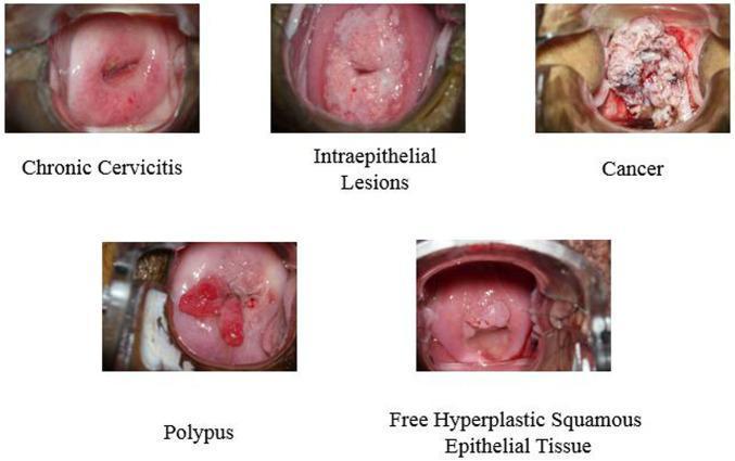
This composite of clinical colposcopy photographs illustrates five distinct pathological states of the human cervix. 1. Chronic Cervicitis: Shows a relatively smooth, erythematous ectocervix with a linear lesion and punctate hemorrhages around the external os. 2. Intraepithelial Lesions: Features a thickened, opaque, whitish acetowhite-like area covering the transformation zone, suggesting dysplastic changes. 3. Cervical Cancer: Displays an advanced neoplastic state characterized by a large, exophytic, friable mass with an irregular, cauliflower-like texture and variegated color (whitish and necrotic red areas) obscuring normal anatomy. 4. Polypus (Cervical Polyp): Exhibits a distinct, pedunculated, smooth, fleshy red growth protruding from the cervical canal. 5. Free Hyperplastic Squamous Epithelial Tissue: Shows a more subtle, uniform, pinkish elevation of the squamous epithelium without the severe disruption seen in malignancy. These images serve as educational references for gynecological oncology and colposcopic diagnosis, highlighting key morphological differences in texture, color, and tissue architecture used to distinguish benign inflammation from precancerous lesions and invasive carcinoma.
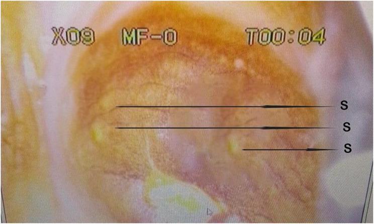
This colpophotograph displays the cervical mucosa following the application of Lugol’s iodine. The image captures a classic clinical presentation of colpitis macularis, commonly referred to as 'strawberry cervix.' The cervical surface shows a predominantly reddish-orange hue with an irregular texture. Key features, labeled with 'S', are small, discrete, raised papules that appear as pale or yellowish punctate spots against the background tissue. These spots represent focal areas of inflammation and capillary dilation or hemorrhage that do not take up the iodine stain (Schiller's test). While this finding is highly characteristic of Trichomonas vaginalis infection, the clinical context indicates a coinfection with female genital schistosomiasis. This diagnostic image is an essential teaching tool for identifying sexually transmitted infections and parasitic manifestations in gynecological examinations, highlighting the importance of colposcopy in visualizing subtle mucosal changes that may not be evident during a standard speculum exam.
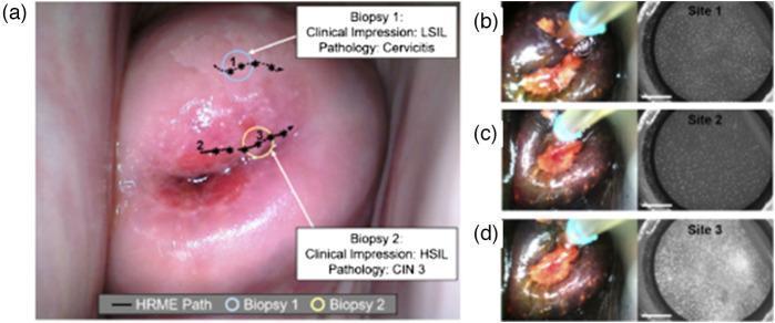
A multi-panel clinical and diagnostic figure demonstrating cervical cancer screening via multimodal colposcopy and high-resolution microendoscopy (HRME). (a) Macroscopic clinical photograph of the cervix following acetic acid application. The image displays a central area of acetowhite epithelium and hyperemic regions near the cervical os. Annotated features include an 'HRME Path' (dashed line) and two biopsy locations: Biopsy 1 (clinical impression of LSIL; pathology: Cervicitis) and Biopsy 2 (clinical impression of HSIL; pathology: CIN 3). (b, c, d) Sequential pairs of macroscopic cervical images and corresponding proflavine-stained HRME fluorescence micro-images from three distinct sites. 'Site 1' and 'Site 2' show dense, uniformly distributed punctate nuclei typical of low-grade or normal tissue. 'Site 3' (associated with HSIL/CIN 3) demonstrates altered nuclear morphology and distribution, characterized by increased nuclear-to-cytoplasmic ratios and disordered cellular arrangement. This figure illustrates the integration of wide-field colposcopy with point-of-care fluorescence microendoscopy for localized diagnostic validation.
Vaginitis vs. Cervicitis - A Complete Comparison
PART A: VAGINITIS
1. Bacterial Vaginosis (BV)
- Thin, homogeneous, white-grey discharge uniformly coating the vaginal walls
- Characteristic fishy (amine) odour, worsened after unprotected sex (alkaline semen raises pH)
- Minimal to no vulvar irritation or itching
- Often asymptomatic
- Increased white homogeneous discharge
- Vaginal pH >4.5
- Positive Whiff test (fishy odour with 10% KOH)
- Clue cells on saline wet mount (vaginal epithelial cells with obscured margins due to adherent coccobacilli)
- Metronidazole 500 mg orally twice daily × 7 days (preferred)
- Metronidazole gel 0.75% intravaginally daily × 5 days
- Clindamycin cream 2% intravaginally nightly × 7 nights
- Secnidazole 2 g orally single dose
- Tinidazole 1 g daily × 5 days
- Clindamycin 300 mg orally twice daily × 7 days
- Treating male partners does not reduce recurrence
2. Vulvovaginal Candidiasis (VVC)
- Vulvar pruritus - the most common and specific symptom
- Discharge: varies from watery to thick "cottage cheese-like" (classic but not always present)
- External dysuria, dyspareunia
- Vulvar erythema and edema; vaginal erythema
- No odour - if odour is present, favours BV instead
- Exacerbation commonly in the week before menses or after coitus (more alkaline pH)
- Normal vaginal pH (4.0-4.5) - most useful discriminating feature
- Uncomplicated: sporadic, mild-moderate symptoms, C. albicans, non-pregnant immunocompetent host
- Complicated: ≥4 episodes/year (recurrent), severe, non-albicans species, or abnormal host (diabetes, immunodeficiency, pregnancy)
- Normal pH (<4.5)
- Saline wet mount: budding yeast ± hyphae (sensitivity 40-60%)
- 10% KOH prep: dissolves epithelial cells, leaves pseudohyphae/buds visible (sensitivity 80%, specificity ~100%)
- Culture for recurrent/atypical cases
- Uncomplicated: intravaginal azoles (clotrimazole, miconazole, butoconazole, terconazole) for 1-7 days; OR fluconazole 150 mg orally × 1 dose
- Severe: fluconazole 150 mg every 72 h × 2-3 doses; intravaginal azoles × 7-14 days
- Recurrent: fluconazole 100-200 mg every 72 h for 7-14 days, then once weekly × 6 months; otesoconazole (150 mg daily × 7 days then once weekly × 11 weeks)
- Treat for DM or HIV if recurrent; treating sexual partners confers no benefit
3. Trichomonas Vaginitis
- Profuse, frothy, greenish-yellow, malodorous discharge
- Vaginal pruritus and irritation
- Vulvar/vaginal erythema and tenderness
- "Strawberry cervix" - petechial hemorrhages on the cervix (best seen on colposcopy)
- Symptoms develop 5-28 days post-exposure; many infections are asymptomatic
- Vaginal pH >5
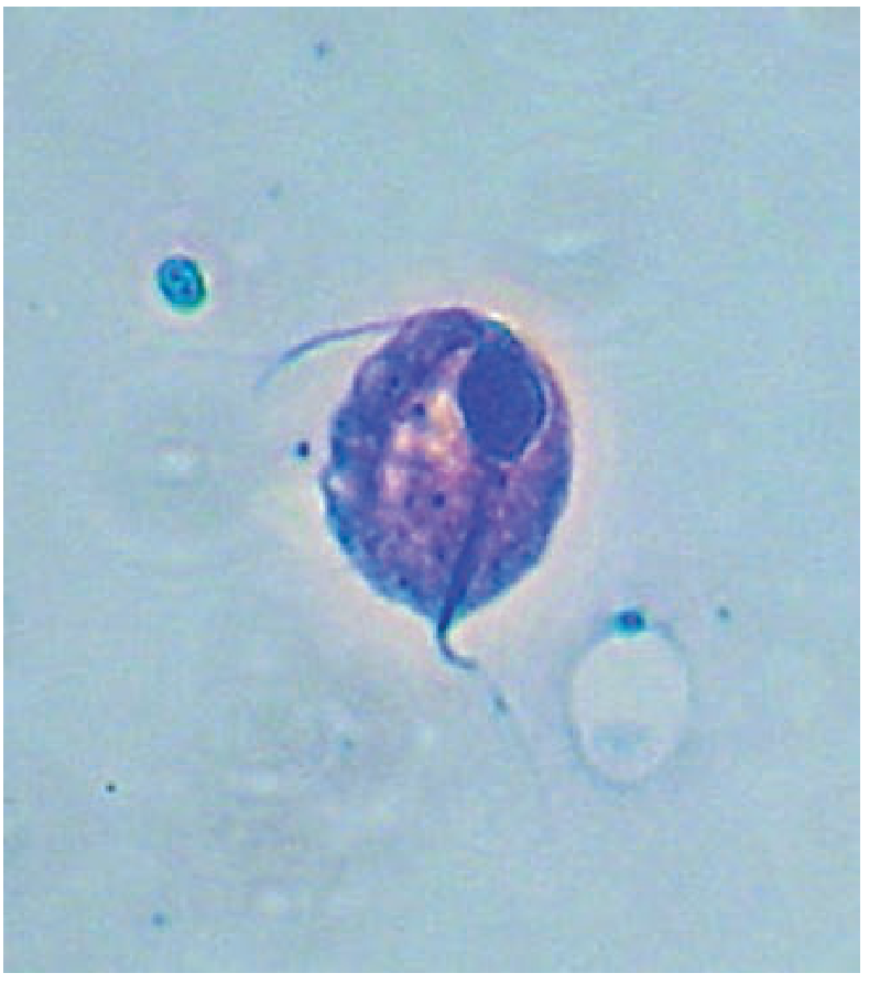
- Saline wet mount: motile trichomonads (sensitivity <50%)
- Culture: 95% sensitive (gold standard) but takes 2-5 days
- NAAT: most sensitive and specific; FDA-approved for vaginal/endocervical/urine specimens
- Nitroimidazoles are the only effective drugs
- Metronidazole 500 mg twice daily × 7 days (preferred; more effective than single dose)
- Tinidazole 2 g orally × 1 dose (fewer GI side effects, useful in metronidazole failure)
- Metronidazole gel is NOT recommended (inadequate tissue levels)
- Treat all sexual partners simultaneously
- Avoid alcohol during treatment and 24 h (metronidazole) or 72 h (tinidazole) after (disulfiram-like reaction)
- If single-dose fails: 7-day course (resistance is quantitative, not absolute)
4. Atrophic Vaginitis (Postmenopausal)
- Vaginal dryness, soreness, itching, dyspareunia
- Thin, scant, yellowish or pinkish discharge; occasionally spotting
- Pale, thin, friable, even ulcerated vaginal epithelium
- Cervix atrophies and may become flush with the vault
- Candida and Trichomonas infections are rare unless using estrogen replacement
- Wet mount: erythrocytes, PMNs, small immature round epithelial cells (parabasal cells)
- Topical vaginal estrogen (cream, pessary, tablet, or vaginal ring) - all are effective
- Systemic estrogen replacement if symptomatic
- Contraindicated in patients with reproductive organ cancer history
- Refer for postmenopausal bleeding to exclude carcinoma
PART B: CERVICITIS
"The ectocervical epithelium can become inflamed by the same organisms responsible for vaginitis. However, N. gonorrhoeae and C. trachomatis infect only the glandular epithelium (endocervix)." - Berek & Novak's Gynecology
Pathogens
| Epithelium Affected | Organisms |
|---|---|
| Ectocervical squamous epithelium | Trichomonas vaginalis, Candida, HSV |
| Endocervical glandular epithelium | N. gonorrhoeae, C. trachomatis, M. genitalium, Ureaplasma urealyticum |
- C. trachomatis accounts for up to 40% of cervicitis seen in STD clinics - Robbins & Kumar
- M. genitalium is found in 10-30% of clinical cervicitis cases - Berek & Novak
- No identifiable organism is found in nearly 50% of cases even with NAAT - Harrison's
Mucopurulent Cervicitis (MPC) - The Clinical Entity
- Yellow mucopurulent discharge from the cervical os (mucopus)
- Endocervical bleeding upon gentle swabbing (friability/ectopy)
- Edematous cervical ectopy
Diagnosis of Cervicitis
- Speculum exam: Inspect endocervical swab against a white/black background - yellow/green colour = mucopus
- Gram stain of mucopus:
-
30 PMNs/high-power field = endocervicitis
- Intracellular Gram-negative diplococci = presumptive gonococcal cervicitis (specific but ≤50% sensitive)
- Gram stain negative for gonococci = presumptive chlamydial cervicitis
-
- NAAT: Mandatory for N. gonorrhoeae and C. trachomatis (most sensitive/specific)
- Concurrent vaginal evaluation for vaginitis is always needed, as BV co-exists with cervicitis commonly - Berek & Novak
Treatment of Cervicitis
| Infection | Regimen |
|---|---|
| Gonococcal cervicitis (first-line) | Ceftriaxone 250-500 mg IM single dose + azithromycin 1 g orally (or doxycycline 100 mg BD × 7 days) |
| Chlamydial cervicitis | Doxycycline 100 mg twice daily × 7 days OR azithromycin 1 g orally × 1 dose |
| M. genitalium | Azithromycin 1 g orally; moxifloxacin if azithromycin-resistant |
| Dual therapy (if gonorrhoea cannot be excluded) | Ceftriaxone + doxycycline/azithromycin |
- Fluoroquinolones are no longer recommended for gonococcal cervicitis due to widespread resistance - Berek & Novak
- Cefixime is no longer first-line for gonorrhoea
- Treat all sexual partners with a similar antibiotic regimen
- Co-existing BV must be treated - if left untreated, cervicitis symptoms and signs persist
COMPARISON TABLE: Vaginitis vs. Cervicitis
| Feature | Vaginitis | Cervicitis |
|---|---|---|
| Site of pathology | Vaginal mucosa | Cervical epithelium |
| Main presenting symptom | Vaginal discharge, itching, odour | Mucopurulent cervical discharge, often asymptomatic |
| Discharge origin | Vaginal walls | Cervical os |
| Organisms | Candida, Trichomonas, Gardnerella/anaerobes | C. trachomatis, N. gonorrhoeae, M. genitalium, HSV |
| pH | Varies by type (see above) | Endocervical pH not used diagnostically |
| Microscopy | Clue cells, pseudohyphae, motile trichomonads | PMNs in cervical mucus; intracellular diplococci |
| STI link | Trichomoniasis (STI); BV and candida are not classical STIs | Almost always STI-related |
| Risk of upper tract spread | BV associated with PID risk | MPC directly precedes PID |
| Partner treatment needed? | Yes for trichomoniasis; not for BV or candida | Always - treat partners |
| Pregnancy risk | BV: preterm birth | MPC: obstetric complications, neonatal herpes (HSV) |
Give seprate information of vaginitis and cervicitis
VAGINITIS
Definition
Anatomy Relevant to Vaginitis
- Lactobacillus species producing lactic acid and hydrogen peroxide
- Vaginal pH normally <4.5 (acidic)
- Oestrogen-dependent glycogen in vaginal epithelial cells (fuel for Lactobacillus)
Types of Vaginitis
1. Bacterial Vaginosis (BV)
- Recent unprotected vaginal intercourse
- Multiple sexual partners / female sex partner
- Vaginal douching
- Antibiotic use
- IUD use (some association)
- Malodorous vaginal discharge (fishy smell)
- Discharge increases after unprotected sex (semen raises pH further)
- Minimal to no vulvar itching or irritation (distinguishes BV from candidiasis)
- Often completely asymptomatic
- Thin, homogeneous, white-grey discharge uniformly coating vaginal walls
- pH >4.5
- No significant vaginal erythema
- Thin white-grey homogeneous discharge
- Vaginal pH >4.5
- Positive Whiff test - fishy amine odour on adding 10% KOH to discharge
- Clue cells on saline wet mount - vaginal epithelial cells coated with coccobacilli, obscured borders, granular appearance
- Increased susceptibility to HIV, C. trachomatis, N. gonorrhoeae
- Preterm birth and spontaneous abortion in pregnancy
- Pelvic inflammatory disease
- Post-operative gynaecological infections
| Drug | Regimen |
|---|---|
| Metronidazole 500 mg oral | Twice daily × 7 days (preferred) |
| Metronidazole gel 0.75% | 5 g intravaginally daily × 5 days |
| Clindamycin cream 2% | 5 g intravaginally nightly × 7 nights |
| Secnidazole | 2 g orally, single dose |
| Tinidazole | 1 g daily × 5 days, OR 2 g daily × 3 days |
| Clindamycin | 300 mg orally twice daily × 7 days |
| Clindamycin ovules | 100 mg intravaginally × 3 nights |
- Treating male partners does not reduce recurrence
- Recurrence is very common; suppressive twice-weekly intravaginal metronidazole gel reduces recurrence
- Vaginal Lactobacillus crispatus CTV-05 (LACTIN-V) reduced recurrence by ~1/3 in a randomised trial - Harrison's
- Pregnancy: Routine screening and treatment does not reduce preterm birth (USPSTF advises against routine screening of asymptomatic pregnant women)
2. Vulvovaginal Candidiasis (VVC)
- Inhibition of normal flora (e.g., systemic antibiotics)
- Reduction of glycogen in vaginal epithelial cells (e.g., diabetes, pregnancy, OCP use)
- Increase in vaginal pH (e.g., menstrual blood, semen)
- Uncomplicated: sporadic, mild-moderate, C. albicans, non-pregnant immunocompetent host
- Complicated: ≥4 episodes/year (recurrent), severe, non-albicans Candida, or abnormal host (uncontrolled DM, immunosuppression, pregnancy)
- Vulvar pruritus - most common and most specific symptom
- Discharge (watery to cottage-cheese-like; can be minimal)
- External dysuria (urine contacts inflamed vulva)
- Dyspareunia
- Exacerbation in the week before menses or after coitus
- Odour is absent - presence of odour favours BV
- Vulvar erythema, oedema, fissures
- Vaginal erythema
- White, curdy or curd-like discharge adhering to vaginal walls
- Normal vaginal pH (4.0-4.5) - key discriminating sign
- Clinical: vaginal pH <4.5 + typical symptoms
- Saline wet mount: budding yeast ± hyphae (sensitivity only 40-60%)
- 10% KOH prep: pseudohyphae and budding yeast visible (sensitivity 80%, specificity ~100%)
- Culture: indicated for recurrent/atypical cases (especially to identify non-albicans species)
- A woman with classic presentation should be treated even if wet mount is negative (low sensitivity)
| Type | Regimen |
|---|---|
| Uncomplicated | Intravaginal azoles (clotrimazole 2% cream × 3 days; miconazole 4% cream × 3 days) OR fluconazole 150 mg orally × 1 |
| Severe | Fluconazole 150 mg every 72 h × 2-3 doses; intravaginal azoles × 7-14 days |
| Recurrent (≥4/year) | Fluconazole 100-200 mg every 72 h × 7-14 days, then once weekly × 6 months |
| Newer option (recurrent) | Otesoconazole 150 mg daily × 7 days, then once weekly × 11 weeks |
- Sexual partner treatment confers no benefit
- Investigate for underlying DM or HIV in recurrent cases
- Candida is not an STI though it can be transmitted sexually
3. Trichomonas Vaginitis
- Profuse vaginal discharge with pruritus and irritation
- Vaginal and vulvar discomfort
- Symptoms develop 5-28 days after exposure
- Many infections are completely asymptomatic (reservoir for transmission)
- Dysuria (urethral involvement)
- Frothy, yellow-green, malodorous discharge (classic)
- Vaginal and vulvar erythema, oedema, tenderness
- "Strawberry cervix" - punctate petechial haemorrhages on the cervix (best seen at colposcopy); pathognomonic when present
- Vaginal pH >5
- Preterm birth and low birth weight
- PID
- Increased risk of HIV acquisition
- Reinfection is common and may mimic treatment failure
- Saline wet mount: motile trichomonads (sensitivity <50% - most common method but misses many)
- Culture: 95% sensitive; gold standard but takes 2-5 days
- NAAT: most sensitive and specific; FDA-approved for vaginal, endocervical, and urine specimens
- Nitroimidazoles are the only effective drug class
- Metronidazole 500 mg twice daily × 7 days (preferred over single dose - more effective)
- Tinidazole 2 g orally × 1 dose (longer half-life, fewer GI side effects; good for metronidazole failure)
- Metronidazole gel is NOT effective (insufficient tissue concentration)
- Treat all sexual partners simultaneously (standard of care)
- Abstain from sex until both partners complete treatment and are asymptomatic
- Avoid alcohol for 24 h (metronidazole) or 72 h (tinidazole) after last dose - disulfiram-like reaction
- If single-dose fails: use 7-day regimen (resistance is quantitative, not absolute)
- CDC hotline for suspected nitroimidazole resistance: 414-718-4141 - Tintinalli's
4. Atrophic Vaginitis
- Vaginal dryness, soreness, itching
- Dyspareunia (most common complaint)
- Occasional spotting or thin yellowish-pink discharge
- Pale, thin, friable, attenuated vaginal mucosa - even ulcerated
- Loss of rugae
- Cervix may atrophy and become flush with vault
- Vaginal pH 5.5-7.0
- Topical vaginal oestrogen: cream, pessary, tablet, or vaginal ring - all effective
- Systemic oestrogen replacement therapy (also prevents atrophic vaginitis)
- Contraindicated in patients with history of reproductive organ cancer
- Any postmenopausal bleeding: refer to gynaecology to exclude carcinoma
Quick Reference - Vaginitis Summary Table
| BV | Candidiasis | Trichomoniasis | Atrophic | |
|---|---|---|---|---|
| Discharge | Thin, white-grey, homogeneous | White, curdy/cottage cheese | Frothy, yellow-green | Thin, scanty, yellow |
| Odour | Fishy (amine) | Absent | Malodorous | Minimal |
| Itch/irritation | Minimal | Marked pruritus | Moderate | Dryness, soreness |
| pH | >4.5 | 3.8-4.5 (normal) | >5 | 5.5-7.0 |
| Wet mount | Clue cells | Pseudohyphae, budding yeast | Motile trichomonads | Parabasal cells, PMNs |
| KOH test | Positive Whiff test | Pseudohyphae visible | Negative | Negative |
| STI? | No | No | Yes | No |
| Treat partner? | No | No | Yes (mandatory) | N/A |
CERVICITIS
Definition
"The majority of cervical lesions are inflammatory (cervicitis). Cervicitis can be subclassified as infectious or noninfectious, although differentiation is difficult due to the presence of normal vaginal flora." - Robbins & Kumar
Anatomy Relevant to Cervicitis
- Ectocervix: stratified squamous epithelium - continuous with vaginal epithelium
- Endocervix: single-layer columnar glandular epithelium
- Transformation zone (TZ): the junction between the two - most susceptible to infection and neoplasia
- Organisms causing vaginitis (Trichomonas, Candida, HSV) can also inflame the ectocervix (squamous epithelium)
- N. gonorrhoeae and C. trachomatis infect only the glandular (columnar) endocervical epithelium - Berek & Novak's Gynecology
Aetiology / Causative Organisms
| Organism | Notes |
|---|---|
| Chlamydia trachomatis | Most common - accounts for up to 40% of cervicitis in STD clinics |
| Neisseria gonorrhoeae | Second most common STI cause; Gram-negative intracellular diplococci |
| Mycoplasma genitalium | Emerging STI; found in 10-30% of clinical cervicitis cases |
| Ureaplasma urealyticum | Less common |
| HSV-2 | Causes ulcerative cervicitis on both squamous and columnar epithelium |
| Trichomonas vaginalis | Causes ectocervicitis (squamous epithelium) |
- Chemical irritants (douches, spermicides, latex)
- Trauma
- Foreign bodies (pessary, IUD, retained tampon)
- Radiation
Mucopurulent Cervicitis (MPC) - The Main Clinical Syndrome
- Equally common
- Caused by the same organisms
- But harder to detect due to nonspecific symptoms and need for pelvic examination
- Most chlamydial and gonococcal infections of the cervix produce no symptoms - Harrison's
Symptoms
- Abnormal vaginal discharge (different from vaginitis - originates from the cervical os)
- Intermenstrual or post-coital bleeding (due to friable cervix)
- Dysuria (if concurrent urethritis)
- Pelvic pain (if ascending infection)
- Dyspareunia
Signs (Cardinal Signs of MPC)
- Yellow/green mucopurulent discharge from the cervical os ("mucopus") - detected by inserting a small cotton swab into the endocervical canal and inspecting against a white background
- Endocervical friability - spontaneous bleeding or bleeding on gentle swabbing
- Oedematous cervical ectopy - the columnar epithelium appearing inflamed, red, granular at the transformation zone
Cervical Ectopy vs. Cervicitis
| Feature | Cervical Ectopy (physiological) | Infectious Cervicitis |
|---|---|---|
| Also called | "Cervical erosion" (misnomer) | MPC |
| What it is | Columnar epithelium extending onto ectocervix | Inflammation of endocervical columnar epithelium |
| Discharge | Clear/slightly cloudy mucus | Yellow/green mucopus |
| Bleeding on touch | May bleed (fragile columnar cells) | Bleeds on gentle swab |
| Colposcopy | Intact epithelium | Inflammatory changes |
| Treatment | None needed (cauterisation NOT warranted) | Antibiotics |
| Association | OCP use, adolescence, early adulthood | STIs |
| Risk | Increases susceptibility to N. gonorrhoeae, C. trachomatis, HIV | - |
- Harrison's Principles of Internal Medicine 22E
Diagnosis
- Remove ectocervical secretions with a large swab
- Insert small cotton swab into endocervical canal → inspect against white/black background
- Yellow/green colour = mucopus = cervicitis
-
20-30 PMNs per 1000× microscopic field (not contaminated by vaginal cells) = endocervicitis
- Intracellular Gram-negative diplococci = presumptive gonococcal cervicitis (specific but ≤50% sensitive)
- Gram stain negative for gonococci = presumptive chlamydial cervicitis
- Must always test for N. gonorrhoeae AND C. trachomatis
- Most sensitive and specific
- Specimens: endocervical swab, vaginal swab, urine
- Also test for M. genitalium where available
- Always evaluate vaginal discharge (wet mount, pH, KOH) as BV and other vaginitis commonly coexist and must be treated together
Complications
- Pelvic Inflammatory Disease (PID) - infection ascending to endometrium, fallopian tubes, ovaries
- Tubal factor infertility
- Ectopic pregnancy
- Chronic pelvic pain
- Fitz-Hugh-Curtis syndrome - perihepatitis (right upper quadrant pain)
- Neonatal complications (maternal transmission):
- HSV: potentially fatal neonatal systemic herpes
- Gonorrhoea: neonatal ophthalmia (ophthalmia neonatorum)
- Chlamydia: neonatal conjunctivitis and pneumonia
- Obstetric complications in pregnancy (preterm labour, premature rupture of membranes)
Treatment
Gonococcal Cervicitis
- First-line: Ceftriaxone 250-500 mg IM single dose
- Always add coverage for chlamydia unless excluded: + doxycycline 100 mg twice daily × 7 days, OR + azithromycin 1 g orally × 1 dose
- Fluoroquinolones are no longer recommended (widespread resistance in N. gonorrhoeae) - Berek & Novak
- Cefixime is no longer first-line for gonorrhoea
Chlamydial Cervicitis
- Doxycycline 100 mg orally twice daily × 7 days (preferred)
- Azithromycin 1 g orally × 1 dose (alternative)
Mycoplasma genitalium Cervicitis
- Azithromycin 1 g orally × 1 dose
- Moxifloxacin if azithromycin resistance suspected (resistance to azithromycin is now recognised)
HSV Cervicitis
- Aciclovir or valaciclovir (standard antiviral regimens for genital herpes)
Key Principles:
- Treat all sexual partners with the same antibiotic regimen - mandatory
- Always treat concurrent BV - untreated BV causes persistence of cervicitis symptoms and signs
- Counsel on STI prevention - condom use, partner notification
- If no gonorrhoea/chlamydia found on NAAT but symptoms persist: consider M. genitalium testing and azithromycin
Summary: Vaginitis vs. Cervicitis at a Glance
| VAGINITIS | CERVICITIS | |
|---|---|---|
| Site | Vaginal walls (epithelium) | Cervix (ecto- and/or endocervix) |
| Primary organisms | Candida, Trichomonas, anaerobes (BV) | C. trachomatis, N. gonorrhoeae, M. genitalium, HSV |
| Symptoms | Discharge, itch, odour, burning | Often asymptomatic; mucopurulent discharge, post-coital bleeding |
| Discharge character | Varies (curdy, frothy, thin, malodorous) | Yellow/green mucopus from cervical os |
| Vaginal pH | Altered (key diagnostic tool) | Not primarily useful |
| STI? | Only trichomoniasis | Almost always STI-related |
| Partner treatment | Only for trichomoniasis | Always required |
| Upper tract risk | BV associated with PID risk | MPC is a direct precursor to PID |
| Pregnancy risk | BV: preterm birth | Neonatal herpes, ophthalmia neonatorum, obstetric complications |
| Key investigation | Wet mount, pH, KOH prep | NAAT for gonorrhoea + chlamydia (mandatory) |
| Treatment | Metronidazole/clindamycin (BV), azoles (candida), nitroimidazoles (trich) | Ceftriaxone ± doxycycline/azithromycin |