
A child presents with a painful swollen neck mass. Ultrasound suggests necrotic lymph nodes. Questions: 1. What diagnosis should be considered? 2. What investigations are useful? 3. What treatment is indicated
Reading File
Now I'll run parallel searches across the medical library for all three questions simultaneously.
Finding Sources
Finding Sources
Finding Sources
Finding Sources
Finding Sources
Finding Sources
Good - found relevant sections. Now I'll read all key sections in parallel.
Reading File
Reading File
Reading File
Excellent content retrieved. Now let me get the Harrison's and Robbins sections for investigations/pathology, plus do a PubMed search for recent guidelines.
Reading File
Reading File
Searching PubMed
cervical lymphadenitis children diagnosis treatment
I now have comprehensive information to provide a thorough, well-cited answer. Let me also fetch the clinical image of TB lymphadenitis in the child.
Loading Image
I now have all the information needed for a comprehensive answer.
Painful Swollen Neck Mass with Necrotic Lymph Nodes in a Child
1. Diagnoses to Consider
The finding of necrotic (caseating) cervical lymph nodes on ultrasound in a child should prompt a prioritized differential:
Primary Diagnosis: Tuberculous Lymphadenitis (Scrofula)
This is the most common form of extrapulmonary TB and a leading cause of necrotic cervical nodes in children. It accounts for 35% of extrapulmonary TB cases worldwide and >40% of cases in the United States in recent series. Key features:
- Most commonly involves the posterior cervical and supraclavicular nodes
- Early nodes are discrete, rubbery, and freely mobile with normal overlying skin
- Over time, nodes become matted and harder; the overlying skin becomes inflamed
- Fluctuance, abscess, or a sinus tract draining caseous material may develop if the node erodes through the skin
- Systemic symptoms (fever, night sweats, weight loss) are uncommon except in HIV-positive patients
- Associated pulmonary disease is present in fewer than 50% of cases
Harrison's Principles of Internal Medicine 22E, p. 1776
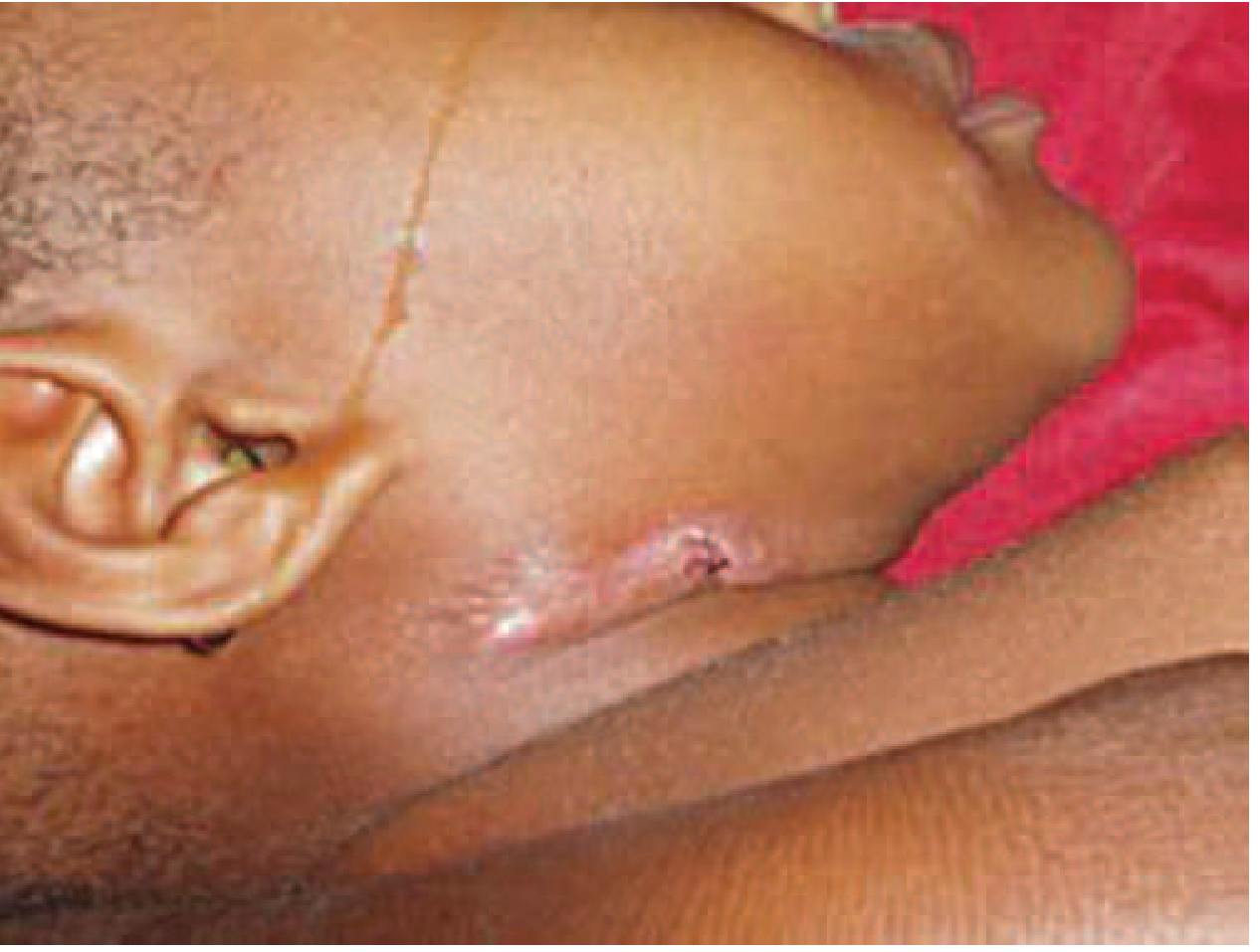
Tuberculous lymphadenitis with sinus tract formation in a young child. From Harrison's Principles of Internal Medicine 22E.
Other Important Differential Diagnoses
| Condition | Key Features |
|---|---|
| Atypical Mycobacteria (NTM) | Very common in young children (<5 years); presents as subcutaneous abscess with violaceous/erythematous overlying skin; systemic symptoms rare |
| Acute Bacterial Lymphadenitis | Staphylococcus aureus, Streptococcus pyogenes; rapid onset, high fever, very tender; may suppurate |
| Cat-Scratch Disease | Bartonella henselae; history of cat exposure; inoculation papule at entry site; usually self-limited |
| Infectious Mononucleosis | EBV; associated with tonsillopharyngitis, fever, malaise; extensive cervical adenopathy |
| Kikuchi-Fujimoto Disease | Necrotizing histiocytic lymphadenitis; young women/children; painful posterior cervical nodes; self-limiting but can mimic TB or lymphoma |
| Lymphoma | Progressive, painless, "rubbery" nodes; B symptoms (fever, night sweats, weight loss >10%); urgent exclusion required |
| Suppurative Bacterial Abscess | Fluctuant, erythematous; may require drainage |
Bailey and Love's Short Practice of Surgery 28th Ed., p. 1158; Textbook of Family Medicine 9e, p. 448; Harrison's 22E
2. Investigations
Imaging
- Ultrasound (already done): Necrotic/caseating nodes have a characteristic appearance with central liquefaction and absent hilum. Doppler may show peripheral vascularity. Can also guide FNA.
- Chest X-ray: Look for pulmonary TB (hilar adenopathy, consolidation, cavitation) - though pulmonary disease is present in <50% of TB lymphadenitis cases.
- CT neck/chest: Better delineates extent, identifies matted nodes, abscess formation, and mediastinal involvement.
Microbiological / Immunological
- Fine-Needle Aspiration (FNA) - the cornerstone diagnostic procedure:
- Send for: AFB smear (positive in ~20% only), mycobacterial culture (positive in 70-80%), cytology/histology
- Overall sensitivity 77%, specificity 93% for TB
- Yield up to 80% in Harrison's series
- Tuberculin Skin Test (TST/Mantoux) or IGRA (Interferon-Gamma Release Assay): Supports TB diagnosis; IGRA preferred in BCG-vaccinated children
- AFB smear and culture on aspirated material
- Nucleic Acid Amplification Test (NAAT/PCR for MTB): Rapid, useful when AFB smear is negative
- GeneXpert MTB/RIF: Rapid TB detection plus rifampicin resistance in <2 hours
Haematological / Biochemical
- CBC with differential: Leukocytosis suggests acute bacterial infection; lymphocytosis/atypical lymphocytes in EBV
- ESR, CRP: Elevated in most infectious causes; markedly elevated in lymphoma
- Monospot / EBV serology: If infectious mononucleosis is suspected
- Bartonella henselae serology: If cat-scratch disease suspected
- LDH, uric acid: If lymphoma is in the differential
Histopathology
- Excisional biopsy (when FNA is non-diagnostic or lymphoma is suspected): Caseating granulomas with Langhans giant cells are characteristic of TB. Kikuchi disease shows necrotizing histiocytic lymphadenitis without granulomas.
Rosen's Emergency Medicine, p. 3263-3265; Harrison's 22E, p. 1776; Textbook of Family Medicine 9e, p. 447
3. Treatment
Tuberculous Lymphadenitis
- Standard anti-TB regimen (same as pulmonary TB):
- Intensive phase (2 months): Isoniazid (H) + Rifampicin (R) + Pyrazinamide (Z) + Ethambutol (E) - 2HRZE
- Continuation phase (4 months): Isoniazid + Rifampicin - 4HR
- Total 6-month regimen (9 months if meningeal or bone TB)
- Nodes not responding to medical treatment should be excised
- Incision and drainage (I&D) is contraindicated - causes permanent sinus formation and prolonged drainage
Rosen's Emergency Medicine, p. 3265; Textbook of Family Medicine 9e, p. 448
Atypical Mycobacteria (NTM)
- Excisional biopsy is both diagnostic and curative - the treatment of choice
- Appropriate antibiotics based on culture sensitivities (e.g., macrolides, rifabutin)
- Incisional drainage is contraindicated - leads to chronic fistulization
Textbook of Family Medicine 9e, p. 445
Acute Bacterial Lymphadenitis
- Antibiotics targeting Staph. aureus and Streptococcus (e.g., amoxicillin-clavulanate, flucloxacillin)
- Treat the primary focus (tonsillitis, dental abscess)
- If fluctuant abscess: incision and drainage is appropriate
- Nodes not responding after 4-6 weeks should be referred for further evaluation
Bailey and Love's 28th Ed., p. 1148; Textbook of Family Medicine 9e, p. 445
Cat-Scratch Disease
- Self-limited; conservative management
- Azithromycin may shorten the course in moderate-severe cases
Key Practical Points
- Do not incise and drain a suspected TB or NTM node - this invariably causes a chronic discharging sinus.
- Necrotic nodes on ultrasound in a child = TB until proven otherwise in high-prevalence settings.
- FNA is the first-line diagnostic step for suspected scrofula - it avoids the need for open surgery in most cases.
- Always rule out lymphoma before attributing chronically enlarged nodes to infection.
- In immunocompromised children (HIV), TB lymphadenitis tends to be generalized with heavier bacterial loads and atypical granulomas.
Sources:
- Harrison's Principles of Internal Medicine 22E, Ch. 183
- Bailey and Love's Short Practice of Surgery 28th Ed., Ch. 52
- Rosen's Emergency Medicine, Ch. 24
- Textbook of Family Medicine 9e, Ch. 18
- Robbins & Kumar Basic Pathology, Ch. 11
Recent evidence: A 2024 systematic review on nontuberculous cervicofacial lymphadenitis (PMID 37659223) emphasizes the diagnostic challenges distinguishing NTM from TB cervical adenitis and supports excisional biopsy as both diagnostic and therapeutic for NTM in children - consistent with the textbook guidance above.
This is a shared conversation. Sign in to Orris to start your own chat.