
Give me structured approach to localisation of stroke, absolutely detail
Reading File
Finding Sources
Finding Sources
Finding Sources
Finding Sources
Finding Sources
Finding Sources
Finding Sources
Finding Sources
Reading File
Finding Sources
Reading File
Reading File
Reading File
Reading File
Reading File
Loading Image
I now have all the material I need. Let me compose the comprehensive, detailed structured answer on stroke localization.
Structured Approach to Localisation of Stroke
Based on Bradley and Daroff's Neurology in Clinical Practice and Localization in Clinical Neurology, 8e
STEP 1 - THE FUNDAMENTAL FRAMEWORK: TWO CIRCULATIONS
Before anything else, classify every stroke into one of two circulations. This single step halves your differential immediately.
| Anterior (Carotid) Circulation | Posterior (Vertebrobasilar) Circulation | |
|---|---|---|
| Vessels | ICA, MCA, ACA, anterior choroidal, ophthalmic | Vertebral, basilar, PICA, AICA, SCA, PCA |
| Territory | Frontal, parietal, temporal (most), basal ganglia, internal capsule | Brainstem, cerebellum, occipital, inferior temporal, thalamus |
| Key clues | Language (left), neglect (right), arm > leg weakness, retinal symptoms | Diplopia, dysphagia, dysarthria, ataxia, crossed deficits, vertigo |
STEP 2 - ANATOMICAL LEVEL: WHERE IN THE NEURAXIS?
Work systematically from cortex down. Each level has a signature.
Level 1 - Cerebral Cortex
Signature features:
- Higher cortical functions affected (aphasia, apraxia, agnosia, neglect, hemineglect)
- Cortical sensory loss (astereognosis, agraphesthesia, two-point discrimination impaired)
- Contralateral homonymous visual field defect (if occipital or parietal-occipital involvement)
- Jacksonian march or focal seizures may occur at onset
- No "crossed" deficits (cranial nerve on one side + limb on the other)
- Face + arm > leg weakness (MCA territory) OR leg > arm weakness (ACA territory)
Differentiating cortical from subcortical: Cortical strokes produce cortical signs (aphasia, neglect, agnosia). Pure motor or pure sensory deficits without cortical signs suggest subcortical or lacunar pathology.
Level 2 - Subcortical White Matter / Internal Capsule / Basal Ganglia
Signature features:
- Pure motor hemiparesis (posterior limb of internal capsule): face + arm + leg equally affected
- No cortical signs (no aphasia, no neglect, no visual field defect)
- Lacunar syndromes dominate (see Step 4)
- Homogeneous, equal weakness - the "capsular stroke" pattern
- Striato-capsular infarction (proximal MCA lenticulostriate branches): large area, may have cortical signs if extensive
Level 3 - Brainstem
Signature features - the crossed deficit:
- Ipsilateral cranial nerve palsy + contralateral limb weakness/sensory loss
- This crossing pattern is pathognomonic of brainstem localization
- Specific level determined by which cranial nerve is involved:
- CN III palsy → midbrain
- CN VI or VII palsy → pons
- CN IX, X, XII palsy → medulla
Other brainstem clues: Horner syndrome (any level), internuclear ophthalmoplegia (medial longitudinal fasciculus, pons), "one-and-a-half" syndrome (pons), bilateral limb signs, impaired consciousness with small lesions.
Level 4 - Cerebellum
Signature features:
- Ipsilateral limb ataxia (cerebellar connections are ipsilateral)
- Gait ataxia, truncal ataxia
- Nystagmus (horizontal or direction-changing)
- Dysarthria (scanning speech)
- NO weakness, NO sensory loss (pure cerebellar lesion)
- Vermis lesions: truncal ataxia, gait ataxia
- Hemispheric lesions: ipsilateral limb dysmetria, dysdiadochokinesia
- Beware: large cerebellar infarcts can cause obstructive hydrocephalus and herniation
STEP 3 - VASCULAR TERRITORY SYNDROMES: ANTERIOR CIRCULATION
3A - Middle Cerebral Artery (MCA) - Most Common Stroke Territory
Anatomy: MCA gives lenticulostriate branches (subcortical) and cortical branches (superior and inferior divisions).
Complete MCA Occlusion (main stem):
- Contralateral hemiplegia (face + arm > leg)
- Contralateral hemisensory loss
- Contralateral homonymous hemianopia
- Left MCA: Broca's aphasia (superior division) or Wernicke's aphasia (inferior division) or global aphasia (complete occlusion)
- Right MCA: Hemispatial neglect, hemi-inattention, constructional apraxia, anosognosia (denial of deficit)
- Gaze deviation toward the lesion (frontal eye field involvement)
- Dysarthria
Superior Division MCA:
- Broca's aphasia (left hemisphere): non-fluent, effortful speech, preserved comprehension
- Contralateral face + arm weakness > leg
- No homonymous hemianopia
Inferior Division MCA:
- Wernicke's aphasia (left): fluent, paraphasic, impaired comprehension
- Contralateral superior homonymous quadrantanopia ("pie in the sky")
- Contralateral neglect (right hemisphere)
MCA Lenticulostriate (Subcortical):
- Contralateral hemiparesis with little sensory involvement
- Pure motor stroke (one cause) - see lacunar section
- Striatocapsular infarction: hemiplegia, hypesthesia, visual field defect, transient aphasia
3B - Anterior Cerebral Artery (ACA)
Territory: Medial surface of the frontal and parietal lobes, anterior corpus callosum, anterior internal capsule.
Syndrome:
- Contralateral leg > arm weakness (homunculus: leg is on the medial surface)
- Contralateral leg > arm sensory loss
- Gait apraxia, urinary incontinence (frontal lobe)
- Bilateral ACA occlusion (rare): Akinetic mutism - patient lies with eyes open, follows visual stimuli but is mute and akinetic; caused by bilateral cingulate gyrus infarction
- Transcortical motor aphasia (left ACA): non-fluent speech, preserved repetition (unlike Broca's)
- Grasp reflex, snout reflex (frontal release signs)
- Callosal disconnection syndrome (anterior corpus callosum): left hand apraxia, alien hand syndrome
3C - Anterior Choroidal Artery
Arises from the ICA just distal to the posterior communicating artery.
Classic triad (incomplete in most cases):
- Contralateral hemiplegia (posterior limb, internal capsule)
- Contralateral hemisensory loss (thalamus, posterior limb)
- Contralateral homonymous hemianopia (lateral geniculate body, optic tract, retrolenticular internal capsule)
3D - Internal Carotid Artery (ICA)
Occlusion findings depend on collateral flow:
- If good collaterals: may be silent
- If poor collaterals: MCA + ACA territory infarction (massive hemispheric stroke)
- Ipsilateral monocular visual loss (amaurosis fugax or permanent): ophthalmic artery involvement - this is a hallmark of ICA disease
- Contralateral hemiplegia, hemisensory loss
- Ipsilateral Horner syndrome (sympathetic fibers travel with ICA)
STEP 4 - LACUNAR SYNDROMES (Small Vessel / Penetrating Artery Occlusion)
Lacunar infarcts are small (< 15 mm), deep, caused by lipohyalinosis or microatheroma of penetrating arteries. Five classic syndromes:
| Syndrome | Location | Features |
|---|---|---|
| Pure Motor Hemiplegia | Posterior limb internal capsule OR pons OR corona radiata | Face + arm + leg equal weakness, NO sensory, NO cortical signs - most common lacunar syndrome |
| Pure Sensory Stroke | Thalamus (ventral posterolateral nucleus) | Contralateral hemisensory loss all modalities, NO motor deficit; described by C.M. Fisher |
| Sensorimotor Stroke | Posterior limb internal capsule + thalamus, or thalamocapsular | Motor + sensory, no cortical signs |
| Ataxic Hemiparesis | Pons OR posterior limb internal capsule OR corona radiata | Ipsilateral cerebellar ataxia + contralateral hemiparesis, leg > arm; "homolateral ataxia and crural paresis" |
| Dysarthria-Clumsy Hand | Pons OR internal capsule genu | Dysarthria + facial weakness + contralateral hand clumsiness/weakness |
Key principle: Lacunar syndromes have NO aphasia, NO visual field defect, NO cognitive change, NO hemineglect - absence of cortical signs with pure motor or sensory deficit points strongly to lacune.
STEP 5 - POSTERIOR CIRCULATION SYNDROMES
5A - Posterior Cerebral Artery (PCA)
Territory: Occipital cortex, inferior temporal lobe, thalamus (penetrating branches), midbrain.
Cortical PCA (P2 segment onwards):
- Contralateral homonymous hemianopia - the signature finding; macular sparing common (macular cortex has MCA collaterals)
- Left PCA: Alexia without agraphia (patient cannot read but can write - disconnection of visual cortex from language areas), anomic aphasia, visual agnosia
- Right PCA: Prosopagnosia (failure to recognize faces), topographical disorientation
- Bilateral PCA occlusion: Cortical blindness - patient is blind but denies it (Anton syndrome = cortical blindness + anosognosia); Balint syndrome (bilateral parieto-occipital) = optic ataxia + ocular apraxia + simultanagnosia
Penetrating PCA (thalamo-perforating branches):
- Aphasia (left pulvinar involvement)
- Global amnesia (bilateral hippocampal-parahippocampal)
- Akinetic mutism
- Dejerine-Roussy syndrome (thalamic pain syndrome): Contralateral sensory loss all modalities + severe contralateral dysesthesias/thalamic pain + vasomotor changes + transient hemiparesis + choreoathetoid movements; caused by thalamogeniculate branch occlusion
5B - Thalamic Infarction Syndromes
The thalamus receives blood from 4 vascular regions:
| Territory | Vessel | Syndrome |
|---|---|---|
| Posterolateral | Thalamogeniculate (P2 segment PCA) | Pure sensory stroke, sensorimotor stroke, Dejerine-Roussy syndrome |
| Anterior | Polar/tuberothalamic artery (posterior communicating) | Amnesia, confusion, personality change, aphasia (left), neglect (right) |
| Paramedian | Thalamo-perforating artery (P1 segment PCA) | Profound amnesia, vertical gaze palsy, somnolence; Bilateral = artery of Percheron occlusion: bilateral paramedian thalamic infarction with coma |
| Dorsal (pulvinar) | Posterior choroidal arteries | Language disturbance (left), neglect (right), visual field defects |
Artery of Percheron: A single thalamoperforating artery arising from the P1 segment supplying both paramedian thalami. Occlusion causes bilateral thalamic infarction with coma or hypersomnolence, profound amnesia, and vertical gaze palsy - all without motor signs.
5C - Vertebrobasilar Territory - Brainstem Syndromes
The "Crossed Deficit" Rule:
Ipsilateral CN palsy + contralateral hemiplegia/hemisensory loss = brainstem lesion, level determined by CN involved.
MIDBRAIN (CN III, IV)
Weber Syndrome (ventral midbrain - cerebral peduncle):
- Ipsilateral CN III palsy (ptosis, dilated pupil, "down and out" eye)
- Contralateral hemiplegia
Benedikt Syndrome (tegmentum midbrain - red nucleus):
- Ipsilateral CN III palsy
- Contralateral tremor, ataxia, choreoathetosis (red nucleus involvement)
Parinaud Syndrome (dorsal midbrain - superior colliculus, pretectal area):
- Upgaze palsy (vertical gaze paresis)
- Convergence-retraction nystagmus on attempted upgaze
- Light-near dissociation of pupils
- Often from posterior thalamo-midbrain perforator or posterior circulation stroke
Claude Syndrome (combination of Benedikt + superior cerebellar peduncle):
- CN III palsy + contralateral cerebellar ataxia
PONS (CN V, VI, VII)
Millard-Gubler Syndrome (ventral pons):
- Ipsilateral CN VI palsy (lateral gaze palsy, diplopia)
- Ipsilateral CN VII palsy (lower motor neuron facial weakness)
- Contralateral hemiplegia
Foville Syndrome (tegmental pons, PPRF + CN VI nucleus):
- Ipsilateral horizontal gaze palsy (eyes deviate away from lesion - in cortical stroke eyes deviate toward lesion)
- Ipsilateral CN VII palsy
- Contralateral hemiplegia
- Key diagnostic point: In pontine strokes, gaze deviation is away from the lesion (PPRF destroyed); in cortical/hemispheric strokes, gaze deviation is toward the lesion (FEF pushes eyes to the opposite side)
One-and-a-Half Syndrome (paramedian pons - PPRF + MLF):
- Ipsilateral gaze palsy (PPRF/CN VI nucleus lesion)
- Ipsilateral internuclear ophthalmoplegia (MLF lesion)
- Net result: only the contralateral eye can abduct; all other horizontal movements are lost
Locked-In Syndrome (bilateral ventral pons - bilateral corticospinal and corticobulbar tracts):
- Complete quadriplegia and anarthria
- Preserved consciousness, preserved vertical eye movements (RIMLF spared)
- Patient communicates only by blinking or vertical eye movements
MEDULLA (CN IX, X, XI, XII)
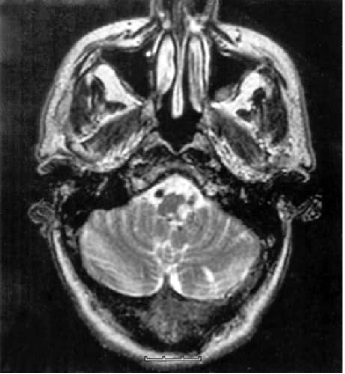
Lateral Medullary Syndrome (Wallenberg Syndrome) - PICA or vertebral artery occlusion:
This is the most clinically important posterior circulation syndrome. Caused by occlusion of the PICA or vertebral artery, producing an infarct in the posterolateral medulla.
| Structure damaged | Feature |
|---|---|
| Spinothalamic tract | Contralateral loss of pain and temperature (limbs and trunk) |
| Descending trigeminal nucleus | Ipsilateral facial loss of pain and temperature |
| Vestibular nuclei | Vertigo, nausea, vomiting, nystagmus |
| Nucleus ambiguus (CN IX, X) | Ipsilateral palatal palsy, dysarthria, dysphagia, hoarseness |
| Descending sympathetic fibers | Ipsilateral Horner syndrome (ptosis, miosis, anhidrosis) |
| Inferior cerebellar peduncle / cerebellum | Ipsilateral limb and gait ataxia |
| Solitary nucleus / dorsal vagal nucleus | Hiccups |
Classic "crossed sensory deficit": Ipsilateral face loss of pain/temperature + contralateral body loss of pain/temperature (thermoanalgesia). Motor power is characteristically SPARED (corticospinal tracts are in the ventral medulla, not affected in lateral medullary syndrome).
Medial Medullary Syndrome (Dejerine's Anterior Bulbar Syndrome) - ASA or vertebral artery branch:
| Structure | Feature |
|---|---|
| Corticospinal tract (pyramids) | Contralateral hemiplegia (spares face) |
| Medial lemniscus | Contralateral loss of discriminative touch, vibration, proprioception |
| CN XII (hypoglossal nerve) | Ipsilateral tongue weakness/atrophy (tongue deviates toward lesion) |
5D - Cerebellar Artery Territories
| Artery | Territory | Key Syndrome |
|---|---|---|
| PICA (Posterior Inferior Cerebellar Artery) | Posteroinferior cerebellum + lateral medulla | Wallenberg syndrome (lateral medullary); cerebellar infarct causing vertigo, ataxia, possible obstructive hydrocephalus |
| AICA (Anterior Inferior Cerebellar Artery) | Anteroinferior cerebellum + lateral caudal pons | Ipsilateral CN VII + VIII involvement (facial palsy + deafness + tinnitus) + lateral pontine syndrome + ipsilateral Horner; AICA infarct should be suspected when lateral pontine features accompany hearing loss |
| SCA (Superior Cerebellar Artery) | Superior cerebellum + lateral rostral pons | Ipsilateral cerebellar ataxia (severe), ipsilateral Horner, ipsilateral CN IV palsy, contralateral hemisensory loss (spinothalamic), ipsilateral CN V partial deficit; contralateral loss of pain/temperature |
STEP 6 - SPECIAL LOCALIZING CLUES: CLINICAL PEARLS
Visual Field Defects - Localizing Value
| Defect | Localization |
|---|---|
| Monocular blindness | Retina or optic nerve (ipsilateral) - anterior to chiasm |
| Bitemporal hemianopia | Optic chiasm |
| Contralateral homonymous hemianopia | Optic tract, LGN, optic radiation, occipital cortex |
| Superior quadrantanopia ("pie in sky") | Inferior optic radiation, temporal lobe (Meyer's loop) |
| Inferior quadrantanopia ("pie in floor") | Superior optic radiation, parietal lobe |
| Macular-sparing hemianopia | Occipital cortex (MCA/PCA boundary zone at occipital pole) |
Gaze Deviation Lateralizing Rule
| Gaze deviation | Meaning |
|---|---|
| Eyes deviate TOWARD the hemiplegia side | Cortical/hemispheric stroke (FEF pushes eyes to the opposite hemisphere's territory, i.e., away from lesion, but patient presents with eyes toward ipsilateral side to lesion) - eyes look "away" from the hemiplegic limbs but "toward" the lesion |
| Eyes deviate AWAY from hemiplegia | Pontine stroke (PPRF destroyed; contralateral PPRF pulls eyes to that side) |
Practical rule: "The eyes look toward a cortical stroke and away from a pontine stroke."
Language as a Lateralizing Tool (Left Hemisphere = Dominant in 90%)
| Aphasia Type | Localization | Key Features |
|---|---|---|
| Broca's (expressive) | Left inferior frontal (Broca's area, BA44/45), superior MCA division | Non-fluent, telegraphic, effortful; comprehension intact; patient aware of deficit |
| Wernicke's (receptive) | Left posterior superior temporal (BA 22), inferior MCA division | Fluent, paraphasic, neologisms; comprehension severely impaired; patient unaware |
| Global | Large left hemisphere (Broca + Wernicke areas both) | Non-fluent + poor comprehension + poor repetition |
| Conduction | Left arcuate fasciculus (connects Broca and Wernicke) | Fluent, good comprehension, severely impaired repetition; paraphasic errors |
| Transcortical Motor | Left frontal (ACA territory, anterior to Broca's) | Non-fluent, but repetition preserved (unlike Broca's) |
| Transcortical Sensory | Left parieto-occipital (posterior watershed) | Fluent, poor comprehension, repetition preserved (echolalia) |
| Alexia without agraphia | Left PCA territory (left occipital cortex + splenium of corpus callosum) | Cannot read, can write; disconnects right visual cortex from language areas |
Hemispatial Neglect
- Right hemisphere (usually right parietal or frontoparietal) causes left hemispatial neglect
- Patient ignores left side of space, left side of body
- Line bisection test: bisects to the right of midline
- Much more severe and persistent with right hemispheric strokes than left
Horner Syndrome Localizing Value in Stroke Context
- Central Horner (hypothalamus to ciliospinal center of Budge, C8-T2): Any stroke affecting the lateral hypothalamus, lateral brainstem (especially lateral medullary), or cervical cord
- In posterior circulation stroke: Ipsilateral Horner + ipsilateral ataxia + ipsilateral CN palsy = lateral brainstem involvement
- Horner syndrome without anhidrosis suggests post-ganglionic; with anhidrosis (face and body) suggests pre-ganglionic or central
STEP 7 - CORTICAL vs. SUBCORTICAL vs. BRAINSTEM DIFFERENTIATION TABLE
| Feature | Cortical | Subcortical/Lacunar | Brainstem | Cerebellar |
|---|---|---|---|---|
| Aphasia | YES (left) | NO | NO (except peduncle lesions) | NO |
| Neglect | YES (right) | NO | NO | NO |
| Visual field defect | YES (hemianopia) | NO (unless thalamocapsular) | YES (rare cortical blindness) | NO |
| Face + arm + leg equal weakness | Rarely | YES (capsular) | YES (with CN palsy) | NO |
| Crossed deficits | NO | NO | YES - hallmark | NO |
| Cerebellar signs | NO | NO | Possible (peduncle) | YES (ipsilateral) |
| Consciousness affected | If large | Rarely | YES if basilar | Late (herniation) |
| Seizures at onset | Possible | Rare | NO | NO |
STEP 8 - THE OCSP (OXFORD/BAMFORD) CLINICAL CLASSIFICATION
A practical bedside classification requiring no imaging:
| Category | Criteria | Vascular Territory |
|---|---|---|
| TACI - Total Anterior Circulation Infarct | All 3: motor/sensory deficit + cortical sign + homonymous hemianopia | Complete MCA or ICA territory |
| PACI - Partial Anterior Circulation Infarct | 2 of 3 above, or cortical sign alone | MCA branch territory |
| LACI - Lacunar Infarct | Pure motor, pure sensory, sensorimotor, or ataxic hemiparesis; NO cortical signs | Small perforating artery |
| POCI - Posterior Circulation Infarct | Any of: brainstem, cerebellar, bilateral motor/sensory, crossed deficit, isolated hemianopia | Vertebrobasilar |
Clinical utility: TACI has worst prognosis (60% dead or dependent at 1 year); LACI best (10-15% dead or dependent). OCSP classification predicts functional outcome, etiology, and recurrence risk.
STEP 9 - INTEGRATING THE APPROACH - BEDSIDE ALGORITHM
SUDDEN FOCAL DEFICIT
        ↓
1. CORTICAL SIGNS? (aphasia, neglect, visual agnosia, cortical sensory loss)
   YES → Cortical involvement → Anterior (MCA/ACA/PCA) or large vessel
   NO  → Subcortical / brainstem / cerebellum
        ↓
2. CROSSED DEFICIT? (ipsilateral CN + contralateral limb)
   YES → BRAINSTEM (identify CN → level: III=midbrain, VI/VII=pons, IX/X/XII=medulla)
   NO  → Not brainstem
        ↓
3. CEREBELLAR SIGNS WITHOUT WEAKNESS?
   YES → Cerebellar (identify artery by co-features: PICA=lateral medulla, AICA=hearing, SCA=high ataxia)
   NO  → Continue
        ↓
4. PURE MOTOR / PURE SENSORY / ATAXIC HEMIPARESIS?
   YES → LACUNAR - no cortical signs confirm
   NO  → Continue
        ↓
5. VISUAL FIELD DEFECT?
   Hemianopia + language → MCA (left) or PCA (left or right)
   Hemianopia alone, macular sparing → PCA
   Monocular → Retinal / optic nerve (carotid territory)
        ↓
6. ARM > LEG WEAKNESS → MCA
   LEG > ARM WEAKNESS → ACA
   Equal face + arm + leg → Internal capsule / pons
STEP 10 - IMAGING CORRELATION
| Syndrome | Expected Imaging Finding |
|---|---|
| MCA territory | CT: wedge hypodensity frontal-parietal-temporal; loss of insular ribbon; MRI DWI: bright cortical + subcortical |
| Lacunar | CT: small deep hypodensity (often missed acutely); MRI: small DWI-bright lesion < 15mm in basal ganglia, capsule, pons, thalamus |
| PCA | CT: occipital hypodensity; MRI DWI: occipital, thalamic, and hippocampal involvement |
| Lateral medullary (Wallenberg) | CT: often normal; MRI T2/DWI: posterolateral medullary hyperintensity; may see vertebral/PICA poor flow void |
| Basilar artery occlusion | CT: "dense basilar" sign; CT angiography: cut-off at basilar; MRI: bilateral pontine, thalamic, occipital DWI changes |
| Artery of Percheron | MRI: butterfly-shaped bilateral paramedian thalamic DWI lesion; may see mesencephalic involvement |
SUMMARY MEMORY AID
- MCA (left): Aphasia + contralateral face/arm hemiplegia + hemianopia
- MCA (right): Neglect + anosognosia + contralateral hemiplegia
- ACA: Leg > arm + frontal release signs + akinetic mutism (bilateral)
- PCA: Hemianopia (macular sparing) + alexia without agraphia (left) + thalamic pain
- Lacune: Pure motor / pure sensory / sensorimotor / ataxic hemiparesis - NO cortical signs
- Wallenberg: Crossed sensory (ipsilateral face / contralateral body) + Horner + dysphagia + ataxia + NO motor weakness
- Medial medulla: Contralateral hemiplegia (spares face) + contralateral dorsal column loss + ipsilateral tongue palsy
- Pons (ventral): Locked-in / one-and-a-half / eyes deviate away from lesion
- Midbrain: CN III palsy + contralateral hemiplegia (Weber) or tremor (Benedikt)
- Thalamus (paramedian, bilateral): Artery of Percheron → coma + amnesia + vertical gaze palsy - NO motor deficit
Sources: Bradley and Daroff's Neurology in Clinical Practice; Localization in Clinical Neurology, 8e (Brazis et al.)
This is a shared conversation. Sign in to Orris to start your own chat.