
Bleeding from penish paediatrics
bleeding from penis paediatric causes management
https://www.ebmedicine.net/topics/genitourinary/pediatric-em…
paraphimosis paediatric penile
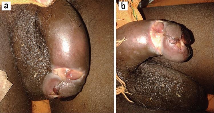
Two-panel clinical photograph (a and b) illustrating a severe case of paraphimosis leading to penile gangrene. Image (a) shows the penis in a state of acute vascular compromise. The glans penis is markedly discolored with a dark purple to black hue, indicative of ischemia. A clear paraphimotic ring is visible where the retracted foreskin is trapped behind the coronal sulcus, causing significant mechanical constriction and proximal shaft edema. Image (b) shows the progression to dry gangrene. The glans exhibits extensive tissue necrosis, sloughing of the skin, and ulceration. The structural integrity is compromised with visible loss of viable tissue and a shriveled, distorted appearance. The anatomical focus is the urogenital system, specifically demonstrating the complications of untreated paraphimosis in an adult male. This visual resource is intended for medical education regarding urological emergencies and the identification of necrotic skin lesions in a clinical setting.
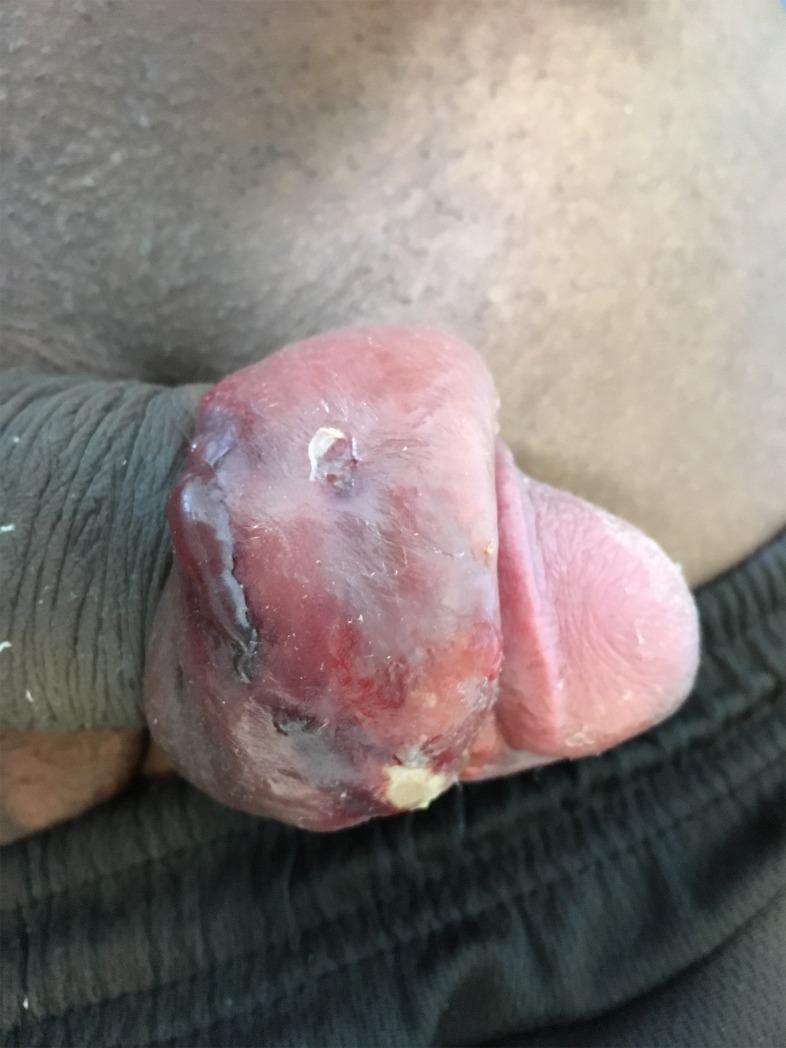
This clinical photograph illustrates a case of infectious paraphimosis, a urological emergency, following the subcutaneous insertion of a penile implant. The image shows the penis with the foreskin severely retracted and trapped behind the glans penis, forming a tight, constricting band. There is marked, circumferential edema and distortion of the prepuce and glans. The tissue exhibits a mottled appearance with varied coloration, including intense erythema and dark reddish-purple areas, suggesting severe inflammation and vascular congestion. Visible signs of infection include whitish purulent exudate on the surface of the swollen tissue and small areas of skin breakdown or ulceration. Superficial skin lacerations are present, potentially serving as the portal of entry for pathogens. This image serves as an educational example of short-term postoperative complications and infectious sequelae associated with non-medical penile modifications.
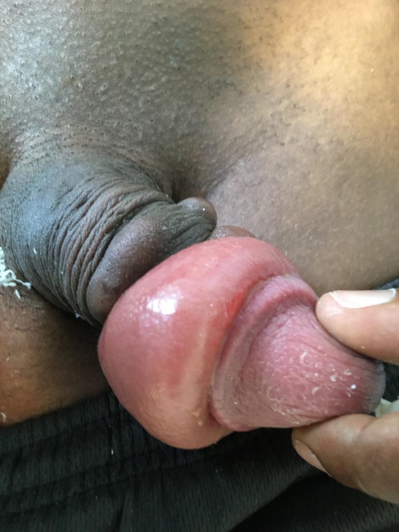
This clinical photograph displays a case of infectious paraphimosis, a urological emergency, occurring as a short-term complication following the insertion of subcutaneous penile implants. The image shows the glans penis in a state of significant edema and erythema. A constricting band of retracted foreskin is visible proximal to the glans, leading to venous and lymphatic congestion. The glans itself exhibits a shiny, tense surface texture with visible inflammation. Distally, the tissue appears somewhat desiccated with small amounts of white, flaky discharge or epithelial desquamation present. The surrounding scrotal skin and lower abdomen are visible, and the clinician's gloved hand is seen retracting the tissue to demonstrate the extent of the swelling and the characteristic ‘doughnut’ appearance of the edematous prepuce. This visual serves as an educational example of post-procedural infectious complications and the clinical presentation of paraphimosis in the context of foreign body insertion.
balanitis xerotica obliterans BXO child
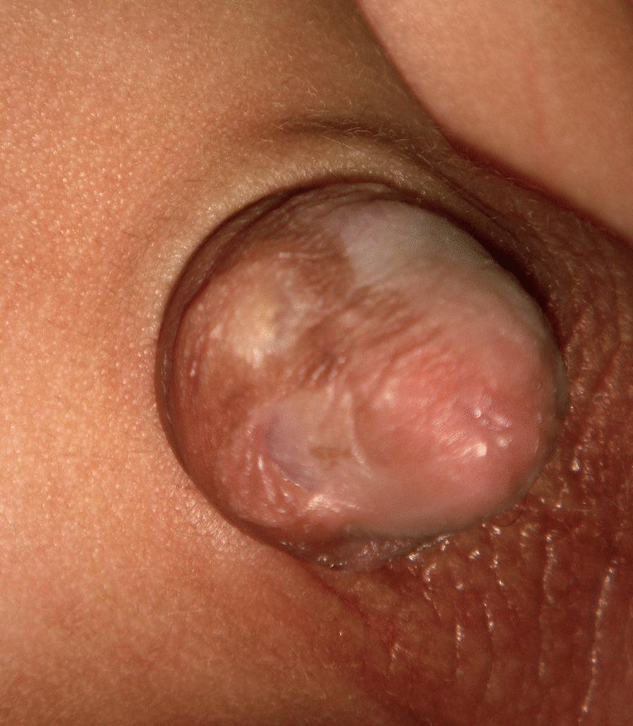
This clinical photograph displays the glans penis of a circumcised patient, illustrating sequelae of balanitis xerotica obliterans (BXO), also known as lichen sclerosus. The glans exhibits a variegated or mottled appearance characterized by areas of hyperpigmentation and atrophic hypopigmentation (whitening). The surface texture is notably altered, showing cicatricial scarring, thickening, and loss of normal mucosal smoothness. Distinctive features include a circular hyperpigmented scar near the coronal sulcus and evidence of skin bridges—fibrous adhesions connecting the penile shaft skin to the glans. The urinary meatus area appears involved with sclerotic changes, typical of chronic inflammatory progression in BXO. This image serves as an educational example of post-circumcision pigmentary and structural complications related to chronic inflammatory dermatoses of the male genitalia, highlighting the visual hallmarks of glandular scarring and dyspigmentation.
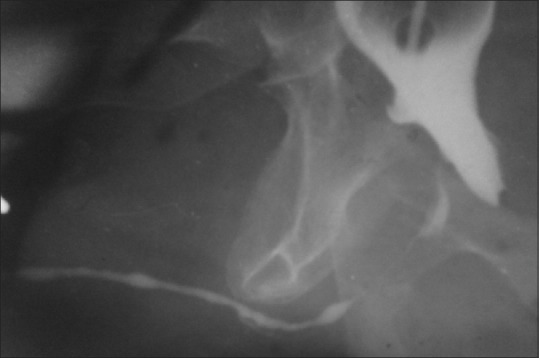
**Imaging Modality:** Retrograde urethrogram (RUG). **Anatomical Region:** Lower urinary tract, specifically the male urethra and bladder base. **Observed Pathology:** The image demonstrates a significant urethral stricture consistent with Balanitis Xerotica Obliterans (BXO) involvement. There is a long-segment, irregular narrowing of the anterior urethra, particularly involving the pendulous and distal segments. **Characteristic Visual Features:** - **Stricture Morphology:** A high-grade, elongated, and irregular filiform narrowing of the urethral lumen is visible. - **Contrast Distribution:** Radiopaque contrast medium depicts a markedly attenuated urethral caliber with areas of bead-like pooling and proximal segments appearing relatively unaffected compared to the distal involvement. - **Landmarks:** The contrast-filled bladder is partially visible superiorly, showing irregular contours that may suggest secondary changes such as bladder wall thickening or trabeculation due to chronic outlet obstruction. **Diagnostic Features:** The diffuse and long-segment nature of the anterior urethral narrowing in the clinical context of BXO (Lichen Sclerosus) distinguishes this from localized post-traumatic or iatrogenic strictures. The presence of significant luminal reduction indicates severe obstructive uropathy.
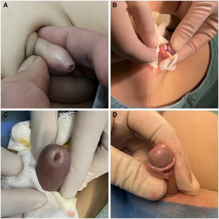
A composite of four clinical photographs (A-D) demonstrating urological conditions of the prepuce and glans penis in a pediatric/surgical context. Panel A illustrates physiologic or pathologic phimosis, showing a narrowed preputial orifice that prevents retraction of the foreskin. Panel B captures an intraoperative view where white, cheesy lumps of smegma are visible around the coronal sulcus upon partial retraction, associated with local inflammation. Panel C displays the characteristic features of Balanitis xerotica obliterans (BXO), also known as lichen sclerosus, presenting with a reddish-purple, shiny glans and evidence of secondary stenosis. Panel D shows advanced BXO manifestations, including white sclerotic scarring and thickening of the foreskin tissue with associated pallor of the glans. The images are relevant for medical education concerning male genital pathology, diagnostic signs of chronic inflammation, and indications for surgical intervention such as circumcision.
Bleeding from the Penis - Paediatric Approach
1. Causes by Category
A. Post-Circumcision Bleeding (Most Common)
- Skin edge bleeding
- Frenular artery bleeding
- Inadequate haemostasis at the time of the procedure
- Direct pressure for 5-10 minutes
- Silver nitrate cautery or absorbable suture ligation
- If persistent or significant: surgical re-exploration under general anaesthesia
- Check clotting screen - may unmask a bleeding diathesis (e.g. haemophilia, von Willebrand disease)
B. Phimosis and Foreskin-Related Bleeding
- Tears/fissures at the preputial ring - visible fresh blood
- Secondary scarring that leads to pathological phimosis
- Chronic cicatrising skin condition, rare under 5 years
- Presents at puberty most commonly
- Symptoms: bleeding, irritation, local infection, discomfort on micturition, urinary retention
- White, indurated plaques on glans and foreskin
- May involve meatus and urethra causing stricture
- Treatment: Circumcision (first-line for phimosis/BXO); topical steroids may help early; urethroplasty for urethral involvement
C. Paraphimosis
- Foreskin retracted and trapped behind the glans - venous/lymphatic congestion leads to swelling, ischaemia
- Swollen glans may develop surface erosions/bleeding
- Urological emergency - risk of gangrene if untreated
- Manual reduction with compression (ice, sugar/osmotic agents to reduce oedema)
- Dorsal slit or emergency circumcision if reduction fails
- Immediate urology referral
D. Balanitis / Balanoposthitis
- Infection/inflammation of the glans (balanitis) or glans + foreskin (balanoposthitis)
- Bloody/purulent discharge from preputial opening
- Common in uncircumcised boys
- Organisms: mixed bacteria, Candida, GAS (Group A Strep)
- Management: Warm soaks, topical antifungal (if Candida) or oral antibiotics (amoxicillin-clavulanate if bacterial), avoid forceful retraction
E. Trauma
- Fall onto hard surface (bicycle bar, fence)
- Crush injury to perineum/penis between the object and the pubic arch
- Typically involves urethra / perineal soft tissue
- Haematoma, swelling, urethral bleeding (blood at meatus)
- Management: urinary catheter only if bladder is palpable; urology referral for urethrogram if haematuria or blood at meatus
- Penile skin caught in zipper - laceration and bleeding
- Management: cut the zipper bridge with bone cutters or unzip carefully; local anaesthesia
- A hair or thread wrapped tightly around the penile shaft in infants
- Causes oedema, discolouration, ischaemia - bleeding from strangulated skin
- Emergency - hair may be buried and invisible under the swollen tissue
- Treatment: cut/remove the hair; depilatory cream (Nair) can dissolve hair under swollen tissue; surgical incision if severe
- Always consider in prepubertal children with genital injuries inconsistent with history
- Mandates safeguarding referral and forensic examination
F. Urethral Causes - Blood at Meatus
| Cause | Key Features |
|---|---|
| Meatal stenosis | Post-circumcision; upward deflected stream, dysuria; rarely bleeds - more obstruction |
| Urethritis | STI in adolescents (gonococcal, chlamydial); purulent ± bloody discharge |
| Urethral prolapse | Mainly prepubertal girls but can mimic in boys; circumferential mucosal eversion |
| Urinary tract infection with haematuria | Dysuria, fever, cloudy urine; less commonly presents with visible urethral bleeding |
| Haemangioma / vascular malformation | Rare; bright red painless bleeding |
| Wilms' tumour / renal causes | Frank haematuria, abdominal mass - not penile per se |
G. Haematological / Systemic Causes
- Haemophilia A or B
- Von Willebrand disease (most common inherited bleeding disorder)
- Thrombocytopaenia (ITP, leukaemia)
- Liver disease (coagulopathy)
2. Structured Assessment Approach
History
- Age, circumcision status
- Site: at meatus vs. from skin/foreskin
- Associated: dysuria, fever, discharge, swelling, injury history
- Bleeding history (family and personal)
- Safeguarding: is the history consistent with the injury?
Examination
- Inspect the glans, meatus, foreskin, shaft skin
- Note: skin colour (ischaemia?), discharge (pus vs. blood), visible lacerations
- Do NOT forcibly retract foreskin
Investigations
- Urine dipstick and MC&S
- Clotting screen, FBC, platelets (if bleeding is unexplained or significant)
- Wound swab if infection suspected
- Urethrogram if blood at meatus + history of trauma
3. Red Flags - When to Refer Urgently
| Red Flag | Action |
|---|---|
| Paraphimosis | Emergency urology |
| Hair tourniquet | Emergency urology |
| Blood at meatus + trauma | Emergency urology (urethrogram needed) |
| Penile ischaemia/gangrene | Emergency surgery |
| Uncontrolled post-circumcision bleeding | Emergency urology |
| Suspected sexual abuse | Safeguarding + forensic paeds |
| Haematological cause suspected | Paediatric haematology |
4. Summary Table
| Age Group | Most Common Cause |
|---|---|
| Neonate/infant | Post-circumcision bleeding, hair tourniquet, nappy rash/excoriation |
| Toddler/young child | Balanoposthitis, trauma (straddle), forceful retraction fissure |
| School-age | BXO/phimosis, balanitis, trauma |
| Adolescent | Paraphimosis, urethritis (STI), trauma, penile fracture (rare) |
- Bleeding after circumcision is the single most common cause overall
- Always screen for a bleeding diathesis when bleeding is out of proportion to the trigger
- Never forcibly retract the foreskin in young children
- Consider non-accidental injury in any genital injury with an inconsistent history
- BXO requires circumcision; topical steroids alone are insufficient once cicatrisation has occurred