
Medical management for pneumonia for exam of medicine
pneumonia CURB-65 severity scoring treatment algorithm
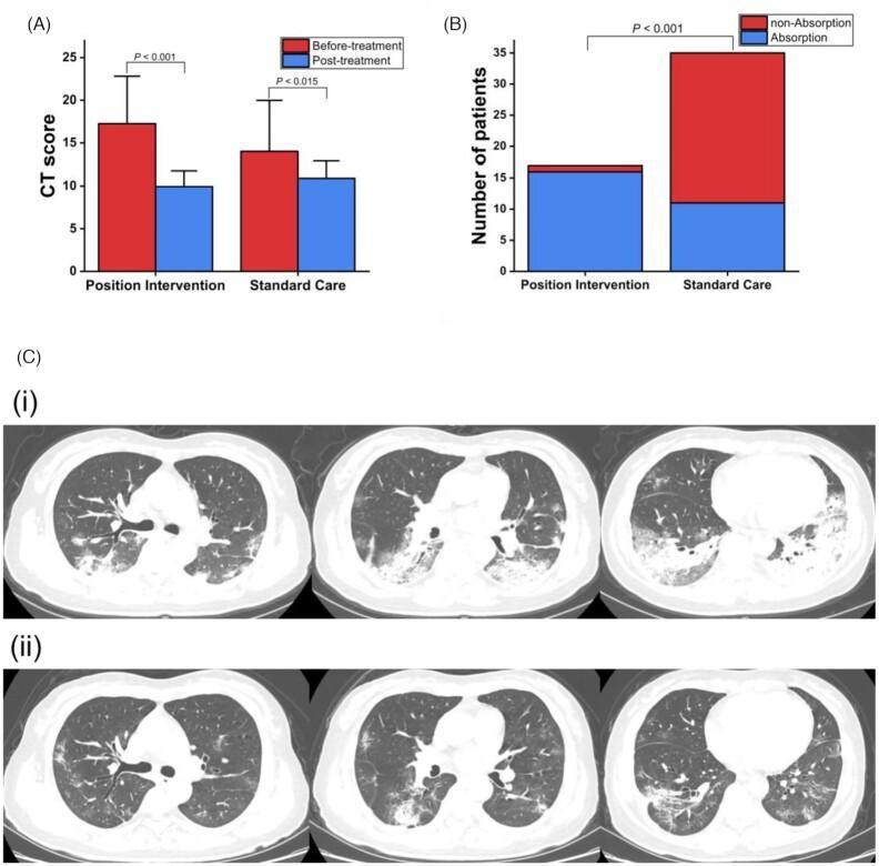
This composite educational graphic illustrates the radiographic evolution of COVID-19 pneumonia following clinical intervention. (A) Bar graph comparing Mean CT scores between 'Position Intervention' and 'Standard Care' groups, showing a statistically significant decrease in lung lesion severity post-treatment in both cohorts (P<0.001 and P<0.015). (B) Stacked bar chart showing a higher frequency of lesion absorption (>30%) in the position intervention group compared to standard care (P<0.001). (C) Longitudinal axial chest CT scans (lung window) of a 52-year-old patient. Row (i) represents the baseline state, showing bilateral, multifocal, predominantly posterior and subpleural ground-glass opacities (GGO) and consolidation. Row (ii) demonstrates follow-up imaging nine days later after prone position therapy, showing significant interval absorption and resolution of the previous consolidation and GGOs, reflecting improved pulmonary aeration. This visual resource is intended for medical education regarding diagnostic imaging in viral pneumonia and the assessment of therapeutic efficacy through quantitative CT scoring and longitudinal radiological monitoring.
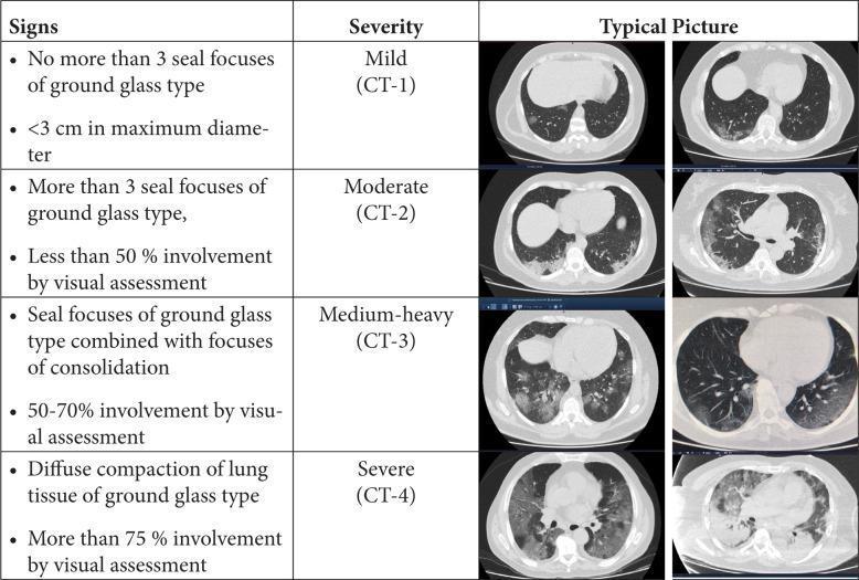
A comparison chart and diagnostic imaging series illustrating a radiological scoring system for pneumonia severity based on axial chest CT scans. The content is categorized into four stages of lung involvement: CT-1 (Mild), CT-2 (Moderate), CT-3 (Medium-heavy), and CT-4 (Severe). CT-1 is characterized by minimal focal ground-glass opacities (GGOs) under 3 cm. CT-2 shows more than three GGOs with less than 50% parenchymal involvement. CT-3 demonstrates a combination of GGOs and pulmonary consolidation, involving 50-70% of the lung volume. CT-4 represents the most severe stage, with diffuse compaction of lung tissue and GGOs or consolidation affecting over 75% of the lung. Representative CT images for each stage highlight the progression from peripheral, patchy opacities to widespread, bilateral, and dense consolidation. This resource serves as a clinical assessment tool for grading the extent of inflammatory lung disease, commonly utilized in the context of viral pneumonias such as COVID-19.
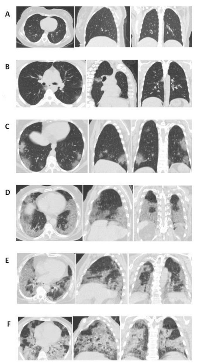
This chest CT compilation illustrates a semi-quantitative severity scoring system for lung involvement, typically applied in the context of viral pneumonia such as COVID-19. The image presents eighteen panels organized into six rows (A-F), showing axial, sagittal, and coronal planes for each severity score level (0 to 5). (A) Score 0: Normal lung parenchyma with no visible abnormalities. (B) Score 1: Less than 5% involvement, showing isolated, faint ground-glass opacities (GGOs). (C) Score 2: 5–25% involvement, with multi-focal GGOs appearing more distinct in peripheral zones. (D) Score 3: 26–49% involvement, demonstrating extensive GGOs and early consolidative patches distributed across middle and lower zones. (E) Score 4: 50–75% involvement, showing dense, confluent consolidations and GGOs involving the majority of the lung volume. (F) Score 5: Over 75% involvement, characterized by diffuse, high-density consolidation and parenchymal architectural distortion across all lobes. This progression visualizes the transition from focal ground-glass density to widespread consolidation, serving as a radiological reference for disease staging and severity assessment.
Medical Management of Pneumonia - Exam Guide
1. Classification (Know This First)
| Type | Definition |
|---|---|
| CAP (Community-Acquired Pneumonia) | Pneumonia in community-dwelling individual |
| HAP (Hospital-Acquired Pneumonia) | Pneumonia ≥48 h after hospital admission |
| VAP (Ventilator-Associated Pneumonia) | Pneumonia ≥48 h after endotracheal intubation |
| HCAP | Removed from 2019 IDSA/ATS guidelines (no longer used) |
2. Common Pathogens by Setting
| Setting | Key Organisms |
|---|---|
| Outpatients | S. pneumoniae, M. pneumoniae, H. influenzae, C. pneumoniae, respiratory viruses |
| Non-ICU inpatients | S. pneumoniae, M. pneumoniae, H. influenzae, Legionella, respiratory viruses |
| ICU (severe CAP) | S. pneumoniae, S. aureus (MRSA), Legionella spp., Gram-negative bacilli, H. influenzae |
3. Severity Assessment - KEY EXAM SCORES
CURB-65 Criteria (1 point each)
| Letter | Criterion |
|---|---|
| C | Confusion (new-onset) |
| U | Urea >7 mmol/L (BUN ≥20 mg/dL) |
| R | Respiratory rate ≥30/min |
| B | Blood pressure systolic <90 or diastolic ≤60 mmHg |
| 65 | Age ≥65 years |
- Score 0-1: Outpatient (30-day mortality ~1.5%)
- Score 2: Consider hospitalization (or close outpatient follow-up)
- Score ≥3: Hospitalize; mortality ~22%; consider ICU if ≥4-5
Pneumonia Severity Index (PSI) / PORT Score
- Uses 20 variables; stratifies into Class I-V
- More validated than CURB-65 but harder to calculate
- Class I-II: Outpatient; Class III: Observation; Class IV-V: Hospitalize
IDSA/ATS Criteria for ICU Admission (2019 guidelines)
- Respiratory failure requiring invasive mechanical ventilation
- Septic shock requiring vasopressors
- RR ≥30/min
- PaO₂/FiO₂ ratio ≤250
- Multilobar infiltrates
- Confusion/disorientation
- BUN ≥20 mg/dL
- WBC <4,000 cells/µL (leukopenia)
- Platelets <100,000/µL
- Hypothermia (core temp <36°C)
- Hypotension requiring aggressive fluid resuscitation
4. Antibiotic Treatment (2019 IDSA/ATS Guidelines) - HIGH YIELD
A. Outpatient CAP
| Patient Group | Preferred Regimen |
|---|---|
| Healthy, no comorbidities | Amoxicillin OR Doxycycline OR Macrolide* (azithromycin/clarithromycin if local resistance <25%) |
| With comorbidities (COPD, diabetes, renal/liver disease, heart failure, malignancy, alcohol use, asplenia) | Amoxicillin-clavulanate or cephalosporin + macrolide or doxycycline OR Respiratory fluoroquinolone monotherapy (levofloxacin 750 mg/d, moxifloxacin 400 mg/d) |
B. Inpatient, Non-Severe CAP (No MRSA/Pseudomonas Risk Factors)
| Option | Drugs |
|---|---|
| Combination (preferred) | β-lactam + macrolide |
| Monotherapy | Respiratory fluoroquinolone |
| Alternative | β-lactam + doxycycline (if macrolide and FQ are contraindicated) |
C. Severe CAP (ICU)
| Option | Drugs |
|---|---|
| Standard | β-lactam + macrolide (azithromycin 500 mg/d) |
| Alternative | β-lactam + respiratory fluoroquinolone |
D. When to Add MRSA or Pseudomonas Coverage
- Prior respiratory culture positive for MRSA
- Recent hospitalization + IV antibiotics in last 90 days (+ local validation)
- Prior respiratory culture positive for P. aeruginosa
- Recent hospitalization + IV antibiotics (± local validation)
5. HAP/VAP Management
- HAP: ≥48h after admission, not intubated
- VAP: ≥48h after endotracheal intubation
- Piperacillin-tazobactam, cefepime, ceftriaxone, or levofloxacin
- Risk factors for MDR organisms
- IV antibiotic use in prior 90 days
- Septic shock at time of VAP
6. Duration of Antibiotic Therapy
- CAP (inpatients): 5-7 days (minimum 5 days; patient must be afebrile and clinically stable)
- Severe CAP: may extend to 7-10 days
- Legionella: 7-10 days
- VAP/HAP: 7-8 days (short course preferred; no benefit to longer courses)
- S. aureus or Pseudomonas VAP: may require longer
7. Special Situations
Aspiration Pneumonia
- Typically involves anaerobes + Gram-negative rods
- Treatment: Amoxicillin-clavulanate OR clindamycin + β-lactam OR pip-tazo for hospitalized patients
- Note: Aspiration pneumonitis (chemical, from gastric acid) does NOT require antibiotics unless infection is confirmed
Atypical Pneumonia
- Mycoplasma, Chlamydia, Legionella
- First-line: Macrolides or doxycycline (outpatient); fluoroquinolones for Legionella
- β-lactams alone are NOT effective (no cell wall)
- Legionella: urinary antigen testing; levofloxacin or azithromycin
8. Supportive Treatment
- Oxygen therapy: Target SpO₂ >92%; use high-flow nasal oxygen or NIV for hypoxic respiratory failure
- Corticosteroids: Not routinely recommended; consider in refractory septic shock or severe CAP with high inflammatory markers (methylprednisolone)
- Fluid resuscitation: Crystalloids for sepsis; avoid fluid overload
- Analgesia: NSAIDs or acetaminophen for pleuritic pain
- DVT prophylaxis: Heparin for hospitalized patients
- Nutrition: Early enteral nutrition in ICU patients
9. Procalcitonin - What to Know
- Released in bacterial (not viral) infection via TNF-α, IL-1β, IL-6
- 2019 IDSA/ATS guidelines: strong recommendation AGAINST withholding antibiotics based on procalcitonin in clinically suspected, radiographically confirmed CAP
- Threshold of <0.25 ng/mL sometimes used to guide antibiotic de-escalation in clinically stable patients, but NOT to withhold initial treatment
- Mycoplasma and Chlamydia pneumonia do NOT raise procalcitonin - a known pitfall
10. Follow-Up and Response to Therapy
- Clinical improvement expected within 48-72 hours of starting antibiotics
- Chest X-ray findings often worsen initially during treatment - not a sign of failure
- If no improvement by 72 h: consider drug-resistant pathogen, complication (empyema, abscess, parapneumonic effusion), wrong diagnosis, or immunocompromised host
11. Complications
| Complication | Features |
|---|---|
| Parapneumonic effusion | Exudative; may become empyema if infected |
| Empyema | Pus in pleural space; requires drainage (chest tube) + antibiotics |
| Lung abscess | Cavitation; longer antibiotic course needed (clindamycin/amoxicillin-clavulanate) |
| Sepsis/Septic shock | ICU admission; vasopressors |
| ARDS | Mechanical ventilation; lung-protective strategy |
| Necrotizing pneumonia | S. aureus PVL-positive, Klebsiella; high mortality |
12. Prevention
- Pneumococcal vaccine: PCV15 or PCV20 for adults ≥65 and high-risk individuals
- Influenza vaccine: Annual; reduces secondary bacterial pneumonia
- VAP bundle: HOB elevation ≥30-45°, daily sedation awakening, weaning protocols, oral chlorhexidine, avoid unnecessary antibiotics, hand hygiene
Bronchiectasis
bronchiectasis CT scan chest HRCT dilated bronchi tram track
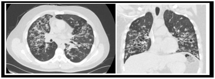
This diagnostic image displays axial (left) and coronal (right) views of a High-Resolution Computed Tomography (HRCT) scan of the chest, focusing on the lung parenchyma. The imaging reveals bilateral, extensive cystic bronchiectasis characterized by severe, permanent dilation of the bronchi, extending to the lung periphery. Visible 'ring-shadow' and 'tram-track' signs are present, indicating thickened bronchial walls. Multiple areas of high-attenuation opacities within the dilated airways suggest mucoid impaction (finger-in-glove sign). The lung parenchyma also exhibits heterogeneous lucencies and cystic changes, characteristic of advanced end-stage lung disease. These findings are clinically significant for the evaluation of severe chronic obstructive and inflammatory processes, such as those found in advanced cystic fibrosis (CF). The image serves as an educational example of progressive bronchiectatic remodeling and mucoid plugging requiring potential surgical intervention like lung transplantation.
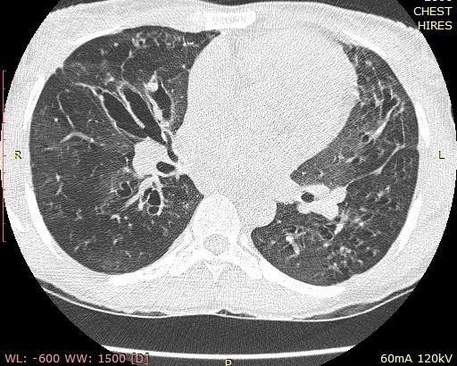
Axial high-resolution computed tomography (HRCT) of the chest demonstrating classic features of bronchiectasis. The primary findings include significant cystic and cylindrical bronchial dilatation, most prominent in the central lung fields. Notable bronchial wall thickening is present throughout, creating a 'tram-track' appearance in longitudinal sections and 'signet-ring' signs where dilated bronchi exceed the diameter of their accompanying pulmonary arteries. The lung parenchyma exhibits a mosaic attenuation pattern, characterized by patchy areas of ground-glass opacity interspersed with regions of decreased lung density, likely representing air trapping or mosaic perfusion. Additionally, multiple ill-defined pulmonary nodules of varying sizes are visible in a random distribution across both lung fields. These findings are clinically significant for chronic inflammatory airway disease, often seen in post-infectious bronchiectasis or cystic fibrosis.
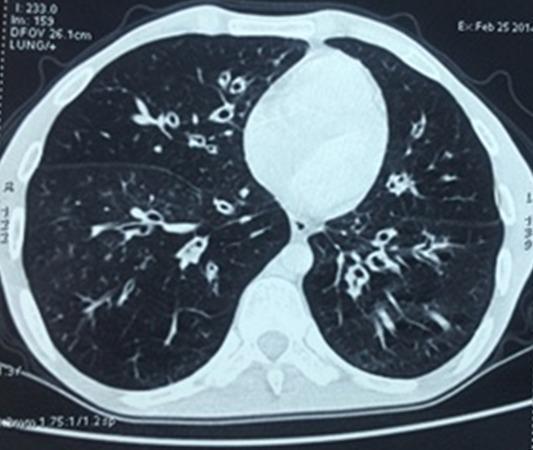
This diagnostic image is an axial chest CT scan using a lung parenchyma window, demonstrating significant pulmonary pathology. The primary finding is widespread, bilateral cylindrical bronchiectasis. Multiple dilated, thick-walled bronchi are visible throughout both lung fields, appearing as 'ring shadows' when viewed in cross-section and 'tram-track' signs where the airways are oriented parallel to the scan plane. The bronchial diameters clearly exceed those of their accompanying pulmonary arteries, a hallmark of bronchiectasis. The distribution is diffuse, affecting multiple lobes bilaterally. There are also subtle areas of bronchial wall thickening and small mucous plugs. These findings are characteristic of chronic obstructive or inflammatory airway disease, often associated with recurrent infections. In this clinical context, the structural damage is a secondary manifestation of Common Variable Immunodeficiency (CVID), reflecting the impact of humoral immune deficiency on long-term respiratory health and the development of permanent airway remodeling.
Medical management of bronchiectasis
bronchiectasis HRCT chest CT scan tram track ring shadow
This diagnostic image displays axial (left) and coronal (right) views of a High-Resolution Computed Tomography (HRCT) scan of the chest, focusing on the lung parenchyma. The imaging reveals bilateral, extensive cystic bronchiectasis characterized by severe, permanent dilation of the bronchi, extending to the lung periphery. Visible 'ring-shadow' and 'tram-track' signs are present, indicating thickened bronchial walls. Multiple areas of high-attenuation opacities within the dilated airways suggest mucoid impaction (finger-in-glove sign). The lung parenchyma also exhibits heterogeneous lucencies and cystic changes, characteristic of advanced end-stage lung disease. These findings are clinically significant for the evaluation of severe chronic obstructive and inflammatory processes, such as those found in advanced cystic fibrosis (CF). The image serves as an educational example of progressive bronchiectatic remodeling and mucoid plugging requiring potential surgical intervention like lung transplantation.
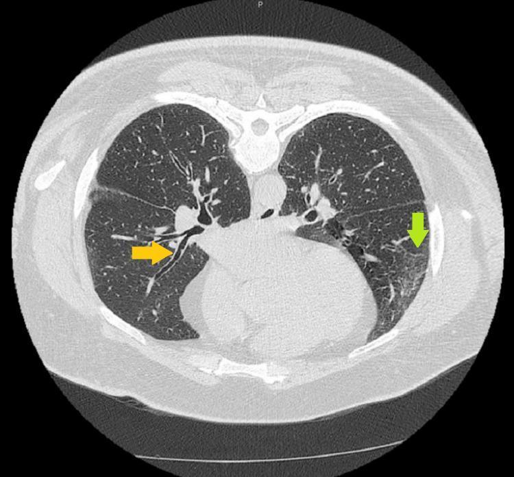
High-resolution axial CT (HRCT) scan of the thorax demonstrating clinical features of bronchiectasis and interstitial lung changes. An orange arrow identifies the 'tram track sign' in the right lower lobe, characterized by parallel, thickened bronchial walls resulting from abnormal, irreversible bronchial dilation. A green arrow in the left lower lobe indicates a region of micronodular opacities and subtle, hazy ground-glass opacities (GGOs), representing increased lung parenchyma density without obscuring the underlying vasculature. These diagnostic findings, in the context of mediastinal lymphadenopathy and systemic granulomatous disease, are classically associated with pulmonary sarcoidosis. The image serves as an educational reference for identifying secondary signs of chronic airway inflammation and interstitial involvement in multi-systemic granulomatous disorders.
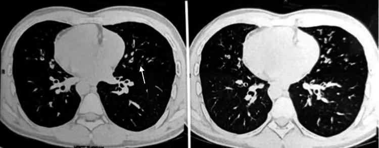
High-resolution computed tomography (HRCT) of the chest in axial view demonstrating findings consistent with bronchiectasis and bronchiolitis. The diagnostic image shows bilateral pulmonary abnormalities, including prominent peribronchial wall thickening and bronchial dilatation. Key radiological signs include the 'signet ring' sign (dilated bronchus larger than its accompanying pulmonary artery) and 'tram-track' opacities (parallel thickened bronchial walls). Small, centrilobular nodular opacities are visible in a 'tree-in-bud' pattern, particularly in the mid and lower lung zones, suggesting small airway impaction or infectious bronchiolitis. Additionally, the images reveal dextrocardia, with the cardiac apex and heart shadow oriented toward the right hemithorax, indicative of situs inversus. This combination of bronchiectasis, sinusitis (contextual), and situs inversus totalis is highly suggestive of Kartagener syndrome. The content is suitable for medical education regarding congenital ciliary dyskinesia and chronic inflammatory lung disease.
Medical Management of Bronchiectasis
1. Definition and Pathophysiology
Mucosal injury → impaired mucociliary clearance → bacterial colonization → neutrophilic inflammation → further airway damage → more colonization → progressive bronchiectasis
2. Causes (Etiology) - HIGH YIELD
| Category | Examples |
|---|---|
| Post-infectious (most common) | S. aureus, Klebsiella, TB (post-TB bronchiectasis), measles, pertussis, Pseudomonas |
| Cystic Fibrosis | Viscid mucus + recurrent infection; presents in childhood/young adulthood |
| Primary Ciliary Dyskinesia | Autosomal recessive; immobile cilia → impaired clearance; Kartagener syndrome (bronchiectasis + situs inversus + sinusitis) |
| Immunodeficiency | Hypogammaglobulinemia (IgA, IgG, IgM deficiency), Common Variable Immune Deficiency (CVID) → recurrent bacterial infections |
| ABPA (Allergic Bronchopulmonary Aspergillosis) | Proximal/central bronchiectasis; in asthmatics and CF patients |
| Alpha-1 antitrypsin deficiency | Associated diffuse bronchiectasis |
| Nontuberculous mycobacteria (NTM) | MAC infection; typically postmenopausal women; lingular and right middle lobe |
| Connective tissue diseases | Rheumatoid arthritis (8-35%), Sjögren syndrome, SLE, ankylosing spondylitis |
| Obstruction | Foreign body, tumor, mucus impaction → localized bronchiectasis |
| Yellow nail syndrome | Triad: yellow nails + lymphedema + pleural effusion; recurrent infections |
| Traction bronchiectasis | End-stage fibrotic lung disease pulling airway walls apart (not true bronchiectasis) |
| Idiopathic | ~50% of non-CF cases have no identified cause |
3. Clinical Features
- Chronic productive cough - the hallmark; ≥30 mL/day mucopurulent or mucopurulent tenacious sputum (three-layer sputum: frothy top, mucoid middle, purulent bottom)
- Hemoptysis - may be massive (from bronchial artery erosion)
- Dyspnea
- Recurrent chest infections / exacerbations
- Rhinosinusitis (associated, especially in PCD)
- "Dry bronchiectasis" - cough without sputum in some (e.g., upper lobe, post-TB)
- Clubbing (finger)
- Coarse crepitations (crackles), sometimes wheeze
- Features of underlying cause (e.g., dextrocardia in Kartagener)
- Advanced disease: cor pulmonale, cyanosis, respiratory failure
4. Investigations
Imaging
- HRCT chest - investigation of choice; shows:
- Tram-track sign (parallel walls of dilated bronchus on cross-section)
- Signet ring sign (dilated bronchus larger than its accompanying pulmonary artery in cross-section)
- Ring shadow (end-on view)
- Tree-in-bud pattern (small airway involvement)
- Mucoid impaction ("finger-in-glove" sign)
- Chest X-ray: bronchial wall thickening, "tram tracks," "ring shadows" - less sensitive
Workup for Underlying Cause
- CBC + differential (eosinophilia → ABPA)
- Quantitative immunoglobulins: IgA, IgG, IgM (immunodeficiency)
- Serum IgE, Aspergillus precipitins, skin prick test (ABPA screen)
- Sputum Gram stain, culture (routine + AFB + fungal)
- Sweat chloride test / CFTR gene mutation (if CF suspected)
- Nasal nitric oxide + ciliary biopsy / electron microscopy (PCD)
- Alpha-1 antitrypsin levels
- Spirometry / PFTs: obstructive pattern
5. Sputum Microbiology (Common Pathogens)
| Disease Stage | Common Organisms |
|---|---|
| Mild / early | Haemophilus influenzae (most common, ~47%) |
| Moderate | Streptococcus pneumoniae, Moraxella catarrhalis |
| Severe / advanced | Pseudomonas aeruginosa (~12%; biofilm former; marker of worse disease) |
| CF-bronchiectasis | Pseudomonas aeruginosa, MRSA, Burkholderia cepacia |
| NTM-bronchiectasis | M. avium complex (MAC), M. abscessus |
6. Medical Management - CORE
A. Airway Clearance / Airway Hygiene
- Postural drainage - positioning to drain by gravity (affected lobe dependent); most traditional technique
- Chest physiotherapy (CPT) - percussion + vibration over chest wall
- Active Cycle of Breathing Technique (ACBT) - breathing control + thoracic expansion + forced expiratory technique (FET/huff)
- Oscillating PEP devices: Flutter valve, Acapella valve, Aerobika valve
- High-frequency chest wall oscillation (HFCWO) vest
- Autogenic drainage - self-drainage by controlled breathing
- Nebulized hypertonic saline (7%) - draws water into airway lumen, reduces sputum viscosity; standard of care
- Mannitol (inhaled) - osmotic agent; approved in some countries for non-CF bronchiectasis
- Dornase alfa (rhDNase) - recommended in CF bronchiectasis; NOT routinely used in non-CF bronchiectasis (may worsen outcomes)
- N-acetylcysteine - mucolytic; limited evidence in bronchiectasis
- Bromhexine - may help mucociliary clearance
B. Antimicrobial Treatment
i. Acute Exacerbations
| Pathogen | Antibiotic of Choice |
|---|---|
| H. influenzae (most common) | Amoxicillin 500 mg TDS or amoxicillin-clavulanate |
| S. pneumoniae | Amoxicillin or respiratory fluoroquinolone |
| P. aeruginosa | Oral: Ciprofloxacin 500-750 mg BD; IV: pip-tazo, cefepime, meropenem, aztreonam + aminoglycoside (double coverage) |
| MRSA | IV vancomycin or linezolid |
| Empirical (no culture) | Amoxicillin-clavulanate or respiratory fluoroquinolone (levofloxacin) |
ii. Eradication Therapy (new P. aeruginosa isolation)
- Ciprofloxacin oral 500-750 mg BD x 3 weeks, THEN
- Inhaled colistin or inhaled tobramycin x 3 months
- Goal: eradicate before chronic colonization establishes
iii. Long-term Suppressive / Maintenance Antibiotics
- Long-term macrolides - most evidence-based option:
- Azithromycin 250-500 mg 3x/week (or daily)
- Erythromycin 400 mg BD
- Mechanism: Anti-inflammatory + anti-biofilm + immunomodulatory (not just antimicrobial)
- Evidence: reduces exacerbation frequency, improves lung function and QOL
- Caution: Check NTM status first (macrolides alone worsen NTM disease and cause resistance); audiology testing (ototoxicity); cardiac QTc monitoring
- Inhaled antibiotics (chronic P. aeruginosa colonization):
- Inhaled tobramycin (TOBI) - alternating 28 days on/off
- Inhaled colistin (polymyxin E)
- Inhaled aztreonam - reduces exacerbations in P. aeruginosa colonized patients
- Inhaled ciprofloxacin (dry powder) - increasingly used
C. Anti-inflammatory Therapy
- Inhaled corticosteroids (ICS): Not routinely recommended unless coexisting asthma or COPD; may reduce sputum volume and airway inflammation; use with caution (infection risk)
- Oral corticosteroids: Short course for ABPA exacerbations; not routine
- Statins: Some evidence for anti-inflammatory benefit; investigational
- Ibuprofen (high-dose): Used in CF to slow lung function decline; not standard in non-CF
D. Bronchodilators
- Beta-2 agonists (SABA/LABA): Improve mucociliary clearance; relieve associated bronchoconstriction; use before chest physiotherapy
- Anticholinergics (ipratropium/tiotropium): If obstructive component or COPD overlap
- No strong RCT evidence specific to bronchiectasis, but widely used clinically
E. Treatment of Underlying Cause (Disease-Specific)
| Cause | Specific Treatment |
|---|---|
| CF | CFTR modulators (ivacaftor, lumacaftor-ivacaftor, elexacaftor-tezacaftor-ivacaftor based on mutation) |
| ABPA | Oral prednisolone + itraconazole or voriconazole |
| Hypogammaglobulinemia (CVID) | IV immunoglobulin (IVIG) replacement therapy |
| Alpha-1 antitrypsin deficiency | AAT augmentation therapy (IV weekly) |
| NTM (MAC) | Macrolide (azithromycin/clarithromycin) + rifampicin + ethambutol x 12+ months |
| Obstructive (FB, tumor) | Bronchoscopic removal / surgery |
7. Management of Hemoptysis
- Mild-moderate: Antibiotics + airway clearance; tranexamic acid
- Massive hemoptysis (>300-600 mL/24h): Resuscitation → bronchial artery embolization (BAE) (first-line for massive hemoptysis) → surgery if embolization fails
- Stop anticoagulants / NSAIDs if possible
8. Pulmonary Rehabilitation
- Exercise training + education program
- Improves exercise tolerance, reduces dyspnea, improves quality of life
- Recommended for all symptomatic patients
9. Miscellaneous / Preventive Measures
- Smoking cessation - mandatory
- Pneumococcal vaccine + annual influenza vaccine
- Vitamin D supplementation - deficiency associated with more P. aeruginosa colonization, worse exacerbation frequency, faster FEV1 decline
- Treatment of GERD - reduces microaspiration trigger
- Nasal saline + intranasal steroids for associated rhinosinusitis
- Nutritional support - weight loss is common; high caloric needs
10. Surgery
- Localized bronchiectasis (single lobe/segment) not responding to medical treatment
- Massive hemoptysis uncontrolled by embolization
- Resection of destroyed lobe (e.g., post-TB upper lobe)
11. Severity Scoring (Prognosis)
| Score | Components |
|---|---|
| BSI (Bronchiectasis Severity Index) | FEV1, prior hospitalization, exacerbation frequency, P. aeruginosa colonization, age, dyspnea, radiographic extent |
| FACED Score | FEV1, Age, Chronic colonization, radiographic Extension, Dyspnea |
12. Key Exam Points
- Dornase alfa - used in CF only; harmful in non-CF bronchiectasis
- Macrolides - anti-inflammatory role; check NTM status before starting
- Inhaled hypertonic saline - key mucoactive agent in non-CF bronchiectasis
- Ciprofloxacin - only oral antibiotic with adequate Pseudomonas activity
- BAE - first-line for massive hemoptysis
- Bilateral (not single) lung transplant - due to suppurative nature
- Tram-track / signet ring sign on HRCT = diagnostic hallmarks