
From now whatever questions i ask i need answers that fetch me full marks for my medicine proffs exams in mbbs
Quick answer
- Start with a crisp definition
- Use standard exam headings and subheadings
- Give classifications in tables or flowcharts where useful
- Include etiopathogenesis, clinical features, investigations, complications, and management in sequence
- Mention important diagnostic criteria, drug doses, and emergency steps when relevant
- Add differentiating points, viva pearls, and common examiner-favourite facts
- Write in a format suitable for 5-mark, 10-mark, or long-answer questions
- Include mnemonics only when they genuinely help recall
- Keep terminology aligned with standard undergraduate medicine texts and current guidance where applicable
Acute Mi
acute myocardial infarction management diagnosis ESC guideline 2023
myocardial infarction AND management
- Definition and universal diagnostic criteria
- Etiology, risk factors, and pathogenesis
- STEMI versus NSTEMI
- Clinical features and examination
- ECG localization and evolution
- Cardiac biomarkers and other investigations
- Immediate treatment and reperfusion strategy
- Complications with time course and management
- Secondary prevention and discharge advice
Which proff format should I write first?
- Start with a crisp definition
- Use standard exam headings and subheadings
- Give classifications in tables or flowcharts where useful
- Include etiopathogenesis, clinical features, investigations, complications, and management in sequence
- Mention important diagnostic criteria, drug doses, and emergency steps when relevant
- Add differentiating points, viva pearls, and common examiner-favourite facts
- Write in a format suitable for 5-mark, 10-mark, or long-answer questions
- Include mnemonics only when they genuinely help recall
- Keep terminology aligned with standard undergraduate medicine texts and current guidance where applicable
- Definition and universal diagnostic criteria
- Etiology, risk factors, and pathogenesis
- STEMI versus NSTEMI
- Clinical features and examination
- ECG localization and evolution
- Cardiac biomarkers and other investigations
- Immediate treatment and reperfusion strategy
- Complications with time course and management
- Secondary prevention and discharge advice
Definition
- Ischemic symptoms
- New ischemic ECG changes
- Development of pathological Q waves
- New regional wall-motion abnormality or loss of viable myocardium on imaging
- Coronary thrombus on angiography/autopsy
Classification
1. According to ECG
| STEMI | NSTEMI |
|---|---|
| Persistent ST-segment elevation or new/presumed new LBBB with appropriate clinical setting | No persistent ST elevation |
| Usually due to complete coronary occlusion | Usually due to partial/transient occlusion |
| Requires immediate reperfusion | Requires risk-based early invasive strategy |
2. Universal definition of MI
| Type | Cause |
|---|---|
| Type 1 | Spontaneous atherothrombotic coronary plaque rupture/erosion |
| Type 2 | Oxygen supply-demand mismatch, for example anemia, tachyarrhythmia, shock, coronary spasm |
| Type 3 | Sudden cardiac death with suspected MI before biomarkers can be obtained |
| Type 4a | PCI-related MI |
| Type 4b | Stent thrombosis |
| Type 4c | Restenosis-related MI |
| Type 5 | CABG-related MI |
Etiology and Risk Factors
Usual mechanism
- Platelet adhesion and activation
- Platelet aggregation
- Activation of coagulation cascade
- Formation of occlusive coronary thrombus
- Myocardial ischemia and necrosis
Non-modifiable risk factors
- Increasing age
- Male sex, or postmenopausal woman
- Family history of premature CAD
- Genetic predisposition
Modifiable risk factors
- Smoking
- Diabetes mellitus
- Hypertension
- Dyslipidemia
- Obesity and sedentary lifestyle
- Unhealthy diet
- Psychosocial stress
- Cocaine/amphetamine use
Less common causes
- Coronary vasospasm, for example cocaine-induced
- Coronary embolism
- Spontaneous coronary artery dissection
- Coronary arteritis/vasculitis
- Severe anemia, shock, hypoxemia, sepsis, tachyarrhythmia: Type 2 MI
Pathogenesis
Sequence
- ATP depletion begins within seconds.
- Loss of contractility occurs within about 1 minute.
- Irreversible myocyte injury begins after approximately 20-30 minutes of severe ischemia.
- Necrosis progresses from subendocardium to subepicardium over several hours: the wavefront phenomenon.
- Early reperfusion salvages ischemic but viable myocardium: “Time is muscle.”
Coronary artery commonly involved
| Artery | Approximate frequency | Area infarcted |
|---|---|---|
| Left anterior descending artery | 40-50% | Anterior wall, septum, apex |
| Right coronary artery | 30-40% | Inferior wall, RV, posterior septum; SA/AV node often supplied |
| Left circumflex artery | 15-20% | Lateral wall |
Clinical Features
Symptoms
Typical presentation
- Severe central/retrosternal constricting or crushing chest pain
- Lasts >20 minutes
- May radiate to left shoulder, left arm, neck, jaw, epigastrium, interscapular area
- Not completely relieved by rest or sublingual nitrate
- Sweating, anxiety, restlessness, sense of impending doom
- Dyspnea
- Palpitations
- Nausea and vomiting, especially in inferior-wall MI
- Syncope, especially with arrhythmia or cardiogenic shock
Atypical or silent MI
- Elderly patients
- Women
- Diabetes mellitus
- Chronic kidney disease
- Dyspnea
- Syncope
- Acute pulmonary edema
- Unexplained weakness
- Epigastric discomfort
- Confusion
- Sudden collapse
Signs
- Pallor, sweating, cold clammy extremities
- Tachycardia or bradycardia
- Hypotension
- Raised JVP in right ventricular infarction
- S4 gallop: reduced LV compliance
- S3 gallop: LV failure
- Basal crepitations/pulmonary edema
- New pansystolic murmur:
- Acute MR due to papillary muscle rupture
- VSD due to interventricular septal rupture
- Pericardial rub in acute pericarditis
Diagnosis
1. ECG - obtain within 10 minutes of first medical contact
STEMI criteria
- Generally ≥1 mm in all leads other than V2-V3
- In V2-V3, higher cutoffs apply:
- Men ≥40 years: ≥2 mm
- Men <40 years: ≥2.5 mm
- Women: ≥1.5 mm
ECG localization
| Infarct location | ST elevation | Reciprocal changes | Likely artery |
|---|---|---|---|
| Inferior wall | II, III, aVF | I, aVL | RCA, occasionally LCx |
| Anterior wall | V1-V4 | Inferior leads | LAD |
| Anteroseptal | V1-V4 | Inferior leads | LAD |
| Extensive anterior | I, aVL, V1-V6 | II, III, aVF | Proximal LAD |
| Lateral wall | I, aVL, V5-V6 | II, III, aVF | LCx/diagonal branch |
| Posterior wall | ST depression and tall R waves in V1-V3 | ST elevation in V7-V9 | RCA/LCx |
| Right ventricular MI | ST elevation in V4R | Usually with inferior MI | Proximal RCA |
Evolution of ECG changes
- Hyperacute tall T waves
- ST elevation
- Development of pathological Q waves
- T-wave inversion
- ST segment returns to baseline over days
- Q waves may persist permanently
2. Cardiac biomarkers
| Marker | Rise | Peak | Returns to normal | Use |
|---|---|---|---|---|
| High-sensitivity troponin I/T | 1-3 h | 12-24 h | 7-14 days | Most sensitive and specific |
| CK-MB | 3-6 h | 18-24 h | 48-72 h | Useful for reinfarction |
| Myoglobin | 1-2 h | 6-9 h | 24 h | Early but non-specific |
Important point
3. Other investigations
- Serial ECGs
- CBC: anemia, leukocytosis
- Blood glucose and HbA1c
- Renal function and electrolytes
- Lipid profile, preferably within 24 hours
- Coagulation profile
- Chest X-ray: pulmonary edema, exclude other causes
- Echocardiography:
- Regional wall-motion abnormality
- LV function/ejection fraction
- Mechanical complications
- LV thrombus
- Coronary angiography: defines culprit vessel and permits PCI
Management
Initial emergency management
Immediate steps
- Admit to coronary care unit.
- ABC approach.
- Cardiac monitor and defibrillator readiness.
- Two IV lines.
- Obtain 12-lead ECG within 10 minutes.
- Measure troponin, but do not delay reperfusion in clear STEMI.
- Assess hemodynamic status and complications.
Immediate medications
| Treatment | Regimen/principle |
|---|---|
| Aspirin | Chew 162-325 mg immediately, then 75-162 mg daily |
| P2Y12 inhibitor | Ticagrelor/prasugrel/clopidogrel according to PCI or fibrinolysis plan |
| Anticoagulation | UFH, enoxaparin, or fondaparinux according to strategy |
| Nitrates | Sublingual GTN for pain if not hypotensive and no RV infarction/PDE-5 inhibitor use |
| Morphine | IV for severe ongoing pain, with antiemetic if required |
| Oxygen | Only if SpO₂ <90%, respiratory distress, or hypoxemia |
| High-intensity statin | Atorvastatin 80 mg or rosuvastatin 20-40 mg |
| Beta-blocker | Oral beta-blocker in stable patient without shock, bradycardia, AV block, or acute HF |
Avoid/withhold nitrates if
- Systolic BP <90 mmHg
- Suspected right ventricular infarction
- Severe aortic stenosis
- Recent phosphodiesterase-5 inhibitor use:
- Sildenafil/vardenafil within 24 h
- Tadalafil within 48 h
Reperfusion Therapy in STEMI
Primary PCI - preferred reperfusion method
Indications
- STEMI within 12 hours of symptom onset
- Ongoing ischemia, shock, severe heart failure, or life-threatening arrhythmia even if presentation is late
- Contraindication to fibrinolysis
Advantages
- Higher rate of patency of infarct artery
- Lower risk of intracranial hemorrhage
- Can identify anatomy and treat culprit lesion
- Useful in cardiogenic shock and after failed thrombolysis
Fibrinolysis
- STEMI within 12 hours of onset, and
- Primary PCI cannot be performed within 120 minutes, and
- No contraindication exists.
Drugs
- Tenecteplase
- Alteplase
- Reteplase
- Streptokinase, where other agents are unavailable
Absolute contraindications to fibrinolysis
- Previous intracranial hemorrhage
- Known intracranial vascular lesion
- Known malignant intracranial tumor
- Ischemic stroke within previous 3 months
- Suspected aortic dissection
- Active bleeding/bleeding diathesis
- Significant closed head/facial trauma within 3 months
- Recent intracranial/intraspinal surgery
Signs of successful thrombolysis
- Relief of chest pain
-
50% reduction in ST elevation at 60-90 minutes
- Reperfusion arrhythmias, especially accelerated idioventricular rhythm
- Early peak of cardiac enzymes
Failed fibrinolysis
NSTEMI Management
- Aspirin + P2Y12 inhibitor
- Anticoagulation
- High-intensity statin
- Anti-ischemic treatment
- Risk stratification using GRACE score
- Early invasive coronary angiography in high-risk patients
Immediate invasive strategy
- Hemodynamic instability/cardiogenic shock
- Recurrent or refractory chest pain
- Life-threatening arrhythmia or cardiac arrest
- Acute heart failure due to ongoing ischemia
- Mechanical complication
- Recurrent dynamic ST/T changes
Early invasive strategy
Complications of AMI
1. Electrical complications
| Complication | Typical timing | Key points |
|---|---|---|
| Ventricular tachycardia/ventricular fibrillation | First 24-48 h | Major cause of early death |
| Sinus bradycardia | Inferior MI | Often vagal or nodal ischemia |
| AV block | Inferior or anterior MI | Inferior usually transient; anterior indicates extensive infarction |
| Atrial fibrillation | Any time | Treat according to hemodynamic status |
| Bundle branch block | Especially anterior MI | May indicate extensive septal involvement |
2. Pump failure
| Condition | Features | Management principle |
|---|---|---|
| LV failure/pulmonary edema | Dyspnea, crepitations, S3, hypoxemia | Oxygen if hypoxemic, IV diuretic, vasodilator if BP permits, urgent reperfusion |
| Cardiogenic shock | Hypotension, cold extremities, oliguria, altered sensorium | Urgent PCI, vasopressors/inotropes, treat mechanical cause |
3. Mechanical complications
| Complication | Typical timing | Classical presentation |
|---|---|---|
| Papillary muscle rupture | 2-7 days | Acute severe MR, pulmonary edema, new apical pansystolic murmur |
| Ventricular septal rupture | 3-5 days | Sudden deterioration, harsh pansystolic murmur at left sternal border, thrill |
| Free-wall rupture | 5-14 days | Hemopericardium, tamponade, pulseless electrical activity, sudden death |
| LV pseudoaneurysm | Contained free-wall rupture | Risk of rupture |
| True LV aneurysm | Weeks to months | Persistent ST elevation, HF, arrhythmia, mural thrombus |
4. Inflammatory complications
- Early fibrinous pericarditis: 1-3 days; pleuritic chest pain and pericardial rub
- Dressler syndrome: weeks to months later; autoimmune fever, pericarditis, pleuropericardial effusion
5. Thromboembolic complications
- LV mural thrombus, particularly with large anterior MI
- Systemic embolism: stroke, limb ischemia, renal infarction
Secondary Prevention After MI
Drug therapy
- Aspirin indefinitely
- P2Y12 inhibitor usually for 12 months after ACS, adjusted for bleeding risk
- High-intensity statin
- Beta-blocker, especially with LV dysfunction, angina, arrhythmias
- ACE inhibitor/ARB, especially with LVEF ≤40%, diabetes, hypertension, CKD, or heart failure
- Mineralocorticoid receptor antagonist if LVEF ≤40% with heart failure or diabetes, provided renal function and potassium permit
Lifestyle and risk-factor control
- Complete smoking cessation
- Cardiac rehabilitation
- Regular graded exercise
- Weight reduction if overweight
- Diabetes and BP control
- Mediterranean-style heart-healthy diet
- Limit alcohol and avoid cocaine/amphetamines
- Influenza vaccination where appropriate
- Education regarding adherence and recurrent symptoms
One-Minute Proff Viva Summary
Acute MI is myocardial necrosis caused by acute ischemia, most commonly due to rupture or erosion of an atherosclerotic plaque followed by coronary thrombosis. Diagnosis requires a rise/fall of cardiac troponin with evidence of ischemia. STEMI requires urgent reperfusion, preferably primary PCI within 120 minutes of first medical contact; otherwise, fibrinolysis is used if no contraindication exists. Initial treatment includes aspirin, a P2Y12 inhibitor, anticoagulation, statin, nitrates where appropriate, oxygen only for hypoxemia, and management of pain and complications. Major complications are arrhythmias, LV failure, cardiogenic shock, papillary muscle rupture, VSD, free-wall rupture, pericarditis, mural thrombus, and ventricular aneurysm.
Acute coronary syndrome
2025 2026 acute coronary syndrome guideline diagnosis management ESC ACC
acute coronary syndrome AND management
Acute Coronary Syndrome (ACS)
Definition
- ST-elevation myocardial infarction (STEMI)
- Non-ST-elevation myocardial infarction (NSTEMI)
- Unstable angina (UA)
Classification
| Feature | STEMI | NSTEMI | Unstable angina |
|---|---|---|---|
| Coronary obstruction | Usually complete and persistent | Usually partial/transient | Partial/transient |
| Myocardial necrosis | Present | Present | Absent |
| Troponin | Elevated with rise/fall | Elevated with rise/fall | Normal |
| ECG | Persistent ST elevation in contiguous leads or STEMI equivalent | ST depression, T-wave inversion, or normal ECG | ST depression, T-wave inversion, or normal ECG |
| Reperfusion | Immediate primary PCI, or fibrinolysis if PCI unavailable in time | No fibrinolysis. Risk-guided early angiography/PCI | No fibrinolysis. Risk-guided management |
Key distinction: NSTEMI and unstable angina together are called NSTE-ACS. Troponin elevation differentiates NSTEMI from unstable angina.
Etiopathogenesis
Usual mechanism
Pathological sequence
- Atherosclerotic plaque becomes unstable due to inflammation and a thin fibrous cap.
- Plaque rupture or superficial erosion exposes collagen and tissue factor.
- Platelets adhere, activate, and aggregate.
- Thrombin is generated and fibrin-rich thrombus forms.
- Coronary vasoconstriction worsens obstruction.
- Myocardial ischemia develops.
- Persistent severe ischemia causes irreversible myocyte necrosis, that is, MI.
Other causes
- Coronary artery vasospasm, including cocaine-induced
- Coronary embolism
- Spontaneous coronary artery dissection
- Coronary vasculitis
- Severe anemia, hypoxemia, sepsis, tachyarrhythmia, or shock causing supply-demand mismatch, that is, Type 2 MI
Risk Factors
Non-modifiable
- Increasing age
- Male sex or postmenopausal women
- Family history of premature coronary artery disease
- Genetic predisposition
Modifiable
- Smoking
- Diabetes mellitus
- Hypertension
- Dyslipidemia
- Obesity
- Sedentary lifestyle
- Unhealthy diet
- Psychosocial stress
- Chronic kidney disease
- Cocaine or amphetamine use
Clinical Features
Symptoms
Typical symptoms
- Severe central or retrosternal constricting chest pain
- Pain lasting more than 20 minutes
- Occurs at rest or with minimal exertion
- Radiation to left arm, shoulder, neck, jaw, back, or epigastrium
- Pain not completely relieved by rest or sublingual nitrate
- Sweating, nausea, vomiting
- Dyspnea
- Palpitations
- Giddiness or syncope
- Anxiety and sense of impending doom
Atypical presentation
- Isolated dyspnea
- Fatigue
- Epigastric discomfort
- Syncope
- Acute confusion
- Acute pulmonary edema
- Silent MI
Signs
- Pallor, sweating, cold clammy extremities
- Tachycardia or bradycardia
- Hypotension
- S4 gallop due to stiff ischemic ventricle
- S3 gallop in LV dysfunction
- Basal crepitations in LV failure
- Raised JVP in right ventricular infarction
- New murmur:
- Acute mitral regurgitation due to papillary muscle rupture
- Ventricular septal rupture
- Pericardial rub in post-MI pericarditis
Diagnosis
1. ECG
STEMI
ECG localization
| Location of infarction | Leads with ST elevation | Usual culprit artery |
|---|---|---|
| Inferior wall | II, III, aVF | Right coronary artery |
| Anterior wall | V1-V4 | Left anterior descending artery |
| Anteroseptal | V1-V4 | LAD |
| Extensive anterior | I, aVL, V1-V6 | Proximal LAD |
| Lateral wall | I, aVL, V5-V6 | Left circumflex artery |
| Posterior wall | ST depression with tall R waves in V1-V3; ST elevation in V7-V9 | RCA or LCx |
| Right ventricular MI | ST elevation in V4R | Proximal RCA |
ECG findings in NSTE-ACS
- ST-segment depression
- Transient ST elevation
- T-wave inversion
- Non-specific ST-T changes
- ECG may be normal
2. Cardiac Biomarkers
High-sensitivity cardiac troponin I or T
- Preferred biomarker for ACS
- Elevated in NSTEMI and STEMI
- Normal in unstable angina
- Diagnosis of MI requires a rise and/or fall in troponin with at least one value above the 99th-percentile upper reference limit, plus clinical evidence of ischemia.
| Biomarker | Rise | Peak | Returns to normal | Importance |
|---|---|---|---|---|
| High-sensitivity troponin | 1-3 hours | 12-24 hours | 7-14 days | Most sensitive and specific |
| CK-MB | 3-6 hours | 18-24 hours | 2-3 days | Useful for reinfarction |
| Myoglobin | 1-2 hours | 6-9 hours | 24 hours | Earliest but non-specific |
3. Other Investigations
- CBC
- Blood glucose and HbA1c
- Renal function, electrolytes
- Lipid profile
- Coagulation profile
- Chest X-ray
- Echocardiography:
- Regional wall-motion abnormality
- LV ejection fraction
- Mechanical complications
- LV thrombus
- Coronary angiography:
- Defines coronary anatomy
- Identifies culprit artery
- Enables PCI
Management of ACS
A. Initial Management for All Patients
Immediate measures
- Admit to coronary care unit.
- ABC assessment.
- Cardiac monitoring and defibrillator availability.
- Two IV access lines.
- Obtain ECG within 10 minutes.
- Send cardiac biomarkers, but do not delay STEMI reperfusion while waiting for troponin.
- Assess for heart failure, shock, arrhythmia, or mechanical complication.
Initial drugs
| Drug/measure | Role |
|---|---|
| Aspirin | Antiplatelet therapy |
| P2Y12 inhibitor | Dual antiplatelet therapy |
| Anticoagulant | Prevents thrombus propagation |
| High-intensity statin | Plaque stabilization and secondary prevention |
| Nitrate | Relief of ischemic pain if hemodynamically stable |
| Morphine | Severe persistent pain |
| Oxygen | Only in hypoxemia, SpO₂ <90%, respiratory distress, or shock |
| Beta-blocker | In stable patients without acute HF, shock, severe bradycardia, or AV block |
Common initial doses for proff exams
- Aspirin: 162-325 mg chewed immediately, then 75-150 mg/day lifelong.
- Clopidogrel: 300-600 mg loading dose, then 75 mg/day, or use ticagrelor/prasugrel where suitable.
- Atorvastatin: 80 mg orally as early as possible.
- GTN: 0.4 mg sublingual, repeated every 5 minutes up to 3 doses if blood pressure permits.
Do not give nitrates in
- Hypotension
- Right ventricular infarction
- Severe aortic stenosis
- Recent PDE-5 inhibitor use, for example sildenafil within 24 hours or tadalafil within 48 hours
B. STEMI Management
Principle: Immediate reperfusion
Primary PCI
- STEMI within 12 hours of symptom onset
- Persistent ischemic symptoms
- Cardiogenic shock
- Acute heart failure
- Life-threatening arrhythmias
- Failed fibrinolysis
Fibrinolysis
- STEMI is diagnosed within 12 hours of symptom onset
- PCI cannot be done within the recommended time
- There is no contraindication
Fibrinolytic drugs
- Tenecteplase
- Alteplase
- Reteplase
- Streptokinase
Absolute contraindications
- Previous intracranial hemorrhage
- Ischemic stroke within previous 3 months
- Known intracranial neoplasm or AV malformation
- Suspected aortic dissection
- Active bleeding
- Significant head injury within 3 months
- Recent intracranial or spinal surgery
Indicators of successful fibrinolysis
- Relief of chest pain
- More than 50% resolution of ST elevation at 60-90 minutes
- Reperfusion arrhythmias, especially accelerated idioventricular rhythm
- Early enzyme peak
C. NSTEMI and Unstable Angina Management
Medical treatment
- Aspirin plus P2Y12 inhibitor
- Anticoagulation
- High-intensity statin
- Nitrates for pain, if appropriate
- Beta-blocker if no contraindication
- ACE inhibitor/ARB in LV dysfunction, diabetes, hypertension, or CKD
- Risk stratification using GRACE score
Indications for immediate angiography, usually within 2 hours
- Hemodynamic instability or cardiogenic shock
- Recurrent or refractory chest pain
- Life-threatening arrhythmias or cardiac arrest
- Acute heart failure due to ongoing ischemia
- Mechanical complication
- Recurrent dynamic ST-T changes
Indications for early angiography, usually within 24 hours
- Confirmed NSTEMI
- Dynamic ST-T changes
- GRACE score >140
- Transient ST elevation
Complications of ACS
1. Electrical complications
- Ventricular tachycardia
- Ventricular fibrillation
- Sinus bradycardia
- AV block
- Atrial fibrillation
- Bundle branch block
2. Pump failure
- Acute LV failure
- Pulmonary edema
- Cardiogenic shock
3. Mechanical complications
| Complication | Usual timing | Clinical feature |
|---|---|---|
| Papillary muscle rupture | 2-7 days | Acute severe MR, pulmonary edema, new apical pansystolic murmur |
| Ventricular septal rupture | 3-5 days | Harsh pansystolic murmur at left sternal border, thrill, shock |
| Free-wall rupture | 5-14 days | Cardiac tamponade, pulseless electrical activity, sudden death |
| True ventricular aneurysm | Weeks to months | Persistent ST elevation, HF, arrhythmia, mural thrombus |
| LV mural thrombus | Days to weeks | Systemic embolism, especially stroke |
4. Inflammatory complications
- Early post-infarction pericarditis: 1-3 days
- Dressler syndrome: weeks to months later
Secondary Prevention
- Aspirin lifelong.
- Dual antiplatelet therapy, commonly for 12 months after ACS, depending on bleeding and ischemic risk.
- High-intensity statin.
- Beta-blocker where indicated.
- ACE inhibitor/ARB, especially in LV dysfunction, diabetes, hypertension, CKD, or heart failure.
- Mineralocorticoid receptor antagonist when LVEF ≤40% with heart failure or diabetes, if potassium and renal function permit.
- Smoking cessation.
- Diabetes, BP, and lipid control.
- Exercise-based cardiac rehabilitation.
- Healthy diet, weight reduction, and regular follow-up.
Short Conclusion for Exams
Acute coronary syndrome includes STEMI, NSTEMI, and unstable angina. It most commonly results from atherosclerotic plaque rupture with superimposed coronary thrombosis. ECG and serial high-sensitivity troponin are central to diagnosis. STEMI requires immediate reperfusion by primary PCI or fibrinolysis where PCI is delayed, whereas NSTE-ACS requires antithrombotic therapy and risk-guided early invasive management. Early diagnosis, timely reperfusion, and aggressive secondary prevention reduce mortality and recurrent cardiovascular events.
Meningitis
WHO meningitis diagnosis treatment guideline bacterial meningitis 2025
bacterial meningitis AND treatment
bacterial meningitis cerebrospinal fluid Gram stain diagram clinical signs
Meningitis
Definition
Classification
Based on etiology
- Acute pyogenic/bacterial meningitis
- Viral (aseptic) meningitis
- Tuberculous meningitis (TBM)
- Fungal meningitis, especially cryptococcal meningitis
- Spirochetal, parasitic, autoimmune, drug-induced, or neoplastic meningitis
Based on duration
| Type | Duration | Usual causes |
|---|---|---|
| Acute | Hours to days | Pyogenic bacteria, viruses |
| Subacute | Days to weeks | Tuberculosis, fungi |
| Chronic | >4 weeks | TB, fungal, malignancy, autoimmune disease |
Acute Bacterial Meningitis
Etiology
Common organisms by age and setting
| Group | Important organisms |
|---|---|
| Neonates | Group B Streptococcus, E. coli, Listeria monocytogenes |
| Children and young adults | Neisseria meningitidis, Streptococcus pneumoniae, Haemophilus influenzae type b |
| Adults | Streptococcus pneumoniae, Neisseria meningitidis |
| Age >50 years | S. pneumoniae, Listeria monocytogenes, aerobic gram-negative bacilli |
| Immunocompromised | Listeria, gram-negative bacilli, fungi |
| Post-neurosurgery, head trauma, CSF shunt | Staphylococcus aureus, coagulase-negative staphylococci, Pseudomonas and other gram-negative bacilli |
Predisposing Factors
- Otitis media, sinusitis, mastoiditis
- Pneumonia
- Head injury with CSF rhinorrhea or otorrhea
- Neurosurgery, ventricular shunt
- Immunosuppression, HIV, malignancy
- Diabetes mellitus, alcoholism
- Splenectomy/asplenia
- Extremes of age
- Lack of vaccination
- Close living conditions, for example hostels and military barracks, in meningococcal disease
Pathogenesis
Clinical Features
Symptoms
- Fever
- Headache
- Neck stiffness
- Vomiting
- Photophobia
- Altered sensorium, confusion, drowsiness, or coma
- Seizures
- Myalgia and malaise
- Cranial nerve palsies
- Focal neurological deficit
Signs
- Fever, tachycardia, hypotension
- Nuchal rigidity
- Positive Kernig sign
- Positive Brudzinski sign
- Photophobia
- Altered level of consciousness
- Papilledema, suggesting raised ICP
- Cranial nerve palsies
- Focal neurologic deficits
- Seizures
- Petechial or purpuric rash in meningococcal meningitis
- Features of shock or disseminated intravascular coagulation in meningococcemia
Kernig sign
Brudzinski sign
Investigations
1. Blood investigations
- CBC with differential count
- Blood culture, preferably before antibiotics
- Blood glucose, taken at the time of lumbar puncture
- Renal and liver function tests
- Serum electrolytes
- Coagulation profile
- HIV testing where appropriate
- ABG/lactate in sepsis or shock
2. Lumbar puncture and CSF examination
- Opening pressure
- Appearance
- Cell count and differential count
- Protein
- CSF glucose with simultaneous blood glucose
- Gram stain
- Bacterial culture and sensitivity
- PCR, if available
- AFB smear, CBNAAT/NAAT, and culture if TB is suspected
- Cryptococcal antigen/India ink in immunocompromised patients
CSF patterns in meningitis
| Feature | Pyogenic bacterial | Tuberculous | Viral | Fungal |
|---|---|---|---|---|
| Opening pressure | Raised | Raised | Normal or mildly raised | Raised, often markedly |
| Appearance | Turbid/purulent | Clear or slightly opalescent, may form cobweb clot | Clear | Clear/slightly cloudy |
| Cells | 100-10,000/µL | 10-500/µL | 10-1,000/µL | Usually lymphocytic |
| Predominant cells | Neutrophils | Lymphocytes | Lymphocytes | Lymphocytes |
| Protein | Markedly raised | Markedly raised | Mild-moderately raised | Raised |
| Glucose | Low, CSF:serum glucose ratio <0.4 | Low | Usually normal | Low or normal |
| Gram stain/other tests | Gram stain/culture may be positive | AFB/NAAT/culture may be positive | Viral PCR may be positive | Cryptococcal antigen/fungal culture |
Exam pearl: Neutrophilic pleocytosis with high protein and low glucose strongly suggests pyogenic meningitis. Lymphocytic pleocytosis with very high protein and low glucose suggests TBM.
3. CT/MRI brain
CT brain before lumbar puncture is indicated if there is:
- Immunocompromised state
- New focal neurologic deficit
- Papilledema
- New-onset seizure
- Moderate-severe altered consciousness, for example GCS <10
- Known CNS mass lesion
- Previous CNS disease
Important emergency rule
Take blood cultures and start empirical antibiotics immediately.
Do not delay antibiotics for CT scan or lumbar puncture if meningitis is strongly suspected.
Treatment of Acute Bacterial Meningitis
Initial management
- Admit to ICU/high-dependency area if unstable.
- ABC resuscitation.
- Oxygen if hypoxemic.
- Two IV lines and cardiac monitoring.
- Blood culture and baseline tests.
- Start IV empirical antibiotics immediately.
- Give IV dexamethasone before or with the first dose of antibiotic when indicated.
- Control seizures.
- Manage shock, raised ICP, fluid and electrolyte disturbances.
- Isolate suspected meningococcal meningitis with droplet precautions.
Empirical Antibiotic Therapy in Adults
| Clinical group | Empirical regimen |
|---|---|
| Age 18-50 years, immunocompetent | Ceftriaxone or cefotaxime + vancomycin |
| Age >50 years, alcoholism, pregnancy, or impaired cellular immunity | Ceftriaxone or cefotaxime + vancomycin + ampicillin |
| Post-neurosurgery/head trauma/CSF shunt | Vancomycin + cefepime, ceftazidime, or meropenem |
| Suspected meningococcal disease | Ceftriaxone or cefotaxime |
Common adult doses
- Ceftriaxone: 2 g IV every 12 hours
- Cefotaxime: 2 g IV every 4-6 hours
- Vancomycin: 15-20 mg/kg IV every 8-12 hours, adjusted for renal function and serum levels
- Ampicillin: 2 g IV every 4 hours
- Meropenem: 2 g IV every 8 hours
Organism-specific Therapy
| Organism | Preferred treatment | Typical duration |
|---|---|---|
| N. meningitidis | Ceftriaxone/cefotaxime | 7 days |
| H. influenzae | Ceftriaxone/cefotaxime | 7 days |
| S. pneumoniae | Ceftriaxone/cefotaxime + vancomycin initially | 10-14 days |
| Listeria monocytogenes | Ampicillin ± gentamicin | At least 21 days |
| Group B Streptococcus | Ampicillin/penicillin G | 14-21 days |
| Gram-negative bacilli | Ceftriaxone/ceftazidime/meropenem according to sensitivity | At least 21 days |
| Pseudomonas | Ceftazidime, cefepime, or meropenem | At least 21 days |
Role of Dexamethasone
- Dexamethasone 10 mg IV every 6 hours for 4 days in adults.
- Give before or with the first antibiotic dose.
- It reduces inflammatory complications, especially in pneumococcal meningitis.
- Do not start dexamethasone after antibiotics have already been given.
Supportive Treatment
- Antipyretics
- IV fluids, avoiding both dehydration and fluid overload
- Vasopressors for septic shock
- Anticonvulsants for seizures
- Mannitol or hypertonic saline only when clinically indicated for raised ICP
- Mechanical ventilation if airway is compromised
- Treat SIADH and electrolyte imbalance
- Monitor GCS, pupils, BP, urine output, and neurologic status
Meningococcal Meningitis
Clinical clue
Chemoprophylaxis for close contacts
| Drug | Adult prophylaxis |
|---|---|
| Rifampicin | 600 mg orally twice daily for 2 days |
| Ciprofloxacin | 500 mg orally single dose |
| Ceftriaxone | 250 mg IM single dose |
Tuberculous Meningitis (TBM)
Clinical Features
- Low-grade fever
- Persistent headache
- Vomiting
- Behavioral change, confusion
- Cranial nerve palsies, especially VI nerve
- Neck stiffness
- Focal deficits
- Seizures
- Features of raised ICP
- Hydrocephalus
CSF in TBM
- Raised opening pressure
- Lymphocytic pleocytosis
- High protein
- Low glucose
- AFB smear often negative due to low sensitivity
- CSF NAAT/CBNAAT, culture, and imaging aid diagnosis
Treatment
- Start anti-tubercular treatment promptly.
- Common regimen: 2 months HRZE followed by 10 months HR for TBM, in line with local/national protocols.
- H: Isoniazid
- R: Rifampicin
- Z: Pyrazinamide
- E: Ethambutol
- Add corticosteroid, for example dexamethasone or prednisolone, tapered over 6-8 weeks.
- Manage hydrocephalus with neurosurgical opinion, ventriculoperitoneal shunt, or endoscopic third ventriculostomy where required.
Complications
- Septic shock
- Disseminated intravascular coagulation
- Raised intracranial pressure
- Cerebral edema
- Hydrocephalus
- Seizures and status epilepticus
- Ischemic stroke due to vasculitis
- Cranial nerve palsies
- Sensorineural hearing loss
- Cognitive impairment
- Motor deficits
- Death
Prevention
- Hib vaccine
- Pneumococcal vaccination
- Meningococcal vaccination in indicated groups
- Prompt treatment of otitis, sinusitis, and CSF leaks
- Chemoprophylaxis of close contacts in meningococcal disease
- Infection-control precautions in hospital
5-Mark Short Note: Acute Bacterial Meningitis
Acute bacterial meningitis is an emergency characterized by infection and inflammation of the leptomeninges and CSF. It commonly presents with fever, headache, neck stiffness, vomiting, photophobia, altered sensorium, seizures, and focal deficits. The commonest adult pathogens are Streptococcus pneumoniae and Neisseria meningitidis; Listeria must be considered in patients older than 50 years and immunocompromised persons. Diagnosis is by blood culture and CSF analysis, which shows raised opening pressure, neutrophilic pleocytosis, high protein, and low glucose. Start empirical IV ceftriaxone or cefotaxime with vancomycin immediately; add ampicillin if Listeria is possible. Dexamethasone should be administered before or with the first antibiotic dose. Complications include shock, raised ICP, seizures, stroke, deafness, hydrocephalus, and death.
Heart Failure
2025 ACC AHA heart failure guideline HFrEF HFpEF treatment diagnosis
heart failure AND treatment
heart failure chest x ray pulmonary edema cardiomegaly diagram
Heart Failure
Definition
Classification
1. Based on left-ventricular ejection fraction (LVEF)
| Type | LVEF | Main defect |
|---|---|---|
| HFrEF: heart failure with reduced EF | ≤40% | Predominantly systolic dysfunction |
| HFmrEF: mildly reduced EF | 41-49% | Intermediate phenotype |
| HFpEF: preserved EF | ≥50% | Predominantly diastolic dysfunction, with evidence of raised LV filling pressure |
| HFimpEF: improved EF | Previous EF ≤40%, now >40% | Improvement after treatment, but continue HFrEF treatment |
2. Based on side involved
- Left-sided heart failure
- Right-sided heart failure
- Biventricular failure
3. Based on time course
- Acute heart failure
- Chronic heart failure
- Acute decompensated heart failure
4. Based on cardiac output
- Low-output failure: most common
- High-output failure: anemia, thyrotoxicosis, AV fistula, beriberi, Paget disease, sepsis
5. Functional classification: NYHA
| Class | Functional limitation |
|---|---|
| I | No limitation of ordinary physical activity |
| II | Slight limitation. Symptoms with ordinary activity |
| III | Marked limitation. Symptoms with less-than-ordinary activity |
| IV | Symptoms at rest; unable to undertake physical activity without discomfort |
Etiology
Causes of left-sided HF
Common causes
- Ischemic heart disease and myocardial infarction
- Hypertension
- Valvular heart disease: MR, AR, AS
- Dilated cardiomyopathy
- Myocarditis
- Arrhythmias, especially atrial fibrillation with rapid ventricular rate
- Congenital heart disease
Causes of HFpEF
- Long-standing hypertension with LV hypertrophy
- Ischemic heart disease
- Diabetes mellitus
- Obesity
- Aging
- Hypertrophic cardiomyopathy
- Restrictive cardiomyopathy
- Constrictive pericarditis
Causes of right-sided HF
- Most commonly secondary to left-sided HF
- Cor pulmonale due to chronic lung disease or pulmonary hypertension
- Right ventricular infarction
- Tricuspid or pulmonary valve disease
- Congenital left-to-right shunts
Pathophysiology
Basic mechanism
Sequence
-
Primary cardiac insult
MI, hypertension, valvular disease, cardiomyopathy, etc. -
Reduced stroke volume/cardiac output
-
Compensatory mechanisms
- Sympathetic nervous system activation
- Renin-angiotensin-aldosterone system activation
- ADH release
- Natriuretic peptide release
-
Effects
- Tachycardia
- Peripheral vasoconstriction
- Sodium and water retention
- Increased preload and afterload
- Ventricular hypertrophy and dilatation
-
Initially, compensation maintains perfusion. Later, it produces:
- Pulmonary and systemic congestion
- Increased myocardial oxygen demand
- Myocyte apoptosis/fibrosis
- Adverse ventricular remodeling
- Progressive fall in cardiac output
Role of Frank-Starling mechanism
Clinical Features
Symptoms of left-sided HF
Due to pulmonary venous congestion
- Exertional dyspnea
- Orthopnea
- Paroxysmal nocturnal dyspnea
- Nocturnal cough
- Wheeze, termed cardiac asthma
- Pink frothy sputum in acute pulmonary edema
Due to low cardiac output
- Fatigue
- Weakness
- Reduced exercise tolerance
- Cold extremities
- Dizziness
- Oliguria
- Confusion in severe HF
Symptoms of right-sided HF
- Pedal edema
- Abdominal distension due to ascites
- Right upper quadrant discomfort due to congestive hepatomegaly
- Early satiety
- Weight gain
- Nocturia
Signs
General signs
- Tachycardia
- Tachypnea
- Hypotension in severe failure
- Cold clammy extremities in low-output state
- Cyanosis
- Cachexia in chronic advanced HF
Left-sided HF
- Displaced, diffuse apex beat
- S3 gallop rhythm: suggests LV volume overload and systolic dysfunction
- S4: reduced ventricular compliance
- Basal fine crepitations
- Pleural effusion
- Pulsus alternans in severe LV dysfunction
Right-sided HF
- Raised JVP
- Positive hepatojugular reflux
- Dependent pitting edema
- Tender hepatomegaly
- Ascites
- Pleural effusion, often right-sided
Investigations
1. ECG
- LVH
- Previous MI/Q waves
- Ischemic ST-T changes
- Atrial fibrillation
- Bundle branch block
- Tachyarrhythmias or bradyarrhythmias
2. Chest X-ray
- Upper-lobe venous diversion
- Kerley B lines
- Perihilar “bat-wing” alveolar edema
- Pleural effusions
- Cardiomegaly, especially in chronic HF
3. Echocardiography - key investigation
- LVEF
- Regional wall-motion abnormalities
- LV hypertrophy/dilatation
- Diastolic dysfunction
- Valve lesions
- Pulmonary artery pressure
- RV function
- Pericardial disease
- Intracardiac thrombus
4. Natriuretic peptides
- BNP and NT-proBNP help in diagnosis and prognosis.
- Low levels make HF less likely.
- Levels may be high in renal failure, pulmonary embolism, AF, sepsis, and elderly patients.
- Levels may be falsely low in obesity.
5. Laboratory tests
- CBC: anemia/infection
- Renal function, urea, creatinine
- Serum electrolytes, especially sodium and potassium
- LFT
- Blood glucose and HbA1c
- Thyroid function tests
- Lipid profile
- Urinalysis
- Troponin if ACS suspected
- Iron studies where relevant
Management
A. General Measures
- Explain disease and educate patient/family.
- Salt restriction, usually less than 2 g sodium/day in congested patients.
- Fluid restriction in severe congestion or hyponatremia.
- Daily weight monitoring.
- Stop smoking and avoid alcohol excess.
- Regular supervised exercise/cardiac rehabilitation when stable.
- Vaccination: influenza and pneumococcal vaccines.
- Identify and treat precipitating factors.
- Avoid NSAIDs, unnecessary calcium-channel blockers such as verapamil/diltiazem in HFrEF, and cardiotoxic drugs.
B. Treatment of Chronic HFrEF
Four pillars of guideline-directed medical therapy
| Drug class | Examples | Main benefit |
|---|---|---|
| ARNI, or ACE inhibitor/ARB | Sacubitril-valsartan; enalapril; losartan | Reduces mortality and hospitalization |
| Evidence-based beta-blocker | Carvedilol, bisoprolol, metoprolol succinate | Reduces mortality, arrhythmia, remodeling |
| Mineralocorticoid receptor antagonist | Spironolactone, eplerenone | Reduces mortality and hospitalization |
| SGLT2 inhibitor | Dapagliflozin, empagliflozin | Reduces HF hospitalization and cardiovascular death, even without diabetes |
Important points
- Start all four classes early in stable euvolemic HFrEF, then titrate toward tolerated target doses.
- Monitor BP, pulse, creatinine, eGFR, and potassium.
- Continue therapy even when EF improves in HFimpEF.
Drugs for symptomatic relief
Diuretics
- Furosemide is used for fluid overload, pulmonary edema, and peripheral edema.
- It improves symptoms and reduces congestion but does not independently confer the mortality benefit of the four foundational classes.
- Monitor daily weight, urine output, renal function, sodium, and potassium.
Other drugs in selected HFrEF patients
- Ivabradine: sinus rhythm, LVEF ≤35%, resting heart rate ≥70/min despite maximally tolerated beta-blocker.
- Hydralazine plus isosorbide dinitrate: if ACEi/ARB/ARNI cannot be used, or in selected persistently symptomatic patients.
- Digoxin: may reduce hospitalization and helps rate control in AF. It does not improve survival.
- IV iron: consider if iron deficiency is confirmed.
- Anticoagulation only for AF, LV thrombus, venous thromboembolism, or another clear indication.
C. Management of HFpEF
- Diuretics for congestion.
- SGLT2 inhibitor, unless contraindicated.
- Strict control of hypertension.
- Control AF: rate/rhythm control and anticoagulation when indicated.
- Treat ischemic heart disease.
- Treat obesity, diabetes, CKD, sleep apnea, and valvular disease.
- Exercise training and weight management.
D. Acute Decompensated Heart Failure and Acute Pulmonary Edema
Clinical picture
- Severe breathlessness
- Orthopnea
- Tachypnea
- Hypoxemia
- Widespread crepitations
- Pink frothy sputum
- Sweating and anxiety
- S3 gallop
- May present with hypertension or cardiogenic shock
Immediate Management: LMNOP
| Step | Management |
|---|---|
| L | Loop diuretic: IV furosemide |
| M | Morphine only for severe distress/anxiety, used cautiously |
| N | Nitrates if hypertensive and not hypotensive/RV infarction |
| O | Oxygen only if hypoxemia; consider CPAP/BiPAP in respiratory distress |
| P | Position upright with legs dependent |
Practical acute management
- Sit patient upright.
- ABC approach and cardiac monitoring.
- Oxygen if SpO₂ <90% or respiratory distress.
- Non-invasive ventilation, CPAP/BiPAP, if severe pulmonary edema.
- IV furosemide for congestion.
- IV nitrates in hypertensive acute pulmonary edema.
- Identify and treat trigger: ACS, arrhythmia, infection, hypertensive crisis, anemia, renal failure, drug/dietary non-adherence.
- In hypotension/hypoperfusion or cardiogenic shock, use ICU care and consider inotrope/vasopressor support and urgent revascularization if ACS is the cause.
Do not use routine oxygen in a normoxemic patient.
Devices and Advanced Therapy
ICD
- Symptomatic HFrEF despite optimal therapy
- LVEF ≤35%
- Expected survival >1 year
CRT
- LVEF ≤35%
- Sinus rhythm
- LBBB with broad QRS, particularly ≥150 ms
- Persistent symptoms despite optimal medical therapy
Advanced HF
- LV assist device
- Heart transplantation
- Palliative-care referral when appropriate
Precipitating Factors of Acute Decompensation
- F: Failure of drug/dietary compliance
- A: Arrhythmia, especially AF
- I: Ischemia/infarction
- L: Lifestyle excess, salt, fluid, alcohol
- U: Up-titration failure or use of harmful drugs, especially NSAIDs
- R: Renal failure
- E: Embolism, infection, endocrine causes such as thyrotoxicosis
Complications
- Acute pulmonary edema
- Cardiogenic shock
- Atrial fibrillation and ventricular arrhythmias
- Sudden cardiac death
- Cardiorenal syndrome
- Hyponatremia
- Hepatic congestion/cardiac cirrhosis
- Pleural effusion and ascites
- Thromboembolism
- Progressive multiorgan dysfunction
Exam Conclusion
Heart failure is a clinical syndrome resulting from impaired cardiac pump function or impaired ventricular filling. It is classified by ejection fraction into HFrEF, HFmrEF, HFpEF, and HFimpEF. Diagnosis is based on clinical features, natriuretic peptides, chest X-ray, and echocardiography. HFrEF should receive early foundational therapy with ARNI/ACEi/ARB, evidence-based beta-blocker, MRA, and SGLT2 inhibitor, while diuretics relieve congestion. Acute pulmonary edema requires urgent positioning, oxygen only if hypoxemic, IV loop diuretic, nitrates when suitable, ventilatory support, and prompt treatment of the precipitating cause.
Ischemic heart disease
2024 ESC chronic coronary syndromes guideline diagnosis management stable angina
chronic coronary syndrome AND management
Ischemic Heart Disease (IHD)
Definition
Clinical Spectrum of IHD
-
Chronic coronary syndrome
- Stable angina pectoris
- Silent myocardial ischemia
- Ischemic cardiomyopathy/heart failure
-
Acute coronary syndrome
- Unstable angina
- NSTEMI
- STEMI
-
Sudden cardiac death due to ischemia-induced fatal arrhythmia
Etiology and Risk Factors
Major cause
- Coronary vasospasm, for example Prinzmetal angina
- Coronary embolism
- Spontaneous coronary artery dissection
- Coronary arteritis, for example Kawasaki disease
- Congenital coronary anomalies
- Severe anemia, hypoxemia, or tachyarrhythmia causing supply-demand mismatch
Non-modifiable risk factors
- Increasing age
- Male sex and postmenopausal women
- Family history of premature CAD
- Genetic predisposition
Modifiable risk factors
- Cigarette smoking
- Hypertension
- Diabetes mellitus
- Dyslipidemia, especially raised LDL cholesterol
- Obesity and sedentary lifestyle
- Unhealthy diet
- Psychosocial stress
- Chronic kidney disease
- Cocaine/amphetamine use
Pathogenesis
Atherosclerotic IHD
Steps
- Endothelial dysfunction occurs due to hypertension, smoking, diabetes, and dyslipidemia.
- LDL enters and accumulates in the intima, then undergoes oxidation.
- Monocytes enter the vessel wall and become macrophages.
- Macrophages ingest lipid, becoming foam cells.
- Atherosclerotic plaque develops with a lipid core and fibrous cap.
- The plaque may produce:
- Fixed narrowing causing exertional stable angina
- Rupture or erosion causing platelet activation and thrombus, leading to ACS
Important concept
- A fixed coronary stenosis of approximately 70% or more often causes stable angina during exertion.
- Acute MI may occur after rupture of a previously non-critical but vulnerable plaque.
Types of Angina
| Feature | Stable angina | Unstable angina | Variant (Prinzmetal) angina |
|---|---|---|---|
| Cause | Fixed atherosclerotic narrowing | Plaque rupture/erosion with non-occlusive thrombus | Coronary vasospasm |
| Precipitant | Exertion, emotion, cold, heavy meal | Occurs at rest/minimal exertion; worsening pattern | Usually at rest, often early morning |
| Duration | Usually <20 min | Often >20 min | Episodic |
| Relief | Rest or sublingual nitrate | Incomplete relief with rest/nitrate | Nitrate or CCB |
| ECG during pain | ST depression | ST depression/T inversion, sometimes normal | Transient ST elevation |
| Troponin | Normal | Normal | Normal |
| Significance | Chronic coronary syndrome | ACS, emergency | Vasospastic disorder |
Stable Angina Pectoris
Definition
Classical anginal pain
- Constricting discomfort in chest, neck, jaw, shoulder, or arm
- Provoked by exertion or emotional stress
- Relieved by rest or nitrates within a few minutes
| Type | Criteria met |
|---|---|
| Typical angina | All 3 |
| Atypical angina | Any 2 |
| Non-anginal chest pain | 0 or 1 |
Clinical features
- Retrosternal constricting, tightness, heaviness, or pressure
- Radiation to left arm, both arms, jaw, neck, back, or epigastrium
- Provoked by walking, climbing stairs, cold weather, emotional stress, or heavy meals
- Relieved by rest or sublingual GTN
- Dyspnea may be an anginal equivalent, especially in elderly patients and people with diabetes
Diagnosis of IHD
1. ECG
- Normal ECG at rest
- Previous MI with pathological Q waves
- LVH
- ST depression and T-wave inversion during ischemia
- Arrhythmias or conduction defects
2. Exercise stress test
- Horizontal or downsloping ST depression ≥1 mm
- Reproduction of angina during exercise
- Exercise-induced arrhythmia or hypotension
3. Echocardiography
- Regional wall-motion abnormality
- LV ejection fraction
- Valve disease
- Stress echocardiography can identify inducible ischemia
4. Coronary CT angiography
5. Coronary angiography
- Persistent angina despite optimal medical treatment
- High-risk findings on non-invasive tests
- Suspected left-main or severe multivessel disease
- Acute coronary syndrome
- Consideration for PCI or CABG
6. Laboratory evaluation
- Lipid profile
- Blood glucose and HbA1c
- Renal function
- CBC for anemia
- Thyroid function if clinically indicated
- Troponin only if ACS is suspected
Management of Chronic IHD/Stable Angina
A. Lifestyle and risk-factor modification
- Stop smoking completely.
- Regular aerobic exercise/cardiac rehabilitation as tolerated.
- Weight reduction if overweight.
- Heart-healthy diet: reduced saturated/trans fats, salt, and refined carbohydrates.
- Control hypertension.
- Optimize diabetes management.
- Treat dyslipidemia.
- Avoid cocaine and amphetamines.
- Annual influenza vaccination where appropriate.
B. Drugs that improve prognosis
| Drug | Role |
|---|---|
| Antiplatelet | Aspirin 75-150 mg/day lifelong, if no contraindication. Clopidogrel if aspirin intolerant |
| High-intensity statin | Lower LDL, stabilize plaque, reduce CV events |
| ACE inhibitor/ARB | Especially with hypertension, diabetes, CKD, LV dysfunction, or prior MI |
| Beta-blocker | Improves prognosis after MI and in LV dysfunction |
| SGLT2 inhibitor/GLP-1 receptor agonist | Consider in appropriate patients with diabetes and cardiovascular risk |
Statins
- Atorvastatin 40-80 mg/day
- Rosuvastatin 20-40 mg/day
C. Anti-anginal Drugs
1. Beta-blockers - first line
- Severe bradycardia
- High-grade AV block
- Acute decompensated HF
- Severe bronchospasm/asthma
2. Calcium-channel blockers
- Non-dihydropyridines: verapamil, diltiazem
Reduce heart rate and contractility. Avoid in HFrEF and AV block. - Dihydropyridines: amlodipine
Useful for BP control and in combination with beta-blocker.
3. Nitrates
- Sublingual GTN for acute angina attack.
- Long-acting nitrates for symptom prevention.
- Headache
- Flushing
- Postural hypotension
- Reflex tachycardia
- Tolerance, so provide a daily nitrate-free interval
4. Other drugs
- Ivabradine: lowers sinus rate
- Ranolazine: useful if symptoms persist despite conventional drugs
- Nicorandil: nitrate-like and potassium-channel opening action
- Trimetazidine: metabolic anti-anginal drug used in some settings
Revascularization
Percutaneous coronary intervention (PCI)
Indications
- Persistent limiting angina despite optimal medical therapy
- High-risk anatomy or ischemia
- Acute coronary syndrome
- Suitable focal coronary lesions
Coronary artery bypass grafting (CABG)
- Left-main coronary artery disease
- Complex multivessel CAD, especially with diabetes
- Severe LV dysfunction with suitable viable myocardium
- Anatomy unsuitable for PCI
- Failed PCI
Acute Coronary Syndrome: Red Flags
- Occurs at rest
- Is new-onset and severe
- Is increasing in frequency, severity, or duration
- Lasts >20 minutes
- Is associated with sweating, vomiting, syncope, breathlessness, or hypotension
Complications of IHD
- Acute myocardial infarction
- Arrhythmias, especially VT/VF and atrial fibrillation
- Sudden cardiac death
- Ischemic cardiomyopathy
- Chronic heart failure
- Papillary muscle dysfunction causing MR
- Ventricular aneurysm
- LV mural thrombus and systemic embolism
Exam Conclusion
Ischemic heart disease is most commonly caused by coronary atherosclerosis. It ranges from stable angina due to fixed coronary stenosis to acute coronary syndrome caused by plaque rupture and thrombosis. Management rests on lifestyle modification, aggressive control of risk factors, antiplatelet therapy, statins, anti-anginal drugs, and revascularization by PCI or CABG in suitable patients.
Acute pulmonary edema
acute cardiogenic pulmonary edema management guideline nitrates noninvasive ventilation IV loop diuretics 2025
acute heart failure AND pulmonary edema
acute cardiogenic pulmonary edema chest radiograph bat wing bilateral perihilar opacities
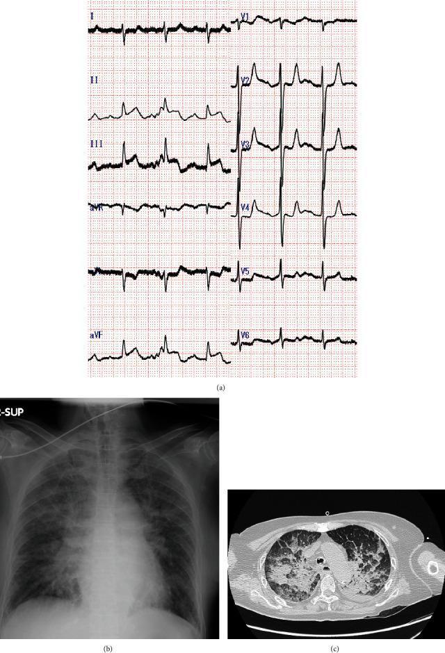
A multi-panel clinical diagnostic set showing a 12-lead electrocardiogram (ECG), a chest radiograph, and a CT scan of the chest. (a) The 12-lead ECG demonstrates sinus tachycardia with marked ST-segment elevation in the inferior leads (II, III, and aVF), consistent with acute inferior myocardial infarction. (b) The anteroposterior chest radiograph shows bilateral, symmetric perihilar opacities in a 'butterfly' or 'bat-wing' distribution, characteristic of acute pulmonary edema and alveolar congestion. (c) The axial chest CT scan (lung window) reveals diffuse, bilateral ground-glass opacities and areas of consolidation, particularly in the dependent and perihilar regions, further supporting the diagnosis of severe pulmonary edema or acute respiratory distress syndrome. These visuals together illustrate a clinical scenario of acute coronary syndrome complicated by acute heart failure and cardiogenic pulmonary edema.
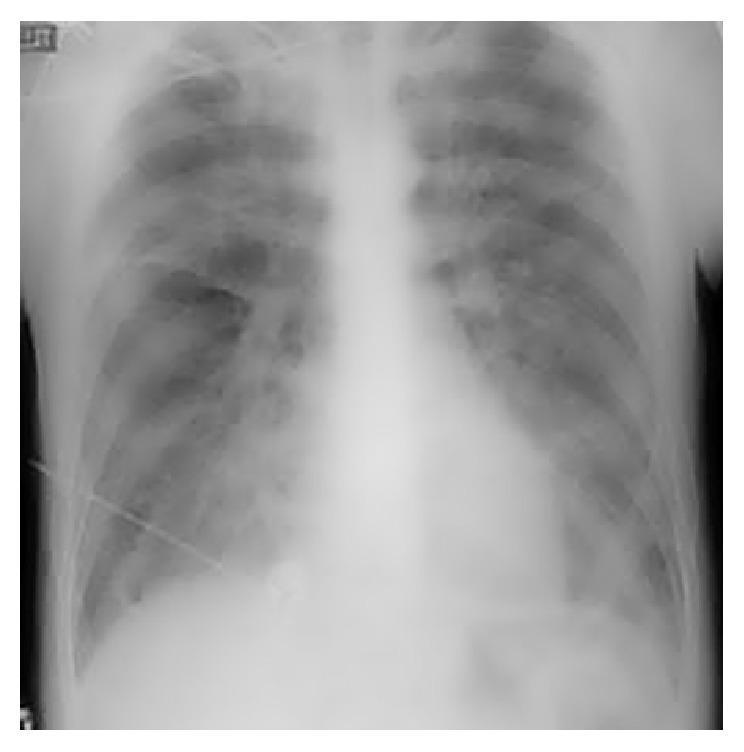
This diagnostic image is a posterior-anterior (PA) or anterior-posterior (AP) chest radiograph. The primary finding is bilateral, diffuse, 'fluffy' opacities throughout both lung fields, consistent with acute pulmonary edema. These opacities are more pronounced in the perihilar regions and mid-to-lower zones, obscuring normal vascular markings and demonstrating a 'bat-wing' or 'angel-wing' distribution pattern. Despite the extensive pulmonary alveolar and interstitial edema, the cardiac silhouette maintains a normal size and shape, without evidence of cardiomegaly or contour abnormalities. This discordance between severe pulmonary edema and a normal cardiac size is often characteristic of non-cardiogenic pulmonary edema or acute cardiogenic failure before ventricular dilation occurs. The mediastinal structures appear central, and the costophrenic angles are partially obscured by the overlying opacities. This image serves as a clinical example of pulmonary congestion and respiratory distress findings in an acute care or emergency medicine context.
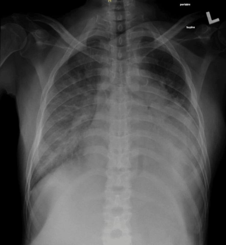
Anteroposterior (AP) supine chest radiograph demonstrating bilateral, diffuse airspace opacities consistent with flash pulmonary edema. The opacification is most pronounced in the perihilar regions, exhibiting a classic 'bat-wing' distribution that obscures the underlying pulmonary vasculature. The heart silhouette appears relatively normal in size, suggesting a non-cardiogenic or acute fluid overload etiology rather than chronic congestive heart failure. Costophrenic angles are partially obscured by the overlying opacities. The mediastinal contours remain within normal limits, and the trachea is midline. This diagnostic image illustrates key radiological findings of acute alveolar edema, often secondary to hypertensive crisis or renal failure, suitable for medical instruction on thoracic imaging and fluid management.
Acute Pulmonary Edema
Definition
Classification
| Type | Mechanism | Examples |
|---|---|---|
| Cardiogenic pulmonary edema | Raised pulmonary capillary hydrostatic pressure | Acute LV failure, acute MI, hypertensive crisis, acute MR |
| Non-cardiogenic pulmonary edema | Increased alveolar-capillary permeability | ARDS, sepsis, aspiration, pancreatitis, high altitude, TRALI |
Etiology of Cardiogenic Acute Pulmonary Edema
Cardiac causes
- Acute myocardial infarction or ongoing ischemia
- Acute decompensated chronic heart failure
- Hypertensive emergency
- Severe mitral regurgitation, including papillary muscle rupture
- Acute aortic regurgitation
- Severe aortic stenosis
- Arrhythmias:
- Rapid atrial fibrillation
- Ventricular tachycardia
- Severe bradycardia/complete heart block
- Myocarditis
- Cardiomyopathy
Precipitating factors in a patient with chronic heart failure
- Drug or dietary non-adherence, particularly salt excess
- Stopping diuretics
- Infection, especially pneumonia
- Anemia
- Renal failure
- Pulmonary embolism
- Thyrotoxicosis
- Drugs: NSAIDs, some calcium-channel blockers, excess IV fluids
Pathophysiology
Stages
-
Interstitial edema
- Peribronchial cuffing
- Kerley B lines
- Reduced lung compliance
-
Alveolar edema
- Flooding of alveoli
- Severe V/Q mismatch and shunt
- Hypoxemia
- Pink frothy sputum in severe cases
Clinical Features
Symptoms
- Sudden severe breathlessness
- Orthopnea
- Paroxysmal nocturnal dyspnea
- Cough with pink frothy sputum
- Wheeze, called cardiac asthma
- Chest pain if ACS is the cause
- Palpitations
- Sweating, fear, and sense of impending doom
Signs
- Patient sits upright, distressed, unable to lie flat
- Tachypnea and use of accessory muscles
- Tachycardia
- Hypoxemia and central cyanosis
- Cold clammy extremities
- Hypertension is common initially, though hypotension suggests cardiogenic shock
- Fine inspiratory crepitations, initially basal then diffuse
- Wheeze
- S3 gallop
- Raised JVP, peripheral edema, or hepatomegaly if there is pre-existing congestive HF
- New murmur may suggest acute MR, VSD, or another valvular lesion
Investigations
Bedside and laboratory tests
- Pulse oximetry and arterial blood gas
- Hypoxemia
- Often respiratory alkalosis initially
- Hypercapnia/acidosis in severe, exhausting respiratory failure
- ECG: ischemia/infarction, LVH, arrhythmia
- CBC: anemia, infection
- Renal function, electrolytes, blood glucose
- Troponin if ACS is suspected
- BNP/NT-proBNP, helpful if diagnosis is uncertain
- Blood cultures if sepsis is suspected
Chest X-ray
- Cardiomegaly in chronic cardiac failure
- Upper-lobe venous diversion
- Kerley B lines
- Peribronchial cuffing
- Bilateral perihilar fluffy alveolar opacities, the “bat-wing” or butterfly pattern
- Pleural effusions
Echocardiography
- LV systolic function and regional wall-motion abnormality
- Acute MR/AR
- Aortic stenosis
- RV function
- Pericardial tamponade
- Mechanical complication of MI
Management
Principle
Treat respiratory failure, relieve pulmonary congestion, reduce preload/afterload where appropriate, and identify and correct the precipitating cause.
Immediate management: ABCDE
1. Position
- Sit patient upright with legs dependent, if BP permits.
- This reduces venous return and improves ventilation.
2. Airway and breathing
- Give oxygen only if hypoxemic, typically SpO₂ <90%, or if there is respiratory distress/shock.
- Target SpO₂ approximately 94-98%, or 88-92% in those at risk of hypercapnic respiratory failure.
- Use CPAP/BiPAP (NIV) early in severe respiratory distress, hypoxemia, hypercapnia, or acidaemia, provided BP is adequate and the patient is cooperative.
- Intubate and mechanically ventilate if there is worsening respiratory failure, reduced consciousness, exhaustion, or inability to protect airway.
3. Circulation and monitoring
- Cardiac monitor, frequent BP measurement, pulse oximetry
- Two IV lines
- Urine output monitoring, often with catheter in severe cases
- Obtain ECG within 10 minutes if ACS is possible
- Look actively for MI, hypertensive crisis, tachyarrhythmia, valvular catastrophe, or infection
Drug Management
| Treatment | Indication and role |
|---|---|
| IV furosemide | For clear fluid overload/congestion. Promotes diuresis and relieves pulmonary/systemic congestion |
| IV nitroglycerin | Particularly useful in hypertensive pulmonary edema, myocardial ischemia, or acute MR, if BP permits |
| Vasodilator support | Nitrates reduce preload and afterload |
| Inotrope | Consider in cardiogenic shock with hypoperfusion, usually in ICU |
| Vasopressor | Norepinephrine is preferred in severe hypotension/shock with low perfusion |
| Morphine | Not routine. May be used cautiously only for extreme anxiety/distress because it can worsen hypotension and respiratory depression |
IV loop diuretic
- Typical initial dose: furosemide 20-40 mg IV, then reassess urine output and clinical response.
- If the patient already takes loop diuretics, a higher IV dose may be necessary.
- Monitor potassium, sodium, renal function, urine output, and BP.
IV nitrates
- Hypotension
- Right ventricular infarction
- Severe aortic stenosis
- Recent PDE-5 inhibitor use
- Marked hypovolemia
Management According to Hemodynamic Profile
| Clinical profile | Features | Main approach |
|---|---|---|
| Warm and wet | Congestion with adequate perfusion, often hypertensive | IV diuretic, nitrates if BP allows, NIV if severe distress |
| Cold and wet | Congestion plus hypoperfusion/shock | ICU, cautious diuresis, inotrope/vasopressor, urgent cause-specific treatment |
| Hypertensive acute pulmonary edema | SBP usually >140 mmHg, marked distress | NIV + IV nitrates, plus diuretic if volume overload |
| ACS-related edema | Chest pain/ECG/troponin changes | ACS protocol and urgent reperfusion where indicated |
| Acute MR/VSD after MI | New murmur, sudden severe edema/shock | Urgent echocardiography, afterload reduction if possible, emergency cardiology/cardiothoracic intervention |
Treat the Cause
| Cause | Specific treatment |
|---|---|
| Acute coronary syndrome | Antiplatelets, anticoagulation as appropriate, urgent PCI/reperfusion |
| Hypertensive emergency | Controlled BP reduction with IV vasodilator, usually nitrates |
| AF with rapid ventricular response | Rate/rhythm control, cardioversion if unstable |
| Ventricular arrhythmia | ACLS-based treatment and correction of ischemia/electrolytes |
| Acute severe MR/VSD | Urgent echo and surgical/interventional management |
| Infection/sepsis | Cultures, prompt antibiotics, careful fluid strategy |
| Renal failure/fluid overload | Diuretic optimization, consider ultrafiltration/dialysis if refractory |
Complications
- Acute hypoxemic respiratory failure
- Hypercapnic respiratory failure
- Cardiogenic shock
- Cardiac arrest
- Acute kidney injury
- Arrhythmias
- Myocardial ischemia/infarction
- Death
Differentiate Cardiogenic from Non-cardiogenic Pulmonary Edema
| Feature | Cardiogenic pulmonary edema | Non-cardiogenic edema/ARDS |
|---|---|---|
| Cause | Elevated pulmonary venous pressure | Increased permeability |
| Heart size | Often enlarged | Usually normal |
| BNP | Often elevated | Usually not markedly elevated |
| JVP/peripheral edema | Often raised/present | Usually absent |
| CXR | Cardiomegaly, vascular redistribution, pleural effusion common | Bilateral infiltrates, usually no cardiomegaly/pleural effusion |
| Echo | LV/valvular abnormality often found | May be normal |
| Treatment | Diuretic, vasodilator if appropriate, treat cardiac cause | Treat cause, lung-protective ventilation, conservative fluid strategy |
5-Mark Exam Answer
Acute pulmonary edema is rapid accumulation of fluid in lung interstitium and alveoli, most commonly due to acute left ventricular failure causing raised pulmonary venous pressure. It presents with sudden severe dyspnea, orthopnea, cough with pink frothy sputum, tachypnea, hypoxemia, diffuse crepitations, wheeze, tachycardia, and often hypertension. Chest X-ray shows pulmonary venous congestion, Kerley B lines, bilateral perihilar bat-wing opacities, and frequently cardiomegaly. Management consists of upright positioning, monitoring, oxygen only for hypoxemia, early CPAP/BiPAP when severe, IV furosemide for congestion, IV nitrates in hypertensive/ischemic patients with adequate BP, and urgent treatment of the precipitating cause such as ACS, arrhythmia, hypertensive emergency, or acute valvular dysfunction.
Infective endocarditis
2023 ESC infective endocarditis guidelines diagnosis treatment blood cultures echocardiography
infective endocarditis AND treatment
infective endocarditis echocardiography vegetation mitral valve clinical signs Janeway Osler
Infective Endocarditis (IE)
Definition
Classification
1. By clinical course
| Type | Usual organism | Course | Typical setting |
|---|---|---|---|
| Acute IE | Staphylococcus aureus | Rapid, destructive, days to weeks | May affect normal valves |
| Subacute IE | Viridans streptococci, enterococci | Indolent, weeks to months | Usually abnormal/damaged valves |
2. By site
- Native-valve endocarditis
- Prosthetic-valve endocarditis
- Device-related IE: pacemaker, ICD leads
- Right-sided IE, usually tricuspid, often in injection-drug use
- Left-sided IE, usually mitral or aortic valve
3. By source
- Community-acquired
- Healthcare-associated
- Injection-drug-use associated
Etiology
Common microorganisms
| Organism | Typical setting |
|---|---|
| Staphylococcus aureus | Acute IE, IV drug use, intravascular catheter, device-related infection |
| Viridans streptococci | Subacute IE, poor dental hygiene, diseased native valve |
| Streptococcus gallolyticus | Association with colonic neoplasia: evaluate colon |
| Enterococci | Elderly, genitourinary or gastrointestinal source |
| Coagulase-negative staphylococci | Prosthetic-valve endocarditis |
| HACEK organisms | Culture-negative or slow-growing IE |
| Fungi, especially Candida | Prosthetic valve, immunosuppression, IV drug use |
| Coxiella burnetii, Bartonella | Blood culture-negative IE |
Predisposing Factors
- Previous infective endocarditis
- Rheumatic or degenerative valvular heart disease
- Congenital heart disease
- Prosthetic heart valve or valve repair material
- Intracardiac devices: pacemaker/ICD
- Intravenous drug use
- Long-term IV cannula, central venous catheter
- Hemodialysis
- Diabetes mellitus
- Immunosuppression
- Poor oral hygiene or dental infection
- Recent invasive procedure, especially in high-risk cardiac patients
Pathogenesis
Sequence
- Turbulent blood flow, valvular disease, prosthetic material, or devices damage the endocardium.
- Platelets and fibrin deposit on the damaged surface, forming sterile non-bacterial thrombotic vegetation.
- Transient bacteremia follows dental, skin, gastrointestinal, genitourinary, or catheter-related infection.
- Organisms adhere to the vegetation and multiply.
- Vegetations enlarge because they are relatively avascular, making antibiotic penetration difficult.
- Complications follow:
- Local valve destruction and regurgitation
- Perivalvular abscess
- Emboli
- Metastatic infection
- Immune-complex injury
Clinical Features
Constitutional features
- Fever, often prolonged
- Chills and rigors
- Malaise, fatigue, anorexia
- Weight loss
- Night sweats
- Arthralgia/myalgia
Cardiac features
- New regurgitant murmur or change in previous murmur
- Features of heart failure due to valvular destruction
- Conduction abnormalities, especially new AV block, suggesting aortic root abscess
- Palpitations/arrhythmias
Peripheral stigmata
| Finding | Description | Mechanism |
|---|---|---|
| Petechiae | Skin, conjunctiva, oral mucosa | Microemboli/vasculitis |
| Splinter hemorrhages | Linear nail-bed hemorrhages | Embolic/vascular injury |
| Osler nodes | Painful, tender nodules on finger/toe pads | Immune-complex vasculitis |
| Janeway lesions | Painless erythematous lesions on palms/soles | Septic emboli |
| Roth spots | Retinal hemorrhages with pale centers | Immune-mediated vascular injury |
| Clubbing | Usually in subacute disease | Chronic inflammation |
| Splenomegaly | May occur in prolonged disease | Reticuloendothelial hyperplasia |
Viva point: Osler nodes are painful, whereas Janeway lesions are painless.
Embolic manifestations
- Stroke or transient ischemic attack
- Splenic infarct/abscess: left upper abdominal pain
- Renal infarct: hematuria/flank pain
- Coronary embolism: MI
- Limb ischemia
- Pulmonary emboli, lung infarcts, or septic pulmonary abscesses in right-sided IE
Diagnosis
1. Blood cultures
- Collect three sets of blood cultures from separate venepuncture sites.
- Ideally take them before starting antibiotics.
- In a critically ill/septic patient, obtain cultures promptly, but do not delay antibiotics unnecessarily.
- Persistent bacteremia supports IE.
- Repeat cultures every 24-48 hours until they are negative.
2. Echocardiography
| Test | Role |
|---|---|
| TTE | First-line investigation in suspected IE |
| TEE | More sensitive for vegetation, abscess, prosthetic-valve IE, and device infection |
- TTE is negative/inconclusive but clinical suspicion remains high
- Prosthetic valve is present
- Cardiac device is present
- There is suspected abscess or complication
- Pre-operative assessment is needed
3. Other investigations
- CBC: normocytic anemia, leukocytosis/leukopenia
- ESR and CRP: elevated
- Renal function, electrolytes, LFT
- Urine routine/microscopy: microscopic hematuria, proteinuria, RBC casts due to glomerulonephritis
- ECG: PR prolongation/new AV block may suggest abscess
- Chest X-ray: pulmonary edema in HF, septic pulmonary emboli in right-sided IE
- CT/MRI brain, chest, abdomen: embolic infarcts/abscesses
- Cardiac CT, FDG-PET/CT or WBC-SPECT/CT: useful particularly in prosthetic-valve/device-related IE when echo is inconclusive
Modified Duke Criteria
Major criteria
1. Positive blood culture for IE
- Typical organism from two separate blood cultures:
- Viridans streptococci
- Streptococcus gallolyticus
- HACEK organisms
- Staphylococcus aureus
- Community-acquired enterococci without primary focus
- Persistently positive cultures:
- Two positive cultures taken >12 hours apart, or
- Three of three or majority of four separate cultures positive
2. Evidence of endocardial involvement
- Echocardiographic vegetation
- Abscess
- New partial dehiscence of prosthetic valve
- New valvular regurgitation
Minor criteria
- Predisposition: cardiac lesion or injection-drug use
- Fever ≥38°C
- Vascular phenomena: arterial emboli, septic pulmonary infarcts, Janeway lesions, intracranial hemorrhage, mycotic aneurysm
- Immunological phenomena: glomerulonephritis, Osler nodes, Roth spots, rheumatoid factor
- Microbiological evidence not fulfilling a major criterion
Interpretation
| Diagnosis | Criteria |
|---|---|
| Definite IE | 2 major, OR 1 major + 3 minor, OR 5 minor |
| Possible IE | 1 major + 1 minor, OR 3 minor |
| Rejected IE | Firm alternative diagnosis or criteria not fulfilled |
Management
Principles
- Admit and manage under a multidisciplinary Endocarditis Team.
- Obtain blood cultures before antibiotics whenever possible.
- Give prolonged, high-dose, bactericidal IV antibiotics.
- Modify therapy according to culture and sensitivity.
- Monitor for heart failure, embolism, renal dysfunction, abscess, and arrhythmia.
- Evaluate early for cardiac surgery where indicated.
Antibiotic choice and doses must be individualized according to organism, susceptibility, renal function, native versus prosthetic valve, and local antimicrobial policy.
Empirical Antibiotic Therapy
- Vancomycin-based regimen, often with ceftriaxone or cefepime depending on local policy and clinical setting.
Organism-Specific Treatment: Exam Outline
| Organism | Common regimen | Usual duration |
|---|---|---|
| Viridans streptococci, penicillin-sensitive | Penicillin G or ceftriaxone | 4 weeks |
| Streptococcus gallolyticus | Penicillin G or ceftriaxone | 4 weeks |
| MSSA native-valve IE | Flucloxacillin/nafcillin/oxacillin | 4-6 weeks |
| MRSA IE | Vancomycin or daptomycin | At least 6 weeks |
| Enterococcal IE | Ampicillin + ceftriaxone, or ampicillin/penicillin + gentamicin in selected cases | Usually 6 weeks |
| HACEK IE | Ceftriaxone | 4 weeks for native valve, 6 weeks for prosthetic valve |
| Fungal IE | Antifungal therapy plus early surgery usually needed | Prolonged |
Important point
- Do not give aminoglycosides casually. They may cause nephrotoxicity and their use depends on the organism and regimen.
- Monitor renal function, serum drug levels where required, CRP, temperature, and serial blood cultures.
Indications for Surgery
H - Heart failure
- Acute severe aortic or mitral regurgitation causing pulmonary edema/HF
- Prosthetic-valve dysfunction causing HF
- Obstruction due to large vegetation
U - Uncontrolled infection
- Abscess, pseudoaneurysm, fistula, or enlarging vegetation
- Persistent positive blood cultures despite appropriate antibiotics
- Infection with fungi or highly resistant organism
- Prosthetic-valve endocarditis with destructive complications
- New conduction block suggesting periannular extension
I - Implant/device infection
- Infected pacemaker/ICD lead or pocket infection generally requires complete device removal plus antibiotics.
E - Embolic prevention
- Recurrent embolization despite treatment
- Large mobile left-sided vegetation, especially after embolic event
- Large vegetation with other surgical indications
Complications
Cardiac
- Acute valvular regurgitation
- Congestive heart failure
- Perivalvular abscess
- Conduction block
- Myocarditis
- Pericarditis
- Prosthetic-valve dehiscence
Embolic/infective
- Ischemic or hemorrhagic stroke
- Cerebral abscess
- Mycotic aneurysm
- Splenic infarct/abscess
- Renal infarct
- Septic pulmonary emboli in tricuspid IE
- Vertebral osteomyelitis/discitis
Immunological
- Immune-complex glomerulonephritis
- Vasculitis
- Rheumatoid-factor positivity
Other
- Sepsis and septic shock
- Acute kidney injury
- Death
Prevention
General measures
- Good dental and skin hygiene
- Early treatment of skin, dental, and bloodstream infections
- Avoid unnecessary intravascular catheters
- Strict asepsis during invasive procedures
- Avoid injection drug use
Antibiotic prophylaxis
- Prosthetic heart valve or prosthetic material used for valve repair
- Previous IE
- Selected congenital heart disease
- Cardiac transplant recipients with valvular disease
5-Mark Short Note
Infective endocarditis is microbial infection of the endocardium, usually the cardiac valves, leading to platelet-fibrin vegetations containing organisms. It commonly presents with prolonged fever, new or changing murmur, anemia, splenomegaly, petechiae, splinter hemorrhages, Osler nodes, Janeway lesions, embolic events, and heart failure. Diagnosis is based on serial blood cultures and echocardiography, interpreted using modified Duke criteria. Treatment consists of prolonged culture-directed bactericidal IV antibiotics, with surgery for acute heart failure due to valve destruction, uncontrolled infection/abscess or persistent bacteremia, prosthetic/device infection, fungi, and high embolic risk.
Valvular heart disease
Valvular Heart Disease: Proff Topic Map
-
General approach
- Definition, classification into stenosis and regurgitation
- Rheumatic versus degenerative versus infective causes
- Symptoms, murmurs, echocardiography, and principles of management
-
Mitral stenosis
- Rheumatic etiology, clinical features, auscultation, ECG/CXR/echo
- Complications: AF, embolism, pulmonary hypertension
- Medical therapy and balloon mitral valvotomy
-
Mitral regurgitation
- Acute versus chronic MR
- Causes, murmur, investigations, complications, and surgery
-
Aortic stenosis
- Degenerative/calcific versus bicuspid valve versus rheumatic AS
- Classical triad: angina, syncope, dyspnea
- Echo severity and valve replacement/TAVI
-
Aortic regurgitation
- Acute versus chronic AR
- Peripheral signs, murmur, investigations, and surgical indications
-
Tricuspid and pulmonary valve disease
- TR, TS, PR, PS, and their classical murmurs
-
Infective endocarditis and rheumatic heart disease
- Important etiologies and complications relevant to valvular lesions
-
Pregnancy and valvular lesions
- High-yield lesion-specific maternal risks and management
High-level overview
Definition → etiology → pathophysiology → symptoms → signs and murmur → investigations, especially echocardiography → complications → medical and definitive management.
- “Describe mitral stenosis and its management.”
- “Discuss aortic stenosis/aortic regurgitation with clinical signs and treatment.”
Rheumatic fever
Rheumatic heart disease
World Heart Federation rheumatic heart disease prevention management 2025 guideline
rheumatic heart disease AND management
Rheumatic Heart Disease (RHD)
Definition
Etiopathogenesis
Basic sequence
Group A streptococcal pharyngitis → acute rheumatic fever → recurrent valvulitis → fibrosis and scarring of valve → chronic stenosis and/or regurgitation.
Mechanism: molecular mimicry
- Streptococcal M protein shares antigenic similarity with cardiac myosin and valvular tissue.
- Antibodies and T-cells generated against streptococcal antigens cross-react with the host heart.
- This is an immune-mediated disease, not direct bacterial infection of the valve.
- Recurrent ARF causes cumulative valvular damage.
Pathology
Acute rheumatic carditis
- Pancarditis: endocarditis, myocarditis, and pericarditis.
- Characteristic lesion: Aschoff body.
- Aschoff bodies contain Anitschkow cells, also called caterpillar cells.
Chronic rheumatic valvular disease
- Leaflet thickening and calcification
- Commissural fusion
- Chordae tendineae thickening, fusion, and shortening
- Valve stenosis and/or regurgitation
Classical mitral-valve appearance
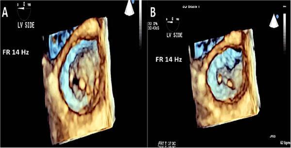
Valves Affected
| Valve lesion | Frequency and usual pattern |
|---|---|
| Mitral valve | Most common |
| Mitral stenosis | Commonest chronic isolated lesion |
| Mitral regurgitation | Common in early disease and younger patients |
| Aortic valve | Usually associated with mitral-valve disease |
| Tricuspid valve | Usually functional TR due to pulmonary hypertension, rarely primary rheumatic involvement |
| Pulmonary valve | Very rarely affected |
Common combinations
- Mitral stenosis with mitral regurgitation
- Mitral stenosis with aortic regurgitation
- Mitral stenosis with aortic stenosis/regurgitation
Clinical Features
General symptoms
- Fatigue
- Reduced exercise tolerance
- Palpitations
- Dyspnea
- Orthopnea and paroxysmal nocturnal dyspnea
- Chest discomfort
- Hemoptysis
- Pedal edema and abdominal distension in right-heart failure
- Fever or constitutional symptoms if recurrent rheumatic activity or infective endocarditis occurs
Rheumatic Mitral Stenosis
Pathophysiology
Mitral obstruction → raised left atrial pressure → pulmonary venous hypertension → pulmonary arterial hypertension → right ventricular hypertrophy/failure → systemic venous congestion.
- Left atrial enlargement
- Atrial fibrillation
- Left atrial thrombus
- Systemic embolism, especially stroke
Symptoms
- Exertional dyspnea
- Orthopnea/PND
- Palpitations due to AF
- Hemoptysis
- Fatigue
- Recurrent bronchitis
- Hoarseness of voice due to left atrial enlargement compressing left recurrent laryngeal nerve: Ortner syndrome
- Thromboembolic stroke
Signs
- Malar flush
- Tapping, localized apex beat
- Loud S1
- Opening snap after A2
- Low-pitched, rumbling mid-diastolic murmur at apex
- Murmur is best heard with bell in left lateral position
- Presystolic accentuation if sinus rhythm is present
- Loud P2 if pulmonary hypertension
- Raised JVP, hepatomegaly, edema in right-heart failure
Rheumatic Mitral Regurgitation
Pathophysiology
MR → LV volume overload + LA volume overload → LA dilatation + LV dilatation → AF, pulmonary congestion, and heart failure.
Clinical features
- Fatigue and exertional dyspnea
- Palpitations
- Orthopnea/PND in severe disease
- Displaced, hyperdynamic apex beat
- Soft S1
- High-pitched, blowing pansystolic murmur at apex radiating to left axilla
- S3 gallop in severe MR
Rheumatic Aortic Stenosis
Clinical features
- Exertional angina
- Exertional syncope
- Exertional dyspnea/heart failure
- Slow-rising low-volume pulse: pulsus parvus et tardus
- Harsh ejection systolic murmur in aortic area radiating to carotids
- Soft/absent A2 in severe AS
Rheumatic Aortic Regurgitation
Clinical features
- Palpitations due to increased stroke volume
- Exertional dyspnea, orthopnea, PND
- Wide pulse pressure
- Collapsing or water-hammer pulse
- Displaced hyperdynamic apex
- High-pitched, early diastolic decrescendo murmur at left sternal edge, best heard with patient sitting forward in end-expiration
- Austin Flint murmur at apex in severe AR
Investigations
1. ECG
- Left atrial enlargement: P mitrale
- Atrial fibrillation
- Right ventricular hypertrophy in pulmonary hypertension
- LVH in MR, AR, or AS
- Conduction abnormalities
2. Chest X-ray
- Cardiomegaly depending on lesion
- Left atrial enlargement, double-density shadow
- Straightening of left heart border in MS
- Pulmonary venous congestion
- Kerley B lines
- Pulmonary edema in severe failure
- Prominent pulmonary arteries in pulmonary hypertension
3. Echocardiography with Doppler - investigation of choice
- Valve morphology and thickening
- Commissural fusion
- Severity of stenosis/regurgitation
- Valve area and transvalvular gradients
- LA/LV/RV size and function
- Pulmonary artery pressure
- Presence of LA thrombus
- Associated valvular lesions
4. Transesophageal echocardiography
- Left atrial appendage thrombus before cardioversion or balloon mitral valvotomy
- Prosthetic-valve dysfunction
- Infective endocarditis vegetation
5. Cardiac catheterization/coronary angiography
- Discrepancy between clinical and echo data
- Assessment before valve intervention in selected older patients or those at risk of CAD
Complications of RHD
- Atrial fibrillation
- Left atrial thrombus and systemic embolism
- Ischemic stroke
- Peripheral arterial embolism
- Pulmonary hypertension
- Right-heart failure
- Congestive cardiac failure
- Infective endocarditis
- Hemoptysis
- Pregnancy-related decompensation
- Sudden deterioration due to tachyarrhythmia, infection, anemia, or embolism
Management
A. General Measures
- Regular cardiology follow-up with periodic echocardiography
- Counsel on adherence to secondary prophylaxis
- Avoid smoking and excess salt in heart failure
- Treat anemia, infection, hypertension, thyroid disease, and arrhythmias
- Pregnancy counselling before conception in women with significant valve lesions
- Maintain good oral and dental hygiene
- Screen and treat household streptococcal infections as appropriate
B. Secondary Prevention of Rheumatic Fever
Preferred regimen
- Benzathine benzylpenicillin G 1.2 million units IM every 3-4 weeks
- Children <27 kg: 600,000 units IM every 3-4 weeks
- Penicillin V orally
- Sulfadiazine orally
- Macrolide in true penicillin allergy
Duration of secondary prophylaxis
| Clinical history | Minimum duration |
|---|---|
| ARF without carditis | 5 years after last attack or until age 21 years, whichever is longer |
| ARF with carditis but no residual valvular lesion | 10 years after last attack or until age 21 years, whichever is longer |
| RHD with persistent valvular lesion | At least 10 years after last attack or until age 40 years, whichever is longer |
| Severe RHD or after valve surgery | Often lifelong, based on specialist/local policy |
C. Treatment According to Valvular Lesion
1. Mitral stenosis
Medical treatment
- Diuretics for pulmonary congestion
- Beta-blocker, verapamil, or diltiazem for rate control in sinus tachycardia/AF if appropriate
- Anticoagulation for:
- AF
- Previous systemic embolism
- Left atrial thrombus
- Digoxin may be used in AF with heart failure
Definitive treatment
- Symptomatic severe rheumatic MS
- Favorable valve morphology
- No left atrial thrombus
- No more than mild MR
- Valve anatomy is unsuitable for PBMV
- Significant associated MR/other valve disease
- Failed PBMV
- Need for concomitant cardiac surgery
2. Mitral regurgitation
- Diuretics for congestion
- Treat AF and anticoagulate when indicated
- Valve repair or replacement for severe symptomatic MR, LV dysfunction/dilatation, or pulmonary hypertension
3. Aortic stenosis
- Definitive therapy is aortic-valve replacement:
- Surgical AVR
- TAVI in selected patients
- Diuretics should be used cautiously if congested
- Avoid excessive vasodilation or dehydration in severe AS
4. Aortic regurgitation
- Diuretics for heart failure
- Control hypertension
- Aortic-valve surgery for severe symptomatic AR or progressive LV dilatation/dysfunction
Anticoagulation in Rheumatic Mitral Stenosis
- Atrial fibrillation
- Previous embolic event
- Left atrial thrombus
Exam pearl: In moderate-severe rheumatic MS with AF, warfarin is preferred. Direct oral anticoagulants are generally not used as a substitute in this setting.
Prevention
1. Primordial prevention
- Reduce overcrowding and poverty
- Improve sanitation, nutrition, healthcare access, and health education
2. Primary prevention
- Early diagnosis and adequate treatment of Group A streptococcal pharyngitis with penicillin
3. Secondary prevention
- Long-term benzathine penicillin to prevent recurrent ARF and progression of RHD
High-Yield Viva Points
- Most commonly affected valve: Mitral valve
- Most common chronic lesion: Mitral stenosis
- Most common cause of MS in young adults in endemic settings: Rheumatic heart disease
- Fish-mouth deformity: Rheumatic mitral stenosis
- Most important complication of MS: Atrial fibrillation with systemic embolism
- Investigation of choice: Echocardiography with Doppler
- Most important long-term preventive measure: Secondary benzathine penicillin prophylaxis
- Definitive intervention for suitable severe rheumatic MS: Percutaneous balloon mitral valvotomy
Short Exam Conclusion
Rheumatic heart disease is chronic valvular damage following acute rheumatic fever caused by an autoimmune response to Group A streptococcal infection. It predominantly affects the mitral valve and commonly causes mitral stenosis, mitral regurgitation, or mixed valvular disease. Diagnosis is clinical and echocardiographic. Management includes secondary penicillin prophylaxis, treatment of heart failure and atrial fibrillation, anticoagulation where indicated, and timely valvotomy, valve repair, or valve replacement.
Rheumatic fever
WHO guideline prevention diagnosis rheumatic fever 2024 Jones criteria treatment benzathine penicillin
rheumatic fever AND treatment
acute rheumatic fever erythema marginatum subcutaneous nodules Sydenham chorea diagram
Acute Rheumatic Fever (ARF)
Definition
Epidemiology
- Commonly affects children aged 5-15 years.
- Rare before 3 years of age.
- More prevalent in settings of overcrowding, poverty, poor sanitation, and poor access to treatment of streptococcal pharyngitis.
- Recurrent attacks produce cumulative valvular damage.
Etiopathogenesis
Causative event
GAS pharyngitis → molecular mimicry → autoimmune inflammation in heart, joints, brain, and skin.
Molecular mimicry
Organ-specific immune injury
| Organ | Mechanism/result |
|---|---|
| Heart | Antibody binding and T-cell infiltration causing pancarditis and valvulitis |
| Joints | Immune complex-mediated sterile inflammation |
| Brain, basal ganglia | Antibody-mediated dysfunction causing Sydenham chorea |
| Skin | Delayed hypersensitivity reaction |
Pathology
Acute rheumatic carditis
-
Endocarditis/valvulitis: most clinically important
- Mitral valve most commonly involved
- Aortic valve next most common
- Acute inflammation causes valvular regurgitation.
-
Myocarditis
- May cause tachycardia, cardiomegaly, heart failure.
-
Pericarditis
- Fibrinous pericarditis with pericardial rub and chest pain.
Characteristic lesion: Aschoff body
- Central fibrinoid necrosis
- Surrounding lymphocytes and plasma cells
- Activated macrophages called Anitschkow cells or caterpillar cells, because of their wavy chromatin.
Clinical Features
Major manifestations: JONES
| Major criterion | Characteristic features |
|---|---|
| J: Joints | Migratory polyarthritis |
| O: Carditis | Pancarditis, valvulitis, new MR/AR, heart failure |
| N: Nodules | Painless subcutaneous nodules |
| E: Erythema marginatum | Evanescent, non-pruritic serpiginous rash |
| S: Sydenham chorea | Involuntary purposeless movements |
1. Migratory polyarthritis
- Most common major manifestation.
- Involves large joints: knees, ankles, elbows, wrists.
- Classically migratory: inflammation subsides in one joint and appears in another.
- Pain is often severe and disproportionate to physical signs.
- Responds dramatically to aspirin or NSAIDs.
- Joint disease is non-destructive and resolves without permanent deformity.
Failure of arthritis to respond to NSAIDs should prompt consideration of an alternative diagnosis.
2. Carditis
Features
- Tachycardia disproportionate to fever
- New murmur:
- Apical pansystolic murmur of mitral regurgitation
- Early diastolic murmur of aortic regurgitation
- Cardiomegaly
- S3 gallop
- Pericardial friction rub
- Heart failure
- ECG: PR prolongation
Important point
3. Sydenham chorea
- Delayed manifestation, often appearing weeks to months after GAS infection.
- Involuntary, irregular, purposeless, non-rhythmic movements involving face, limbs, and trunk.
- May be unilateral.
- Disappears during sleep.
- Associated with emotional lability, hypotonia, clumsiness, poor handwriting, and deteriorating school performance.
4. Erythema marginatum
- Rare but highly specific.
- Pink, non-pruritic, blanching macules or papules.
- Spread centrifugally with central clearing, giving a serpiginous or annular appearance.
- Mainly affects trunk and proximal limbs.
- Usually spares the face.
- Becomes more visible after warmth, such as a hot bath.
5. Subcutaneous nodules
- Firm, painless, freely mobile nodules.
- Usually 0.5-2 cm.
- Occur over extensor surfaces and bony prominences:
- Elbows
- Knees
- Wrists
- Ankles
- Spine
- Occiput
- Often associated with severe carditis.
Minor Jones Criteria
| Minor criterion | Features |
|---|---|
| Fever | Usually low to moderate grade |
| Arthralgia | Joint pain without objective arthritis |
| Raised acute-phase reactants | Raised ESR and CRP |
| Prolonged PR interval on ECG | Only if not explained by carditis |
Diagnosis: Revised Jones Criteria
For an initial episode of ARF
- Two major criteria, OR
- One major + two minor criteria
Plus evidence of preceding GAS infection
- Positive throat culture for GAS
- Positive rapid streptococcal antigen test
- Elevated or rising ASO titre
- Elevated anti-DNase B titre
- Recent scarlet fever
For recurrent ARF
- Two major, OR
- One major + two minor, OR
- Three minor criteria
with supporting evidence of preceding streptococcal infection.
Investigations
To support GAS infection
- Throat culture
- Rapid antigen detection test
- Antistreptolysin O titre: elevated or rising titre
- Anti-DNase B titre
A throat swab may be negative when ARF appears because the pharyngitis occurred several weeks earlier.
To assess inflammation
- CBC: leukocytosis, mild normocytic normochromic anemia
- ESR: raised
- CRP: raised
Cardiac assessment
- ECG:
- Sinus tachycardia
- PR interval prolongation
- Arrhythmia in severe carditis
- Chest X-ray:
- Cardiomegaly
- Pulmonary edema in heart failure
- Echocardiography with Doppler:
- Recommended in suspected or confirmed ARF
- Detects clinical or subclinical carditis
- Shows MR, AR, chamber enlargement, ventricular dysfunction, pericardial effusion
Differential Diagnosis
- Septic arthritis
- Viral arthritis
- Juvenile idiopathic arthritis
- Systemic lupus erythematosus
- Post-streptococcal reactive arthritis
- Infective endocarditis
- Kawasaki disease
- Lyme arthritis
- Viral myocarditis
- Functional chorea, Wilson disease, SLE, drug-induced chorea
Treatment
Principles
- Eradicate residual streptococcal infection.
- Suppress inflammation and relieve symptoms.
- Treat carditis and heart failure.
- Control chorea.
- Start long-term secondary prophylaxis.
- Ensure rest and follow-up.
1. Eradication of GAS
Preferred regimen
- Benzathine benzylpenicillin G IM single dose
- ≥27 kg: 1.2 million units
- <27 kg: 600,000 units
Alternatives
- Penicillin V orally for 10 days
- Amoxicillin orally for 10 days
- Macrolide, for example azithromycin, in true penicillin allergy
2. Anti-inflammatory treatment
Arthritis without severe carditis
- Aspirin or an NSAID, for example naproxen.
- Aspirin has traditionally been used and produces a rapid response in rheumatic polyarthritis.
- Provide gastroprotection where appropriate and monitor for adverse effects.
Moderate/severe carditis or heart failure
- Corticosteroids, typically prednisolone, may be used in significant carditis.
- Treat heart failure conventionally:
- Rest
- Salt restriction
- Diuretics
- ACE inhibitor where suitable
- Digoxin selectively, especially for AF, but use cautiously in acute carditis
Activity restriction
- Bed rest during active carditis/heart failure.
- Gradual return to activity after clinical and inflammatory improvement.
3. Treatment of Sydenham chorea
General measures
- Rest in a quiet environment.
- Reassurance and psychological support.
- Protect patient from injury.
For troublesome chorea
- Sodium valproate
- Carbamazepine
- Haloperidol in selected severe cases
Secondary Prophylaxis
Preferred regimen
- 1.2 million units IM every 3-4 weeks in adults/larger children
- 600,000 units IM every 3-4 weeks in children <27 kg
Duration
| Clinical situation | Continue for |
|---|---|
| ARF without carditis | 5 years after last attack or until age 21, whichever is longer |
| ARF with carditis but no residual valve disease | 10 years after last attack or until age 21, whichever is longer |
| ARF with persistent valvular disease | At least 10 years after last attack or until age 40, whichever is longer |
| Severe RHD or after valve surgery | Often lifelong, based on specialist and local guidance |
Prevention
1. Primordial prevention
- Reduce overcrowding and poverty.
- Improve nutrition, sanitation, healthcare access, and awareness.
2. Primary prevention
- Diagnose and fully treat GAS pharyngitis promptly with appropriate antibiotics.
3. Secondary prevention
- Long-term benzathine penicillin prophylaxis after ARF to prevent recurrence and RHD.
Complications
- Chronic rheumatic heart disease
- Mitral regurgitation in acute carditis
- Mitral stenosis in chronic RHD
- Aortic regurgitation or mixed valvular disease
- Atrial fibrillation
- Heart failure
- Pulmonary hypertension
- Left atrial thrombus and systemic embolism
- Infective endocarditis
High-Yield Viva Points
- Cause: Autoimmune response following GAS pharyngitis.
- Usual age: 5-15 years.
- Latency: 2-4 weeks after pharyngitis.
- Most common major manifestation: Migratory polyarthritis.
- Most dangerous manifestation: Carditis.
- Most commonly involved valve: Mitral valve.
- Pathognomonic lesion: Aschoff body.
- Characteristic cell: Anitschkow or caterpillar cell.
- Painful arthritis with dramatic salicylate response: ARF.
- Chorea disappears during: Sleep.
- Most effective secondary prophylaxis: Benzathine benzylpenicillin G IM every 3-4 weeks.
5-Mark Short Note
Acute rheumatic fever is an immune-mediated, multisystem inflammatory disease occurring 2-4 weeks after Group A beta-hemolytic streptococcal pharyngitis, mainly in children aged 5-15 years. It is diagnosed by revised Jones criteria: two major or one major plus two minor criteria, together with evidence of previous GAS infection. Major manifestations are carditis, migratory polyarthritis, Sydenham chorea, erythema marginatum, and subcutaneous nodules. Management includes eradication of streptococci with penicillin, anti-inflammatory treatment for arthritis/carditis, management of heart failure or chorea, and long-term benzathine penicillin prophylaxis to prevent recurrent ARF and rheumatic heart disease.
Hypertension
2024 ESC hypertension guideline diagnosis treatment blood pressure classification
hypertension AND treatment
hypertensive retinopathy fundus grade 4 papilledema image
Hypertension
Definition
Classification of Blood Pressure in Adults
| Category | Systolic BP (mmHg) | Diastolic BP (mmHg) |
|---|---|---|
| Optimal | <120 | <80 |
| Normal | 120-129 | 80-84 |
| High normal | 130-139 | 85-89 |
| Grade 1 hypertension | 140-159 | 90-99 |
| Grade 2 hypertension | 160-179 | 100-109 |
| Grade 3 hypertension | ≥180 | ≥110 |
| Isolated systolic hypertension | ≥140 | <90 |
Exam point: Diagnose hypertension only after repeated measurements on separate visits, except in very high BP with acute target-organ damage.
Types of Hypertension
1. Primary or essential hypertension
- Accounts for about 90-95% of cases.
- No single identifiable cause.
- Caused by interaction of genetic predisposition and environmental factors.
2. Secondary hypertension
Causes of secondary hypertension: “CRAP-D”
| System | Causes |
|---|---|
| C: Coarctation/Cushing syndrome | Coarctation of aorta, Cushing syndrome |
| R: Renal | CKD, glomerulonephritis, polycystic kidney disease, renal artery stenosis |
| A: Aldosteronism | Primary hyperaldosteronism |
| P: Pheochromocytoma/primary endocrine | Pheochromocytoma, thyroid disease, acromegaly |
| D: Drugs | NSAIDs, steroids, oral contraceptives, decongestants, cyclosporine, tacrolimus, cocaine, amphetamines, erythropoietin |
When should secondary hypertension be suspected?
- Hypertension before age 30 years without family history or risk factors
- Abrupt onset or rapidly worsening hypertension
- Resistant hypertension
- Severe hypertension or hypertensive emergency
- Hypokalemia, especially without diuretic use
- Episodic headache, palpitations, sweating
- Renal bruit
- Rise in creatinine after ACE inhibitor/ARB
- Features of endocrine disease
- Unequal upper and lower limb BP or weak femoral pulse
Risk Factors for Essential Hypertension
Non-modifiable
- Family history
- Increasing age
- Male sex before menopause
- Ethnicity/genetic predisposition
Modifiable
- High dietary salt intake
- Obesity and central obesity
- Sedentary lifestyle
- Excess alcohol
- Smoking
- Stress
- Diabetes mellitus
- Dyslipidemia
- Obstructive sleep apnea
- Unhealthy diet, low fruit and vegetable intake
Pathogenesis of Essential Hypertension
Important mechanisms
- Increased sympathetic nervous system activity
- Increased renin-angiotensin-aldosterone system activity
- Renal sodium and water retention
- Increased peripheral vascular resistance
- Endothelial dysfunction with reduced nitric oxide
- Vascular remodeling and arterial stiffness
- Obesity, insulin resistance, and inflammation
Simple flowchart
Genetic susceptibility + environmental factors
→ sympathetic and RAAS activation
→ vasoconstriction + sodium/water retention
→ increased cardiac output and systemic vascular resistance
→ sustained hypertension
→ target-organ damage
Clinical Features
Symptoms, if present
- Occipital headache, especially in morning
- Giddiness
- Palpitations
- Tinnitus
- Blurring of vision
- Fatigue
- Epistaxis
- Dyspnea or angina due to cardiac involvement
- Neurologic deficits due to stroke/TIA
Examination
General examination
- Measure BP correctly in both arms initially.
- Check pulse rate and rhythm.
- Measure BMI and waist circumference.
- Look for features of Cushing syndrome, acromegaly, thyroid disease, or renal disease.
- Examine for peripheral edema.
Cardiovascular examination
- Displaced/heaving apex: LV hypertrophy
- S4 gallop
- Signs of heart failure
- Carotid, renal, abdominal, and peripheral bruits
- Weak/delayed femoral pulses in coarctation
Fundus examination: hypertensive retinopathy
| Grade | Fundus finding |
|---|---|
| I | Generalized arteriolar narrowing |
| II | AV crossing changes, AV nicking |
| III | Hemorrhages, cotton-wool spots, hard exudates |
| IV | Grade III changes plus papilledema |
Grade IV retinopathy with papilledema suggests malignant hypertension.
Renal and neurologic examination
- Renal bruit suggests renal artery stenosis.
- Assess for focal neurological deficit, encephalopathy, or stroke.
- Look for CKD and fluid overload.
Diagnosis and Correct Measurement of BP
Correct office BP measurement
- Avoid smoking, caffeine, and exercise for at least 30 minutes before measurement.
- Patient should sit quietly for 5 minutes.
- Back supported, feet flat on the floor, legs uncrossed.
- Use an appropriate cuff size.
- Arm should be at heart level.
- Take at least two readings, 1-2 minutes apart.
- Use the average of readings.
- Measure in both arms at first visit and use the arm with the higher reading thereafter.
Out-of-office confirmation
| Method | Hypertension threshold |
|---|---|
| Home BP monitoring average | ≥135/85 mmHg |
| Daytime ambulatory BP average | ≥135/85 mmHg |
| Night-time ambulatory BP average | ≥120/70 mmHg |
| 24-hour ambulatory BP average | ≥130/80 mmHg |
White-coat hypertension
- Office BP high
- Home/ambulatory BP normal
Masked hypertension
- Office BP normal
- Home/ambulatory BP high
Investigations
Routine investigations in all patients
| Test | Purpose |
|---|---|
| Urine routine and urine albumin/protein | Proteinuria, hematuria, renal disease |
| CBC | Anemia, polycythemia |
| Fasting blood glucose/HbA1c | Diabetes screening |
| Lipid profile | Cardiovascular risk assessment |
| Serum creatinine, eGFR, urea | Renal damage and drug selection |
| Serum sodium and potassium | Electrolyte disorder, hyperaldosteronism |
| Uric acid | Baseline risk and diuretic-associated hyperuricemia |
| ECG | LVH, ischemia, AF |
| Fundus examination | Hypertensive retinopathy |
Additional tests when indicated
- Echocardiography: LVH, LV dysfunction
- Renal ultrasound: renal parenchymal disease
- Renal artery Doppler/CT angiography: renal artery stenosis
- Aldosterone-renin ratio: primary hyperaldosteronism
- Plasma-free metanephrines or urinary fractionated metanephrines: pheochromocytoma
- TSH: thyroid disorder
- Sleep study: obstructive sleep apnea
- CT/MRI abdomen: adrenal lesion
- CT angiography/MR angiography: coarctation or renovascular disease
Target-Organ Damage
Heart
- Left ventricular hypertrophy
- Ischemic heart disease/MI
- Heart failure
- Atrial fibrillation
- Sudden cardiac death
Brain
- TIA
- Ischemic stroke
- Intracerebral hemorrhage
- Hypertensive encephalopathy
- Vascular dementia
Kidney
- Albuminuria/proteinuria
- Nephrosclerosis
- Chronic kidney disease
- Renal failure
Eye
- Hypertensive retinopathy
- Retinal hemorrhages/exudates
- Papilledema
Blood vessels
- Aortic aneurysm/dissection
- Peripheral arterial disease
- Accelerated atherosclerosis
Management
Treatment Goals
A. Non-pharmacological Management
| Intervention | Advice |
|---|---|
| Salt restriction | Reduce salt intake to about <5 g/day |
| Weight reduction | Aim for normal BMI and reduced waist circumference |
| Healthy diet | DASH-type diet: fruits, vegetables, whole grains, legumes, low-fat dairy |
| Exercise | At least 150 min/week moderate aerobic activity |
| Alcohol | Avoid or limit intake |
| Smoking | Complete cessation |
| Sleep | Treat obstructive sleep apnea and ensure adequate sleep |
| Stress | Stress reduction, yoga/meditation where useful |
| Avoid drugs | Avoid NSAID overuse, cocaine, amphetamines, and unnecessary sympathomimetics |
B. Drug Treatment
First-line antihypertensive drugs
- ACE inhibitors
- Enalapril, ramipril, lisinopril
- ARBs
- Losartan, telmisartan, valsartan
- Calcium-channel blockers
- Amlodipine, nifedipine
- Thiazide/thiazide-like diuretics
- Hydrochlorothiazide, chlorthalidone, indapamide
Preferred combinations
ACE inhibitor or ARB + CCB
OR
ACE inhibitor or ARB + thiazide/thiazide-like diuretic
ACEi/ARB + CCB + thiazide-like diuretic
Do not combine an ACE inhibitor with an ARB because of increased risk of renal impairment and hyperkalemia.
C. Drug Choice in Specific Conditions
| Condition | Preferred drugs/important point |
|---|---|
| Diabetes with albuminuria/CKD | ACE inhibitor or ARB |
| HFrEF | ACEi/ARB/ARNI, evidence-based beta-blocker, MRA, SGLT2 inhibitor, diuretic as appropriate |
| Post-MI/angina | Beta-blocker + ACEi/ARB |
| Atrial fibrillation | Beta-blocker or diltiazem/verapamil for rate control if appropriate |
| BPH | Alpha-blocker may help symptoms but is not routine first-line BP therapy |
| Pregnancy | Labetalol, nifedipine, methyldopa |
| Resistant hypertension | Add spironolactone if potassium and renal function permit |
| Pheochromocytoma | Alpha-blockade before beta-blockade |
| Primary hyperaldosteronism | Spironolactone/eplerenone or adrenalectomy if unilateral lesion |
Contraindications and adverse effects
| Drug class | Important adverse effect/avoidance |
|---|---|
| ACE inhibitor | Cough, angioedema, hyperkalemia; contraindicated in pregnancy and bilateral renal artery stenosis |
| ARB | Hyperkalemia; contraindicated in pregnancy |
| Thiazide | Hyponatremia, hypokalemia, hyperuricemia, hyperglycemia |
| CCB, dihydropyridine | Pedal edema, flushing, headache |
| Beta-blocker | Bradycardia, bronchospasm, fatigue; caution in asthma and AV block |
| Spironolactone | Hyperkalemia, gynecomastia |
Resistant Hypertension
Definition
Approach
- Confirm correct BP measurement.
- Check adherence.
- Exclude white-coat effect with home/ambulatory BP.
- Review drugs that raise BP, especially NSAIDs and steroids.
- Assess excess salt, alcohol, and obesity.
- Investigate secondary causes.
- Add spironolactone if appropriate.
Hypertensive Crisis
Definition
Hypertensive urgency versus emergency
| Feature | Hypertensive urgency | Hypertensive emergency |
|---|---|---|
| BP | Markedly elevated | Markedly elevated |
| Acute target-organ damage | Absent | Present |
| Examples | Severe asymptomatic BP elevation | Encephalopathy, stroke, ACS, acute pulmonary edema, AKI, aortic dissection |
| Treatment | Oral drugs, gradual reduction over days | ICU, titrated IV drugs, controlled reduction |
High BP alone is not a hypertensive emergency. An emergency requires acute target-organ damage.
Features of hypertensive emergency
- Hypertensive encephalopathy: headache, confusion, seizures, visual disturbance
- Acute ischemic stroke or intracranial hemorrhage
- Acute pulmonary edema/LV failure
- Acute coronary syndrome
- Aortic dissection
- Acute kidney injury
- Eclampsia
- Grade III/IV hypertensive retinopathy
General principles of treatment
- Admit to ICU/high-dependency unit.
- Continuous BP monitoring.
- Use titratable IV drug appropriate to the specific condition.
- Usually lower mean arterial pressure by no more than 20-25% in the first hour.
- Then lower toward about 160/100-110 mmHg over the next 2-6 hours, unless a special situation applies.
- Avoid rapid uncontrolled BP reduction, which can cause cerebral, renal, or coronary ischemia.
Drugs in hypertensive emergency
| Clinical situation | Preferred IV treatment |
|---|---|
| Acute pulmonary edema | Nitroglycerin or nitroprusside plus loop diuretic |
| Acute coronary syndrome | Nitroglycerin, beta-blocker if appropriate |
| Aortic dissection | IV beta-blocker first, such as esmolol/labetalol, then vasodilator if needed |
| Hypertensive encephalopathy | Nicardipine, clevidipine, or labetalol |
| Acute ischemic stroke | Individualized, according to reperfusion eligibility |
| Intracerebral hemorrhage | Controlled IV reduction, commonly with nicardipine/labetalol |
| Eclampsia/preeclampsia | IV labetalol, IV hydralazine, or oral immediate-release nifedipine plus magnesium sulfate |
| Catecholamine excess | Phentolamine or nicardipine; alpha blockade is important |
High-Yield Viva Points
- Most common type: Essential hypertension
- Most common secondary cause in adults: Renal parenchymal disease
- Best test to assess BP outside clinic: Ambulatory BP monitoring
- Investigation of choice for LVH: Echocardiography
- Most important end-organ systems: Heart, brain, kidneys, retina, blood vessels
- First-line drug classes: ACEi/ARB, CCB, thiazide-like diuretic
- Drug of choice in pregnancy: Labetalol, nifedipine, or methyldopa
- Drug contraindicated in pregnancy: ACE inhibitor/ARB
- Best add-on in resistant hypertension: Spironolactone, if suitable
- Hypertensive emergency: Severe BP elevation with acute target-organ damage
- Do not rapidly lower BP in most hypertensive emergencies: Risk of ischemia
5-Mark Short Note
Hypertension is sustained elevation of systemic arterial BP, conventionally diagnosed at persistent office BP of at least 140/90 mmHg. Most cases are essential hypertension, while secondary causes include renal disease, renovascular disease, endocrine disorders, coarctation of aorta, and drugs. It is usually asymptomatic but produces target-organ damage involving the heart, brain, kidneys, retina, and blood vessels. Evaluation includes repeated BP measurement, urine examination, renal function, electrolytes, glucose, lipid profile, ECG, and fundoscopy. Management includes salt restriction, weight control, exercise, smoking cessation, and drug therapy with ACEi/ARB, calcium-channel blocker, or thiazide-like diuretic. Hypertensive emergency requires urgent controlled IV BP reduction because acute target-organ damage is present.