

1000324704.jpg
Give me destined answer for these questions, according to rughs requirement....for 10 marks
I can see the questions from the image. Let me read them carefully and provide detailed answers.
analyse_media
Reading File
The questions are clear. Now let me search the medical library for all three topics simultaneously.
Finding Sources
Finding Sources
Finding Sources
Finding Sources
Finding Sources
Finding Sources
Finding Sources
Finding Sources
Finding Sources
Reading File
Reading File
I now have excellent textbook content. Let me compile the full, detailed answers for all 3 questions.
Fasciculus Answers (10 Marks Each)
Q1. List the Hormones of the Thyroid Gland. Briefly Explain the Functions of Thyroid Hormone. What is Myxedema?
Hormones of the Thyroid Gland
The thyroid gland secretes three hormones:
| Hormone | Full Name | Details |
|---|---|---|
| T4 | Thyroxine (Tetraiodothyronine) | Major secretory product (90%), less active |
| T3 | Triiodothyronine | More potent (3-5x more active than T4), active form |
| Calcitonin | Calcitonin | Secreted by parafollicular (C) cells; regulates calcium |
T4 is converted to T3 in peripheral tissues (liver, kidneys) by deiodination. T3 is the biologically active form.
Functions of Thyroid Hormones (T3 and T4)
1. Metabolic Effects (Calorigenic Action)
- Increase basal metabolic rate (BMR) by stimulating oxygen consumption in nearly all tissues
- Increase heat production (thermogenesis)
- Stimulate Na+/K+ ATPase activity, which increases ATP utilization
2. Effects on Carbohydrate Metabolism
- Increase glucose absorption from the gut
- Promote glycogenolysis and gluconeogenesis
- Increase insulin-dependent glucose uptake by cells
3. Effects on Fat Metabolism
- Stimulate lipolysis - mobilize fatty acids from adipose tissue
- Decrease plasma cholesterol levels (increase LDL receptor expression)
- Deficiency leads to hypercholesterolemia
4. Effects on Protein Metabolism
- Physiological levels: promote protein synthesis (anabolic)
- Excessive levels: cause protein catabolism (catabolic) - leading to muscle wasting
5. Cardiovascular Effects
- Increase heart rate (positive chronotropy)
- Increase cardiac output and stroke volume
- Increase systolic blood pressure
- Decrease peripheral vascular resistance
6. Effects on CNS and Nervous System
- Essential for normal brain development in fetus and neonates (absence → cretinism)
- In adults: maintain alertness, memory, and cognitive function
- Hyperthyroidism → anxiety, restlessness, insomnia
- Hypothyroidism → sluggishness, memory loss
7. Effects on Growth
- Essential for normal growth and skeletal maturation
- Work synergistically with growth hormone (GH)
- Absence in children → stunted growth and delayed bone maturation
8. Effects on Reproductive System
- Required for normal menstrual cycles and fertility
- Hypothyroidism → menorrhagia, anovulation, infertility
9. Effects on the GI Tract
- Stimulate gut motility
- Hyperthyroidism → diarrhea; Hypothyroidism → constipation
Myxedema
Definition: Myxedema is a severe form of hypothyroidism in adults characterized by the accumulation of mucopolysaccharides (hyaluronic acid and chondroitin sulfate) in the interstitial tissues, causing non-pitting edema of the skin and subcutaneous tissues.
Causes:
- Primary hypothyroidism (Hashimoto's thyroiditis - most common)
- Thyroidectomy or radioiodine treatment
- Iodine deficiency
- Secondary/tertiary hypothyroidism (pituitary or hypothalamic failure)
Clinical Features (Symptoms and Signs):
| System | Features |
|---|---|
| General | Fatigue, weight gain, cold intolerance |
| Skin | Dry, coarse, yellowish skin; non-pitting edema; puffy face |
| Hair/Nails | Dry brittle hair, loss of outer third of eyebrow (Queen Anne's sign), brittle nails |
| CVS | Bradycardia, decreased cardiac output, pericardial effusion |
| CNS | Lethargy, depression, memory impairment, delayed relaxation of deep tendon reflexes (hung-up reflex) |
| Metabolic | Decreased BMR, hypercholesterolemia, hyponatremia |
| Reproductive | Menorrhagia, infertility |
| GI | Constipation, megacolon |
| Voice | Hoarse, husky voice |
Myxedema Coma: The extreme form - life-threatening, with hypothermia, altered consciousness, hypoventilation, and cardiovascular collapse. Treated with IV T3/T4 + corticosteroids + supportive care.
Lab Findings:
- High TSH (primary), Low free T4
- Elevated cholesterol
- Normocytic or macrocytic anemia
Q2. Explain the Origin, Course, and Termination of the Corticospinal Tract with a Neat Labelled Diagram. Tabulate Differences between Upper Motor Neuron (UMN) and Lower Motor Neuron (LMN) Lesion.
Corticospinal (Pyramidal) Tract
Origin
The corticospinal tract arises from three areas of the cerebral cortex:
- 30% from the primary motor cortex (Area 4 - precentral gyrus)
- 30% from the premotor and supplementary motor areas (Area 6)
- 40% from the somatosensory cortex (Areas 3, 1, 2 - posterior to central sulcus)
The most important fibers originate from giant Betz cells (pyramidal cells) in the primary motor cortex. There are about 34,000 large Betz cell fibers per tract (mean diameter ~16 µm), out of a total >1 million fibers.
Course
- Fibers from the cortex converge and pass through the posterior limb of the internal capsule (between the caudate nucleus and putamen of the basal ganglia)
- Descend through the crus cerebri (basis pedunculi) of the midbrain
- Pass through the longitudinal fascicles of the pons
- Form the pyramids of the medulla oblongata (hence the alternative name "pyramidal tract")
- At the lower end of the medulla - approximately 85-90% of fibers cross (decussate) to the opposite side at the pyramidal decussation (motor decussation)
- These crossed fibers descend in the lateral corticospinal tract in the lateral funiculus of the spinal cord
- The remaining ~10-15% uncrossed fibers descend ipsilaterally as the anterior (ventral) corticospinal tract - these eventually cross at the level of their termination
Termination
- The lateral corticospinal tract fibers terminate primarily on interneurons in the intermediate zone (Rexed laminae V-VIII) of the spinal cord gray matter
- A small number terminate on sensory relay neurons in the dorsal horn
- A very few terminate directly on anterior horn motor neurons (alpha motor neurons) - important for fine, skilled voluntary movements of distal limbs
- The anterior corticospinal tract also eventually crosses and terminates bilaterally, mainly controlling axial and proximal bilateral postural movements
Labelled Diagram of Corticospinal Tract
  CEREBRAL CORTEX
  (Primary motor, premotor, somatosensory)
           |
     Internal Capsule
     (Posterior limb)
           |
      MIDBRAIN
   (Crus cerebri / Basis pedunculi)
           |
       PONS
   (Longitudinal fascicles)
           |
     MEDULLA OBLONGATA
     [PYRAMIDS]
           |
    ← Motor decussation → (~85-90% cross here)
  (Lower medulla)
   /               \
Lateral              Anterior
Corticospinal        Corticospinal
Tract (crossed)      Tract (uncrossed,
                     crosses later)
   |
Spinal Cord interneurons
& anterior horn cells
(Contralateral muscles)
(Refer to Fig. 56.4 in Guyton & Hall - Corticospinal tract pathway)
Loading Image
Here is the complete labeled diagram from Guyton & Hall:
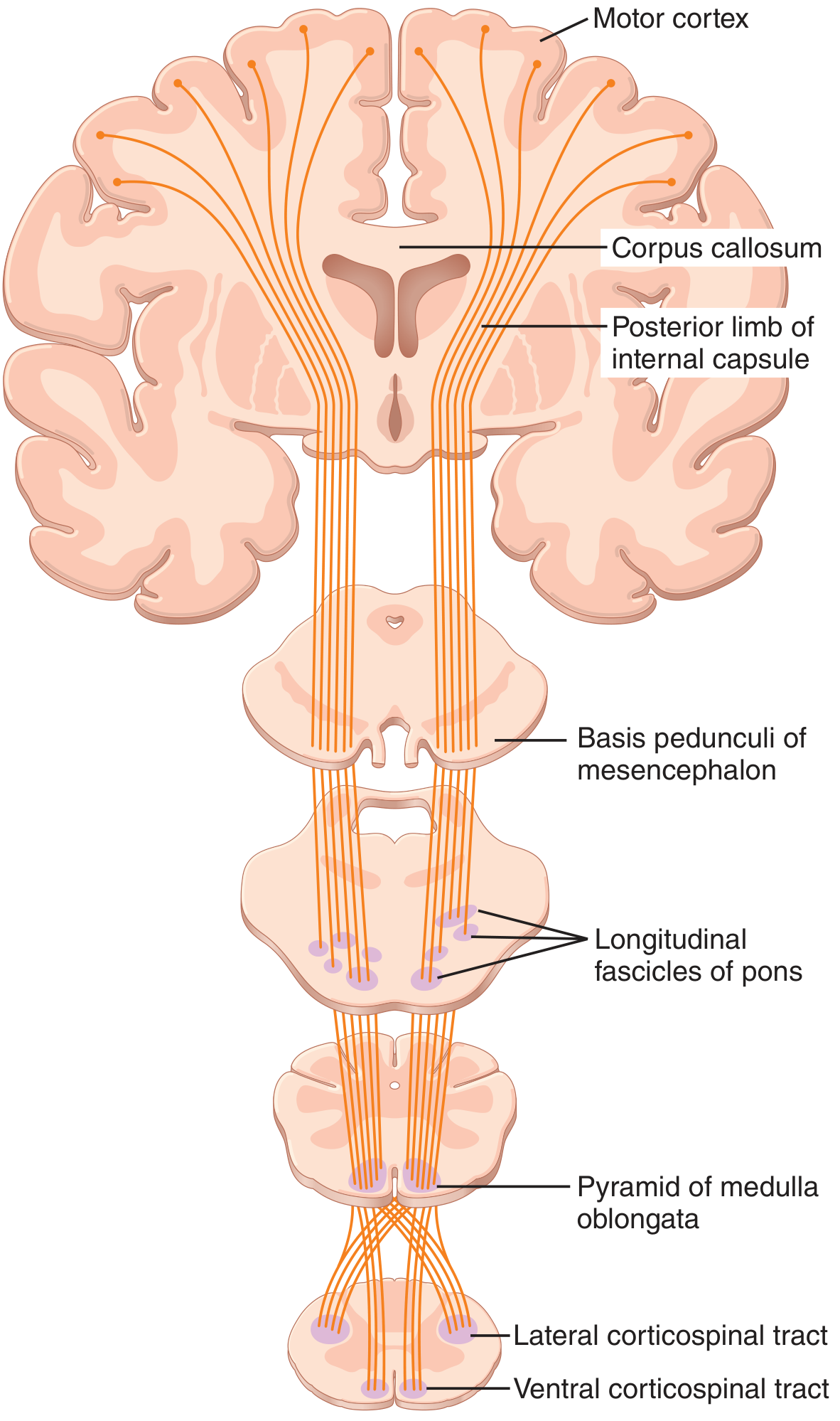
Figure: Corticospinal (pyramidal) tract showing Motor cortex → Corpus callosum → Posterior limb of internal capsule → Basis pedunculi of mesencephalon → Longitudinal fascicles of pons → Pyramid of medulla oblongata → Lateral corticospinal tract + Ventral corticospinal tract (Guyton & Hall, Medical Physiology)
Differences Between Upper Motor Neuron (UMN) and Lower Motor Neuron (LMN) Lesion
| Feature | UMN Lesion | LMN Lesion |
|---|---|---|
| Location of lesion | Cortex, internal capsule, brainstem, or spinal cord (above anterior horn) | Anterior horn cell, ventral root, peripheral nerve, or neuromuscular junction |
| Tone | Increased (spasticity/hypertonia) | Decreased (flaccidity/hypotonia) |
| Power/Weakness | Weakness (paresis/plegia) - affects groups of muscles | Weakness or paralysis of specific muscles supplied |
| Reflexes | Exaggerated / hyperreflexia | Diminished or absent (hyporeflexia / areflexia) |
| Plantar reflex | Extensor (Babinski's sign positive - big toe dorsiflexes) | Flexor (normal) or absent |
| Wasting/Atrophy | Absent or minimal (disuse atrophy only, late) | Marked muscle wasting (early, due to denervation) |
| Fasciculations | Absent | Present (irregular contractions of muscle bundles) |
| Clonus | Present | Absent |
| Distribution | Contralateral (if above decussation); ipsilateral (below); grouped (hemiplegic pattern) | Specific muscles in the distribution of the affected nerve/root |
| Examples | Stroke (capsular hemiplegia), spinal cord compression, MS | Polio, Guillain-Barre Syndrome, nerve injury, MND (anterior horn) |
Q3. Define Arterial Blood Pressure. What is the Normal Blood Pressure? Explain the Regulation of Blood Pressure in Detail.
Definition of Arterial Blood Pressure
Arterial blood pressure is the lateral pressure exerted by the blood on the walls of the arteries per unit area. It is the driving force for blood flow through the systemic circulation. It is measured in mmHg.
Normal Blood Pressure
| Parameter | Normal Value |
|---|---|
| Systolic BP (SBP) | 120 mmHg |
| Diastolic BP (DBP) | 80 mmHg |
| Pulse Pressure | 40 mmHg (SBP - DBP) |
| Mean Arterial Pressure (MAP) | ~93 mmHg (DBP + 1/3 Pulse Pressure) |
- Hypertension: BP > 140/90 mmHg
- Hypotension: BP < 90/60 mmHg
Regulation of Blood Pressure
Blood pressure is regulated by short-term (rapid) and long-term (slow) mechanisms.
A. SHORT-TERM (RAPID) REGULATION
1. Nervous System Mechanisms (Most Rapid - seconds to minutes)
The nervous system can increase arterial pressure within 5-10 seconds to double normal, or decrease it to half within 10-40 seconds.
a) Baroreceptor Reflex (Most Important)
- Baroreceptors (pressoreceptors) are stretch receptors located in the walls of:
- Carotid sinus (at bifurcation of common carotid - CN IX)
- Aortic arch (CN X)
- Mechanism:
- Rise in BP → baroreceptors stretched → increased afferent firing → vasomotor center inhibited → decreased sympathetic + increased parasympathetic output → vasodilation + decreased heart rate/contractility → BP falls back to normal
- Fall in BP → less stretch → decreased firing → vasomotor center activated → increased sympathetic → vasoconstriction + increased HR/contractility → BP rises back to normal
- This is a negative feedback mechanism
- Responds to rapid changes; not effective for long-term BP regulation (baroreceptors reset over 1-2 days)
b) CNS Ischemic Response (Cushing Reflex)
- When cerebral blood flow falls dangerously, the vasomotor center is strongly excited
- Produces massive sympathetic vasoconstriction - can raise BP to 250 mmHg
- Acts as an "emergency last resort" mechanism
c) Chemoreceptor Reflex
- Peripheral chemoreceptors (carotid and aortic bodies) respond to hypoxia, hypercapnia, and acidosis
- Stimulate the vasomotor center → increase BP
- Primary role is respiratory control; BP effect is secondary
d) Atrial and Pulmonary Artery Receptors
- Volume receptors in atria detect low cardiac filling pressure
- Activate sympathetic system via the Bainbridge reflex
- Increase heart rate when atrial pressure rises (to compensate)
2. Hormonal Mechanisms (Minutes to hours)
a) Renin-Angiotensin-Aldosterone System (RAAS)
- Fall in renal perfusion pressure → kidneys release renin
- Renin converts angiotensinogen → Angiotensin I → (ACE) → Angiotensin II
- Angiotensin II: potent vasoconstrictor + stimulates aldosterone release
- Aldosterone: promotes Na+ and water reabsorption in kidney → increases blood volume → increases BP
- This is also an important long-term mechanism
b) Epinephrine and Norepinephrine (Adrenal Medulla)
- Released during sympathetic activation / stress
- Increase heart rate, cardiac output, and cause vasoconstriction
- Raise blood pressure quickly
c) Vasopressin (ADH - Antidiuretic Hormone)
- Released from posterior pituitary in response to decreased BP / increased plasma osmolality
- Causes vasoconstriction + water reabsorption in kidneys → increases blood volume and BP
B. LONG-TERM REGULATION
Renal Fluid Volume Mechanism (Most Powerful Long-Term Regulator)
The kidneys regulate blood pressure by controlling extracellular fluid (ECF) volume and blood volume through the mechanism of pressure natriuresis / pressure diuresis:
- BP rises → kidneys excrete more Na+ and water (pressure diuresis and natriuresis) → blood volume falls → BP returns to normal
- BP falls → kidneys retain Na+ and water → blood volume rises → BP returns to normal
This mechanism is extremely powerful and can normalize BP over hours to days. The RAAS strongly modulates this mechanism:
- High Angiotensin II → shifts the renal function curve to higher pressures (promotes hypertension)
- ACE inhibitors / ARBs block this, restoring BP toward normal
The "infinite gain" of the renal mechanism means that given enough time, the kidneys can correct almost any disturbance in blood pressure, provided the system is intact.
Summary Table: BP Regulation Mechanisms
| Mechanism | Speed | Type |
|---|---|---|
| Baroreceptor reflex | Seconds | Short-term |
| CNS ischemic response | Seconds | Emergency |
| Chemoreceptor reflex | Seconds | Short-term |
| Epinephrine/Norepinephrine | Minutes | Short-term |
| RAAS | Minutes-hours | Short + Long term |
| ADH/Vasopressin | Minutes-hours | Short + Long term |
| Aldosterone | Hours | Long-term |
| Renal fluid volume mechanism | Hours-days | Long-term (most powerful) |
Sources:
- Guyton and Hall Textbook of Medical Physiology - Corticospinal tract (Ch. 56), Arterial pressure regulation (Ch. 18)
- Costanzo Physiology - Negative feedback in arterial pressure regulation
- Medical Physiology - Neural and baroreceptor regulation of arterial pressure
Give me destined answer for these questions, according to rughs requirement....for 10 marks
It looks like you forgot to attach the image with the questions! Could you please share the photo of your question paper so I can provide the detailed answers for you?This is the same set of questions from your previous message — I already answered all three in detail! Here is a clean, organized version for your reference:
Fasciculus Answers (10 Marks Each)
Q1. List the Hormones of the Thyroid Gland. Briefly Explain Functions of Thyroid Hormone. What is Myxedema?
Hormones of the Thyroid Gland
| Hormone | Secreted By | Notes |
|---|---|---|
| Thyroxine (T4) | Follicular cells | Major secretory product (90%); less active form |
| Triiodothyronine (T3) | Follicular cells | More potent (3-5x); active form; T4 converted to T3 in periphery |
| Calcitonin | Parafollicular (C) cells | Lowers blood calcium; not a thyroid hormone per se |
Functions of Thyroid Hormones (T3/T4)
1. Metabolic / Calorigenic Action
- Increase Basal Metabolic Rate (BMR) - stimulate O₂ consumption in almost all tissues
- Increase heat production (thermogenesis)
- Stimulate Na⁺/K⁺ ATPase activity → increased ATP utilization
2. Carbohydrate Metabolism
- Increase glucose absorption from the gut
- Promote glycogenolysis and gluconeogenesis
- Increase insulin-dependent glucose uptake
3. Fat Metabolism
- Stimulate lipolysis - mobilize fatty acids
- Decrease plasma cholesterol (increase LDL receptor expression)
- Deficiency → hypercholesterolemia
4. Protein Metabolism
- Physiological levels → protein synthesis (anabolic)
- Excess levels → protein catabolism (muscle wasting)
5. Cardiovascular Effects
- Increase heart rate (positive chronotropy)
- Increase cardiac output and stroke volume
- Increase systolic BP; decrease peripheral vascular resistance
6. CNS / Nervous System
- Essential for fetal/neonatal brain development (absence → cretinism)
- Adults: maintain alertness, memory, cognition
- Hyperthyroidism → anxiety, restlessness
- Hypothyroidism → sluggishness, memory loss
7. Growth and Development
- Essential for normal skeletal growth and maturation
- Works synergistically with Growth Hormone (GH)
8. Reproductive System
- Required for normal menstrual cycles and fertility
- Hypothyroidism → menorrhagia, anovulation, infertility
9. GI Tract
- Stimulate gut motility
- Hyperthyroidism → diarrhea; Hypothyroidism → constipation
Myxedema
Definition: Myxedema is a severe form of adult hypothyroidism characterized by accumulation of mucopolysaccharides (hyaluronic acid and chondroitin sulfate) in interstitial tissues, producing non-pitting edema of the skin and subcutaneous tissues.
Causes:
- Hashimoto's thyroiditis (most common - autoimmune)
- Thyroidectomy / radioiodine treatment
- Iodine deficiency
- Pituitary/hypothalamic failure (secondary/tertiary)
Clinical Features:
| System | Features |
|---|---|
| General | Fatigue, weight gain, cold intolerance |
| Skin | Dry, coarse, yellowish skin; non-pitting (doughy) edema; puffy face |
| Hair/Nails | Dry brittle hair; loss of outer 1/3 of eyebrow (Queen Anne's sign); brittle nails |
| CVS | Bradycardia, decreased cardiac output, pericardial effusion |
| CNS | Lethargy, depression, memory impairment, hung-up (delayed) deep tendon reflexes |
| Metabolic | Decreased BMR, hypercholesterolemia, hyponatremia |
| Voice | Hoarse, husky voice |
| GI | Constipation, megacolon |
| Reproductive | Menorrhagia, infertility |
Lab Findings:
- TSH: High (primary hypothyroidism)
- Free T4: Low
- Elevated cholesterol; normocytic or macrocytic anemia
Myxedema Coma: Life-threatening extreme form - hypothermia, unconsciousness, hypoventilation, cardiovascular collapse. Treated with IV T3/T4 + corticosteroids + supportive care.
Q2. Explain Origin, Course, and Termination of the Corticospinal Tract with a Neat Labelled Diagram. Tabulate Differences Between UMN and LMN Lesion.
Corticospinal (Pyramidal) Tract
Origin
The corticospinal tract arises from:
- 30% - Primary motor cortex (Area 4 - precentral gyrus) - from Betz cells (giant pyramidal cells)
- 30% - Premotor and supplementary motor areas (Area 6)
- 40% - Somatosensory cortex (Areas 3, 1, 2 - postcentral gyrus)
There are ~34,000 large Betz cell fibers per tract (diameter ~16 µm), out of >1 million total fibers. Betz cell fibers conduct at ~70 m/sec - the fastest signals from brain to cord.
Course
- Fibers converge → pass through posterior limb of internal capsule (between caudate nucleus and putamen)
- Descend through crus cerebri (basis pedunculi) of midbrain
- Pass through longitudinal fascicles of pons
- Form the pyramids of medulla oblongata (hence "pyramidal tract")
- At lower medulla - ~85-90% of fibers cross at the pyramidal (motor) decussation
- Crossed fibers → descend as lateral corticospinal tract (lateral funiculus of spinal cord)
- ~10-15% uncrossed fibers → descend as anterior (ventral) corticospinal tract - cross at spinal cord level later
Termination
- Primarily on interneurons in the intermediate zone (Rexed laminae V-VIII) of spinal cord gray matter
- Small number on sensory relay neurons in the dorsal horn
- Very few directly on anterior horn motor neurons (alpha motor neurons) - for fine skilled movements of distal limbs
- Anterior corticospinal tract terminates bilaterally - controls axial/postural muscles
Labelled Diagram of Corticospinal Tract
Fig: Corticospinal (Pyramidal) Tract - showing Motor cortex → Posterior limb of internal capsule → Basis pedunculi → Longitudinal fascicles of pons → Pyramid of medulla → Lateral and Ventral corticospinal tracts (Guyton & Hall, Medical Physiology)
Differences: UMN vs LMN Lesion
| Feature | UMN Lesion | LMN Lesion |
|---|---|---|
| Site of lesion | Cortex, internal capsule, brainstem, or spinal cord above anterior horn | Anterior horn cell, ventral root, peripheral nerve, or NMJ |
| Tone | Increased - spasticity | Decreased - flaccidity |
| Power | Weakness (paresis/plegia) - groups of muscles | Weakness of specific muscles in nerve/root distribution |
| Reflexes | Exaggerated (hyperreflexia) | Diminished or absent (areflexia) |
| Plantar reflex | Extensor - Babinski sign positive (big toe dorsiflexes) | Flexor (normal) or absent |
| Muscle wasting | Absent or minimal (disuse atrophy only, late) | Marked early wasting (denervation atrophy) |
| Fasciculations | Absent | Present |
| Clonus | Present | Absent |
| Distribution | Contralateral hemiplegia (above decussation); hemiplegic pattern | Localized to specific nerve/root/muscle distribution |
| Examples | Stroke, spinal cord compression, MS | Poliomyelitis, Guillain-Barre, peripheral nerve injury |
Q3. Define Arterial Blood Pressure. What is Normal Blood Pressure? Explain the Regulation of Blood Pressure in Detail.
Definition
Arterial Blood Pressure is the lateral pressure exerted by the blood on the walls of arteries per unit area. It is the driving force for blood circulation and is measured in mmHg.
Normal Blood Pressure
| Parameter | Normal Value |
|---|---|
| Systolic BP (SBP) | 120 mmHg |
| Diastolic BP (DBP) | 80 mmHg |
| Pulse Pressure | 40 mmHg (SBP - DBP) |
| Mean Arterial Pressure (MAP) | ~93 mmHg (DBP + 1/3 Pulse Pressure) |
| Hypertension | > 140/90 mmHg |
| Hypotension | < 90/60 mmHg |
Regulation of Blood Pressure
Blood pressure regulation is divided into Short-term (Rapid) and Long-term mechanisms.
A. SHORT-TERM (RAPID) REGULATION
1. Nervous System Mechanisms (Seconds)
The nervous system can raise BP to double normal within 5-10 seconds, or lower it to half within 10-40 seconds - making it the most rapid regulatory mechanism.
a) Baroreceptor Reflex (Most Important)
- Baroreceptors (stretch receptors/pressoreceptors) located in:
- Carotid sinus - signals via Glossopharyngeal nerve (CN IX)
- Aortic arch - signals via Vagus nerve (CN X)
- Mechanism:
BP rises → Baroreceptors stretched → Increased afferent firing
→ Vasomotor center inhibited → Decreased sympathetic + Increased parasympathetic
→ Vasodilation + Decreased HR + Decreased contractility
→ BP falls back to normal (Negative Feedback)

BP falls → Less stretch → Decreased firing
→ Vasomotor center activated → Increased sympathetic
→ Vasoconstriction + Increased HR/contractility → BP rises
- Responds to rapid, moment-to-moment changes
- Baroreceptors reset over 1-2 days, so not effective for long-term control
b) CNS Ischemic Response (Cushing Reflex)
- When cerebral perfusion falls dangerously → vasomotor center strongly excited
- Produces powerful sympathetic vasoconstriction → BP may rise to 250 mmHg
- Acts as an emergency "last resort" mechanism
c) Chemoreceptor Reflex
- Peripheral chemoreceptors (carotid and aortic bodies) respond to hypoxia, hypercapnia, acidosis
- Stimulate vasomotor center → increase BP
- Primary role is respiratory; BP effect is secondary
2. Hormonal Mechanisms (Minutes to Hours)
a) Renin-Angiotensin-Aldosterone System (RAAS)
Fall in renal perfusion pressure
        ↓
Kidney releases RENIN
        ↓
Angiotensinogen → Angiotensin I → (ACE) → Angiotensin II
        ↓                                        ↓
  Potent vasoconstriction              Stimulates Aldosterone
        ↓                                        ↓
   BP rises                    Na⁺ + H₂O retention → Blood volume ↑ → BP ↑
b) Epinephrine and Norepinephrine (Adrenal Medulla)
- Released during stress/sympathetic activation
- Increase HR, cardiac output, and cause vasoconstriction → raise BP rapidly
c) Antidiuretic Hormone (ADH / Vasopressin)
- Released from posterior pituitary in response to decreased BP or increased plasma osmolality
- Causes vasoconstriction + water reabsorption in kidneys → increases blood volume and BP
B. LONG-TERM REGULATION
Renal Fluid Volume Mechanism (Most Powerful Long-Term Regulator)
The kidneys regulate BP by controlling blood volume through pressure natriuresis/diuresis:
- BP rises → kidneys excrete more Na⁺ and water (pressure diuresis) → blood volume falls → BP normalizes
- BP falls → kidneys retain Na⁺ and water → blood volume rises → BP normalizes
This mechanism has "infinite gain" - given enough time, it can correct almost any sustained BP disturbance. It is modulated strongly by RAAS.
Summary Table: BP Regulation Mechanisms
| Mechanism | Speed | Type |
|---|---|---|
| Baroreceptor reflex | Seconds | Short-term (rapid) |
| CNS ischemic response (Cushing) | Seconds | Emergency |
| Chemoreceptor reflex | Seconds | Short-term |
| Epinephrine/Norepinephrine | Minutes | Short-term |
| RAAS (Angiotensin II) | Minutes-Hours | Short + Long term |
| ADH/Vasopressin | Minutes-Hours | Short + Long term |
| Aldosterone | Hours | Long-term |
| Renal fluid volume mechanism | Hours-Days | Long-term (most powerful) |
Sources: Guyton & Hall Medical Physiology (Corticospinal Tract Ch.56; BP Regulation Ch.18), Costanzo Physiology
This is a shared conversation. Sign in to Orris to start your own chat.