
Trophoblastic tumor
Reading File
Searching sources...
Finding Sources
Searching PubMed
"gestational trophoblastic" AND "neoplasia"
Finding Sources
Reading File
Reading File
Reading File
Reading File
Reading File
Reading File
Now let me get the choriocarcinoma section from the larger Robbins textbook and the PSTT/ETT section from Berek & Novak:
Reading File
Reading File
Reading File
Finding Sources
Finding Sources
Loading Image
Reading File
Loading Image
I now have comprehensive data. Let me also quickly fetch the PSTT and ETT histology images to include in the answer:
Loading Image
Loading Image
I now have all the material needed for a thorough answer. Here is the comprehensive overview:
Gestational Trophoblastic Disease (GTD) / Trophoblastic Tumors
Gestational trophoblastic disease encompasses a spectrum of tumors and tumor-like conditions characterized by proliferation of placental (trophoblastic) tissue. The major entities are: hydatidiform mole (complete and partial), invasive mole, gestational choriocarcinoma, placental site trophoblastic tumor (PSTT), and epithelioid trophoblastic tumor (ETT). All except PSTT/ETT elaborate hCG to varying degrees.
- Robbins & Kumar Basic Pathology, p. 703
Classification
| Entity | Nature | hCG | Malignant? |
|---|---|---|---|
| Complete hydatidiform mole | Benign (premalignant) | Very elevated | No (15% risk of GTN) |
| Partial hydatidiform mole | Benign (premalignant) | Moderately elevated | No (rare progression) |
| Invasive mole | Locally invasive | Elevated | Borderline |
| Choriocarcinoma | Malignant | Very elevated | Yes |
| PSTT | Malignant (rare) | Low (hPL elevated) | Yes |
| ETT | Malignant (rare) | Low | Yes |
1. Hydatidiform Mole
Epidemiology
- Incidence: ~1 in 1,000-2,000 pregnancies in the US; twice as common in Southeast Asia
- Risk higher at extremes of reproductive life (teenagers; age 40-50)
Complete vs. Partial Mole
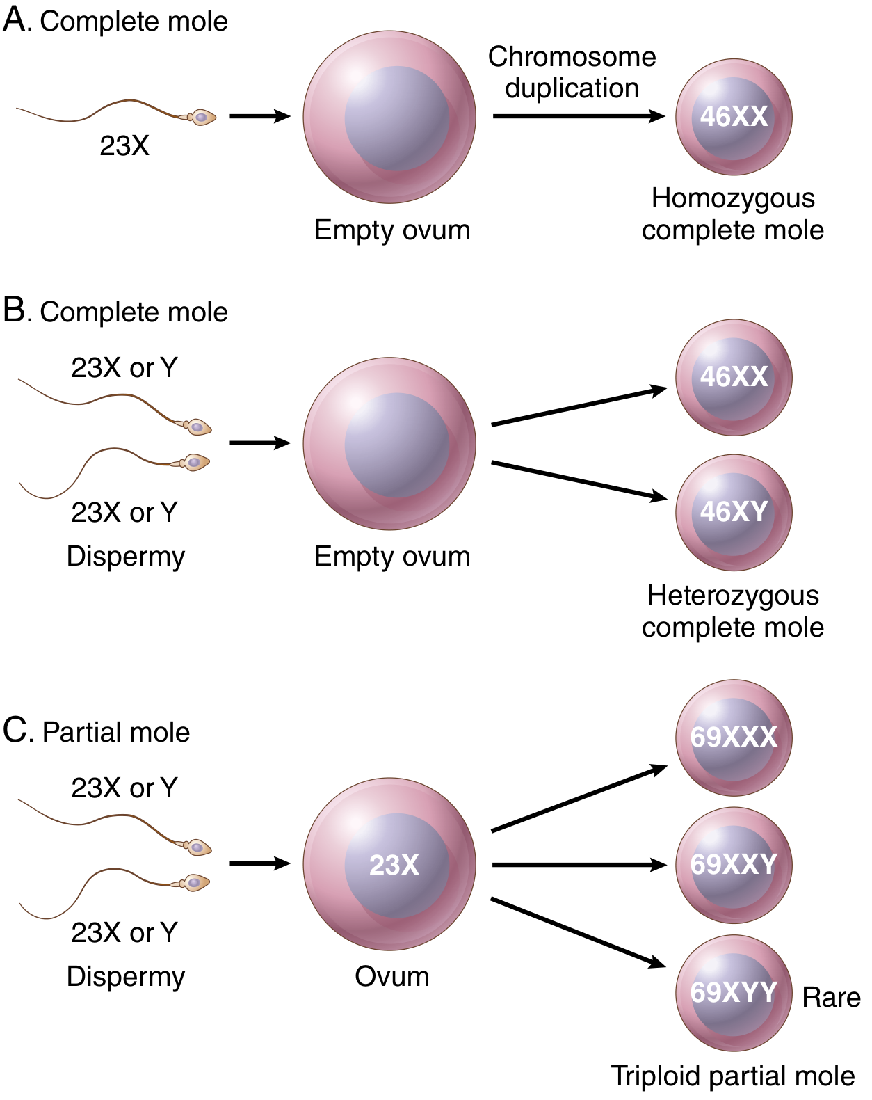
| Feature | Complete Mole | Partial Mole |
|---|---|---|
| Karyotype | Diploid (46,XX most common; or 46,XY) | Triploid (69,XXY most common) |
| Origin | Empty ovum + 1 sperm (androgenesis) or dispermy | Normal ovum + 2 sperm |
| Embryo/fetus | Absent | May be present (with lethal anomalies) |
| Villous edema | All villi | Some villi |
| Trophoblast proliferation | Diffuse, circumferential | Focal, slight |
| Serum hCG | Very elevated (often >100,000 IU) | Less elevated |
| Risk of choriocarcinoma | ~2.5% | Rare |
| Risk of persistent/invasive mole | ~15% | Increased, but less |
Genetics
- Complete mole: entirely paternal in origin - the ovum has lost its maternal DNA. Ninety percent arise from androgenesis (one sperm duplicates) → 46,XX; 10% from dispermy → 46,XX or 46,XY. No embryo forms.
- Partial mole: normal ovum fertilized by 2 sperm → triploid (e.g., 69,XXY). Fetal parts may be present but growth-restricted with CNS and skeletal anomalies (e.g., syndactyly).
Morphology
Gross: markedly distended uterus filled with grape-like, vesicular chorionic villi.
Histology: marked villous enlargement, edema, cisternae formation, trophoblastic hyperplasia.
Clinical Features
- Presents as spontaneous miscarriage or is found on ultrasound
- hCG levels exceed those of a normal pregnancy of the same gestational age
- Treatment: suction curettage
- Post-evacuation surveillance: serial hCG for 6-12 months
- Persistent elevation of hCG → invasive mole or choriocarcinoma (requires chemotherapy)
2. Invasive Mole
- An infiltrative lesion that penetrates, and sometimes perforates, the uterine wall
- Defined diagnostically by a plateau of β-hCG over 4 measurements across 3 weeks, or a rise over 3 consecutive weekly measurements, or hCG elevated for ≥6 months
- Hydropic villi lined by proliferating cyto- and syncytiotrophoblast invade the myometrium
- Emboli to distant sites (lungs, brain) can occur in ~5% of complete moles
- Small lung metastases (<2 cm) often regress spontaneously; larger ones require chemotherapy
- Treatment: curettage, or hysterectomy; chemotherapy if indicated
- Robbins & Kumar Basic Pathology, p. 703; Schwartz's Surgery
3. Gestational Choriocarcinoma
Epidemiology
Arises in 1 in 20,000-30,000 pregnancies in the US.
Antecedent pregnancy:
- 50% follow complete hydatidiform mole
- 25% follow spontaneous abortions
- ~22% follow normal pregnancies
- Remainder: ectopic pregnancies or arise within the placenta
Morphology
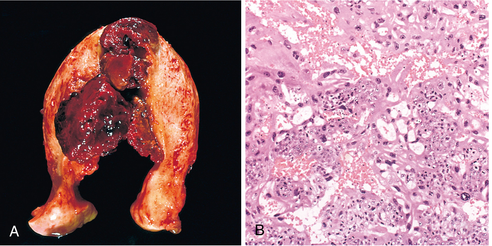
- Soft, fleshy, yellow-white hemorrhagic, necrotic uterine mass
- Histology: proliferating syncytiotrophoblasts and cytotrophoblasts - no chorionic villi (key distinguishing feature)
- Abundant, sometimes abnormal mitoses
- Invades myometrium; penetrates blood vessels
Clinical Features
- Irregular vaginal spotting (bloody, brown discharge)
- Very high hCG levels (higher than hydatidiform mole)
- Early and widespread hematogenous spread (lymphatic invasion is uncommon)
Sites of metastasis (in order of frequency):
- Lungs - 50%
- Vagina - 30-40%
- Brain
- Liver
- Kidney/bone
Treatment & Prognosis
- Nearly 100% remission rate with chemotherapy - one of the most chemosensitive solid tumors known
- Evacuation + chemotherapy; surgery reserved for bleeding emergencies
- Many cured patients go on to have normal subsequent pregnancies
- Non-gestational choriocarcinoma (from gonads/mediastinum) is morphologically identical but carries a worse prognosis and lacks paternally derived DNA
4. Placental Site Trophoblastic Tumor (PSTT)
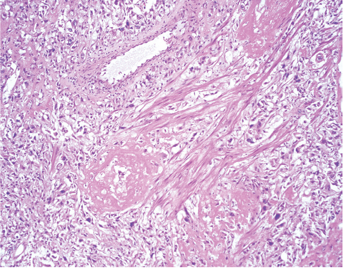
- <2% of gestational trophoblastic neoplasms
- Origin: neoplastic proliferation of extravillous (intermediate) trophoblasts
- Cell marker: produce human placental lactogen (hPL) rather than hCG; serum hCG is characteristically low
- Karyotype: diploid, often 46,XX
- Usually arises months after pregnancy (normal pregnancy in ~50%; also after abortion or hydatidiform mole)
- Presents as uterine mass with abnormal uterine bleeding or amenorrhea
- Invasion without local destruction or hemorrhage (unlike choriocarcinoma)
- Relatively insensitive to chemotherapy - primary treatment is hysterectomy
- Good prognosis if confined to uterus; 10-15% die of disseminated disease
- A 2026 systematic review (PMID 40628611) has examined fertility-sparing options for PSTT
5. Epithelioid Trophoblastic Tumor (ETT)
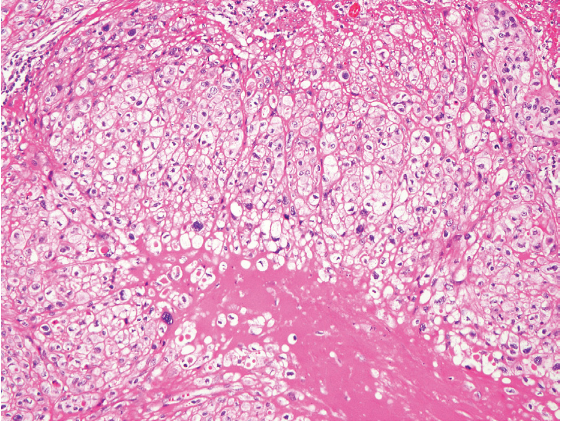
- Rarest form of GTN
- Origin: mononuclear trophoblast cells (intermediate trophoblast), often with abundant clear cytoplasm and associated with pink hyaline material
- Location: usually intrauterine or cervical; extrauterine tumors also occur
- Presents following pregnancy with amenorrhea or palpable mass
- hCG production: low (like PSTT)
- Lacks the overt invasive features of PSTT yet is capable of metastasis (lungs, bone most common)
- ~10-15% with metastatic disease succumb to the tumor
- Less chemotherapy-sensitive; surgery is preferred for localized disease
Staging & Risk Scoring (FIGO/WHO)
GTN (malignant forms) is staged anatomically by FIGO (Stages I-IV) and risk-stratified by the WHO Prognostic Scoring System:
| WHO Score | 0 | 1 | 2 | 4 |
|---|---|---|---|---|
| Age | <40 | >40 | - | - |
| Antecedent pregnancy | Mole | Abortion | Term | - |
| Interval (months) | <4 | 4-7 | 7-13 | >13 |
| Pre-treatment hCG | <1,000 | 1,000-10,000 | 10,000-100,000 | >100,000 |
| Prior chemotherapy | - | - | Single agent | ≥2 agents |
- Score ≤6 = Low risk → single-agent chemotherapy (methotrexate or actinomycin-D)
- Score >6 = High risk → multi-agent chemotherapy (e.g., EMA-CO)
- 5-year survival in high-risk patients: ~90%
- Schwartz's Principles of Surgery, Table 41-15
Key Diagnostic Tumor Markers
| Tumor | Primary Marker |
|---|---|
| Hydatidiform mole | hCG (very high in complete) |
| Invasive mole | hCG (persistently elevated) |
| Choriocarcinoma | hCG (very high) |
| PSTT | hPL (hCG low) |
| ETT | hCG low; hPL variable |
Recent Evidence
A 2025 systematic review and meta-analysis (PMID 40031424) examined pembrolizumab (anti-PD-1) in gestational trophoblastic neoplasia, reflecting growing interest in immunotherapy for chemotherapy-resistant GTN.
Sources: Robbins, Cotran & Kumar Pathologic Basis of Disease | Robbins & Kumar Basic Pathology | Berek & Novak's Gynecology | Schwartz's Principles of Surgery
This is a shared conversation. Sign in to Orris to start your own chat.