
Pyloric stenosis
Reading File
Pyloric stenosis is a condition in infants where the pyloric muscle hypertrophies, causing gastric outlet obstruction. Classic presentation: projectile non-bilious vomiting in a 2-8 week old infant, with a palpable "olive" mass and hypochloremic hypokalemic metabolic alkalosis. Treatment is surgical - Ramstedt pyloromyotomy.
Searching sources...
Finding Sources
Searching PubMed
"pyloric stenosis" infant
Reading File
Reading File
Reading File
Reading File
Reading File
Loading Image
Here is a comprehensive overview of pyloric stenosis based on multiple authoritative textbooks.
Pyloric Stenosis (Hypertrophic Pyloric Stenosis)
Definition & Epidemiology
Hypertrophic pyloric stenosis (HPS) is the most common cause of gastric outlet obstruction in infancy and the most common surgical condition requiring intervention in the first few months of life.
- Incidence: ~1 in 250-300 live births
- Sex: Males affected 4-5x more than females (>80% of cases in males)
- Age of onset: Presents between 3-6 weeks of life (range 2-10 weeks); rare after 12 weeks; peak incidence at ~5 weeks
- Risk factors: First-born child, white race, positive family history, premature birth, young maternal age
- Genetic component: High concordance in monozygotic twins; genome-wide studies have linked several GI development genes; siblings of affected patients have ~6% risk (vs. ~0.2% general population)
- Associated conditions: Turner syndrome, trisomy 18 confer increased risk
- Robbins Pathologic Basis of Disease; Current Surgical Therapy 14e
Pathophysiology
HPS is not a congenital malformation - the pylorus is normal at birth. Hypertrophy of the pyloric muscularis propria (primarily circular muscle, with some longitudinal involvement) develops postnatally and causes progressive gastric outlet obstruction. Mucosal and submucosal edema and inflammation further exacerbate luminal narrowing. The pyloric canal fails to relax and lengthen normally.
Important association: Erythromycin or azithromycin exposure (oral or via breast milk) in the first 2 weeks of life is linked to development of HPS.
- Robbins Pathologic Basis of Disease; The Developing Human (Moore)
Clinical Presentation
| Feature | Detail |
|---|---|
| Vomiting | Projectile, non-bilious (obstruction is pre-ampullary), worsening progressively over days |
| Onset | Begins as mild regurgitation, escalates to forceful projectile emesis |
| Feeding behavior | Infant remains hungry after vomiting ("hungry vomiter") |
| Peristaltic waves | Visible left-to-right peristaltic waves across the upper abdomen just before vomiting |
| Palpable "olive" | Firm, ovoid 1-2 cm epigastric mass - nearly pathognomonic; historically found in up to 90%, but now <30% due to earlier presentation and diagnosis |
| Dehydration | Sunken fontanelle, dry mucous membranes, decreased urine output, poor weight gain/weight loss |
- Current Surgical Therapy 14e; Robbins; Tintinalli's Emergency Medicine; Grainger & Allison's Diagnostic Radiology
Metabolic Abnormalities
Repeated vomiting of gastric contents produces a characteristic electrolyte pattern:
Hypochloremic, hypokalemic, hyponatremic metabolic alkalosis
- Mechanism: Loss of HCl in vomit → chloride depletion → kidneys retain bicarbonate → alkalosis. Potassium is lost both in vomit and via renal exchange for hydrogen ions as the kidney attempts to maintain acid-base balance (paradoxical aciduria in a setting of alkalosis).
- A bicarbonate >30 mEq/L indicates severe alkalosis and is a contraindication to anesthesia (risk of postoperative apnea/respiratory arrest).
Diagnosis
Ultrasound (Gold Standard)
US has replaced barium studies and has sensitivity and specificity up to 98% and 100%, respectively.
Diagnostic criteria on ultrasound:
- Pyloric muscle wall thickness ≥ 4 mm
- Pyloric channel length ≥ 16-17 mm
- Failure of fluid to pass through the pylorus during the exam
Classic US signs:
- "Shoulder sign": Hypertrophic muscle bulging into the gastric antrum
- "Nipple sign": Double-layered hypertrophic mucosa protruding into the stomach
- "Cervix sign": Overall sonographic appearance resembling the uterine cervix
- Abnormal, exaggerated peristaltic waves visible in real time
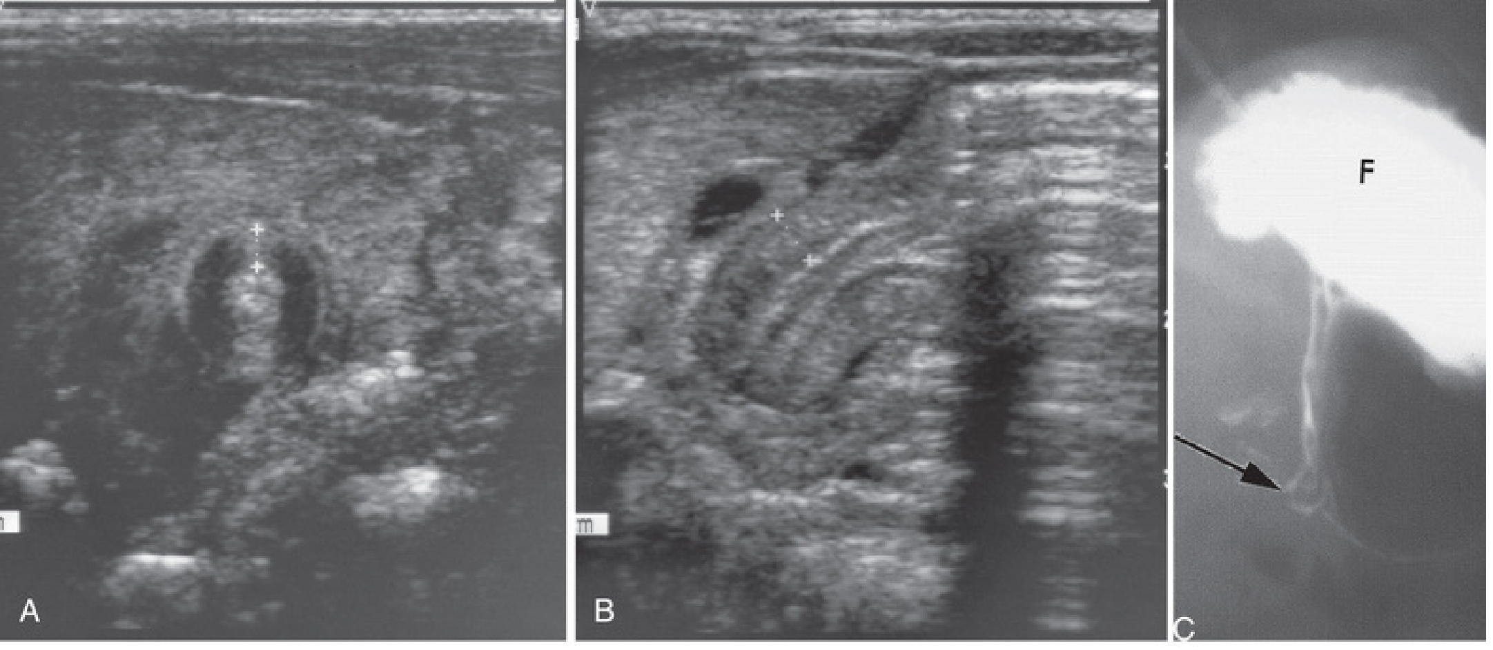
Fig: A - Transverse US showing pyloric muscle wall thickness >4mm. B - Horizontal US showing pyloric channel length >14mm. C - Contrast radiograph showing narrowed pyloric canal (arrow) and distended gastric fundus (F). From The Developing Human, Moore.
Upper GI Series (UGI)
Used when US is equivocal (e.g., when GER or pylorospasm remains in the differential). Classic finding: the "string sign" - thin elongated streak of contrast through the narrowed pylorus; delayed gastric emptying.
- Current Surgical Therapy 14e; Grainger & Allison's Diagnostic Radiology
Management
Step 1 - Resuscitation First (NOT an emergency operation)
Surgery is never a true emergency. The priority is correction of dehydration and metabolic alkalosis:
- IV fluid resuscitation: 1-2 boluses of 20 mL/kg normal saline
- Maintenance fluids: D5/0.45% NaCl with KCl at 1.5x maintenance rate
- Monitor electrolytes every 6-12 hours
Endpoints for safe anesthesia:
- Wet diapers (adequate urine output)
- Serum chloride: 90-100 mEq/L
- Serum bicarbonate: ≤30 mEq/L
Step 2 - Surgical Repair: Ramstedt Pyloromyotomy
Once metabolic derangements are corrected, surgery is performed:
- Laparoscopic pyloromyotomy is now the preferred approach (3 ports: umbilical + two 3-mm stab incisions)
- The pyloric muscle is incised longitudinally and split from stomach to duodenum, leaving mucosa intact
- A leak test (30-60 mL air via OG tube) confirms mucosal integrity
- Post-op: ad lib feeding with 60 mL limit; most infants discharged within 36 hours
Complications of Pyloromyotomy
| Complication | Presentation | Management |
|---|---|---|
| Incomplete myotomy | Ongoing projectile vomiting | Return to OR for revision |
| Mucosal perforation | Can present as sepsis if missed intraoperatively | Closure + omental buttress OR repair + re-rotation of pylorus 180° for repeat myotomy |
- Current Surgical Therapy 14e
Acquired Pyloric Stenosis (Adults)
For completeness: pyloric stenosis can also occur in adults due to:
- Antral gastritis or peptic ulcers near the pylorus
- Carcinoma of the distal stomach or pancreas (fibrosis or malignant infiltration)
Recent Evidence
A 2026 systematic review (PMID 41196118) addressed recurrent HPS, noting it is rare but requires early diagnosis and re-operation. A 2024 meta-analysis (PMID 39503899) evaluated preoperative prophylactic antibiotics for surgical site infection prevention in HPS - this is an area of evolving practice. Note also that macrolide exposure (erythromycin, azithromycin) in the first 2 weeks of life remains a recognized modifiable risk factor.
Summary mnemonic: PHONO - Projectile vomiting, Hypochloremic alkalosis, Olive mass, Non-bilious, Operate after resuscitation.


1000562984.jpg
1000562985.jpg
Give me answer in detail
I can see this is a B.Sc.(N) 6th Semester Child Health Nursing exam paper. The Q.2(ii) asks:
"Define Pyloric stenosis. List the classical features of pyloric stenosis and explain the surgical management of pyloric stenosis. Draw a nursing care plan on an infant with pyloric stenosis."
Let me pull detailed textbook content for a complete exam answer.
Reading File
Finding Sources
Finding Sources
Reading File
Reading File
Reading File
Reading File
Finding Sources
I now have all the content I need from multiple authoritative surgical and emergency medicine textbooks. Here is the complete, detailed exam answer:
Q.2 (ii) — Pyloric Stenosis: Complete Exam Answer
B.Sc.(N) 6th Semester | Child Health Nursing I & II | 15 Marks
1. DEFINITION
Pyloric stenosis (more precisely, Infantile Hypertrophic Pyloric Stenosis - IHPS) is a condition in which there is progressive hypertrophy and hyperplasia of the circular and longitudinal muscles of the pylorus (the distal sphincteric portion of the stomach), resulting in narrowing of the pyloric canal and gastric outlet obstruction in infants.
- It is NOT a congenital defect - the pylorus is normal at birth and enlarges postnatally
- It is the most common cause of surgical GI obstruction beyond the first month of life
- Incidence: 1 in 250-300 live births
- Male-to-female ratio: 4:1 (males predominantly affected)
- Ramstedt pyloromyotomy is the definitive surgical cure
- Robbins Pathologic Basis of Disease; Current Surgical Therapy 14e
2. ETIOLOGY & RISK FACTORS
| Factor | Detail |
|---|---|
| Sex | Males 4-5x more common |
| Birth order | First-born child |
| Race | More common in White infants |
| Family history | ~6% risk in siblings of affected infants (vs 0.2% general population) |
| Genetics | High concordance in monozygotic twins; GWAS linked several GI genes |
| Macrolide antibiotics | Erythromycin/azithromycin exposure in first 2 weeks of life (oral or via breast milk) |
| Prematurity | Premature infants at higher risk |
| Associated syndromes | Turner syndrome, Trisomy 18 |
3. PATHOPHYSIOLOGY
Normal pylorus at birth
↓
Postnatal hypertrophy of pyloric muscularis propria (circular > longitudinal)
↓
Pyloric canal becomes progressively narrowed and elongated
↓
Gastric outlet obstruction
↓
Stomach contents cannot pass → forceful (projectile) vomiting
↓
Loss of HCl in vomit → ↓ Cl⁻, ↓ H⁺
↓
Kidneys conserve H⁺, excrete K⁺ → ↓ K⁺
↓
HYPOCHLOREMIC, HYPOKALEMIC METABOLIC ALKALOSIS
↓
Dehydration, malnutrition, failure to thrive
4. CLASSICAL FEATURES (Clinical Presentation)
A. History
- Age of onset: 3-6 weeks (range 2-10 weeks); rare after 12 weeks
- Progressive, projectile, non-bilious vomiting - begins as mild regurgitation and escalates to forceful vomiting after every feed
- Vomit contains undigested milk, gastric secretions - never green/bilious (obstruction is above the ampulla of Vater)
- "Hungry vomiter" - infant vomits but immediately wants to feed again (ravenous appetite)
- Weight loss, poor weight gain, failure to thrive
B. Physical Examination Signs
| Sign | Description |
|---|---|
| Projectile vomiting | Hallmark sign; vomit may shoot across the room |
| Visible peristaltic waves | Left-to-right peristaltic waves visible across upper abdomen just before vomiting - classic |
| Epigastric "olive" mass | Firm, mobile, ovoid 1-2 cm mass in right epigastrium; nearly pathognomonic; palpated in up to 90% (now <30% due to earlier diagnosis) |
| Dehydration signs | Sunken fontanelle, dry mucous membranes, sunken eyes, decreased skin turgor, decreased urine output |
| Malnutrition | Loose skin folds, wasted appearance in late cases |
C. Laboratory Findings (Electrolytes)
| Parameter | Change | Reason |
|---|---|---|
| Serum Chloride | ↓ (Hypochloremic) | Lost in gastric vomit |
| Serum Potassium | ↓ (Hypokalemic) | Lost in vomit + renal exchange |
| Serum Sodium | ↓ (Hyponatremic) | Volume depletion |
| Blood pH / Bicarbonate | ↑ (Metabolic Alkalosis) | Loss of acid (H⁺) |
| Urine pH | Acidic (paradoxical aciduria) | Kidneys excrete H⁺ to compensate |
D. Ribbon-Like / Currant Jelly Stool?
- Note: In Q.1(viii) of your paper, "ribbon-like stool" is associated with anorectal malformation (not pyloric stenosis). Pyloric stenosis causes vomiting, not stool abnormalities.
5. DIAGNOSIS
A. Ultrasound (Gold Standard)
- Sensitivity and specificity up to 98% and 100%
- Diagnostic criteria:
- Pyloric muscle thickness ≥ 4 mm
- Pyloric channel length ≥ 16-19 mm
- Pyloric diameter > 14 mm
- Failure of fluid to pass through pylorus
- Special US signs: Shoulder sign, Nipple sign, Cervix sign
B. Upper GI Series (Barium Study)
- Used when US is equivocal
- Classic finding: "String sign" - thin streak of barium through narrowed pylorus
- Shows delayed gastric emptying and distended stomach
- Plain X-ray: gas-filled, distended stomach with paucity of gas in distal bowel
C. Complete Metabolic Panel
- Confirms hypochloremic, hypokalemic metabolic alkalosis
- Bicarbonate >30 mEq/L = contraindication to anesthesia (risk of post-op apnea)
6. SURGICAL MANAGEMENT
STEP 1 - PRE-OPERATIVE RESUSCITATION (MANDATORY - NOT a surgical emergency)
Surgery must NEVER be performed before correcting dehydration and metabolic alkalosis.
Fluid Protocol:
- IV Normal Saline (0.9%) bolus: 20 mL/kg (repeat 1-2 times as needed)
- Maintenance fluids: D5/0.45% NaCl + KCl at 1.5x maintenance rate
- Monitor electrolytes every 6-12 hours
- Potassium supplementation: KCl 0.5-1 mEq/kg IV over 1-2 hours if needed
Endpoint / Safe Criteria for Surgery:
- Wet diapers (adequate urine output)
- Serum Chloride: 90-100 mEq/L
- Serum Bicarbonate: ≤ 30 mEq/L
- Potassium: within normal range
Pre-operative gastric decompression:
- Nasogastric tube insertion for gastric suction (reduces aspiration risk at induction)
- Stomach must be suctioned immediately before anesthesia
STEP 2 - SURGICAL PROCEDURE: RAMSTEDT PYLOROMYOTOMY
Ramstedt Pyloromyotomy is the definitive, curative operation.
Approaches available:
- Laparoscopic (currently preferred) - 3 incisions: 1 umbilical port + 2 lateral 3-mm stab incisions
- Open - Right upper quadrant (RUQ) transverse incision or periumbilical incision
Operative steps:
- Infant placed supine under general anesthesia
- Pylorus is identified and stabilized with a grasper
- A longitudinal incision is made through the anterior wall of the pyloric canal (through serosa and muscle only - NOT into mucosa)
- Muscle fibers are split (spread apart) from the stomach end to the duodenal end
- Bulging mucosa should be visible beneath - confirms complete myotomy
- Leak test performed: 30-60 mL air injected via orogastric tube; confirms:
- Air passes freely through pylorus
- No air escapes through mucosa (no perforation)
- Wound closure
Key surgical principle: The mucosa must remain intact - it is NOT incised.
STEP 3 - POST-OPERATIVE CARE
- Feeding protocol: Start with clear liquids/half-strength formula 4-6 hours post-op; advance to full feeds; ad lib feeding with 60 mL limit initially
- Apnea monitoring: Post-op apnea is a known risk (especially if HCO₃ was elevated pre-op); monitor with pulse oximetry and apnea monitor
- Analgesia: Local infiltration of wound + paracetamol (acetaminophen)
- Discharge typically within 36 hours after tolerating at least 2 consecutive feeds
COMPLICATIONS OF PYLOROMYOTOMY
| Complication | Presentation | Management |
|---|---|---|
| Incomplete myotomy | Persistent projectile vomiting post-op | Return to OR for revision |
| Mucosal perforation | Sepsis, peritonitis if missed | Intraoperative: closure + omental buttress OR close myotomy + rotate 180° for new myotomy; If missed: emergency laparotomy + peritoneal washout |
| Wound infection | Wound redness, discharge | Antibiotics, wound care |
| Post-op apnea | Apnea episodes post-extubation | Apnea monitor, oxygen support |
7. NURSING CARE PLAN FOR AN INFANT WITH PYLORIC STENOSIS
NURSING DIAGNOSIS 1: Deficient Fluid Volume
Related to: Persistent vomiting leading to fluid and electrolyte loss
Evidence: Sunken fontanelle, dry mucous membranes, decreased urine output, decreased skin turgor
| Nursing Interventions | Rationale |
|---|---|
| Maintain IV access; administer prescribed IV fluids (NS bolus + D5NS maintenance) | Restores intravascular volume and corrects dehydration |
| Strict intake and output monitoring; weigh wet diapers | Accurate assessment of fluid balance |
| Monitor vital signs (HR, BP, capillary refill) every 1-4 hours | Detects hemodynamic compromise |
| Monitor electrolytes as ordered (every 6-12 hours) | Guides fluid and electrolyte replacement |
| Monitor for signs of overhydration (edema, labored breathing) | Prevents fluid overload |
| Insert NGT as ordered; record aspirate | Decompresses stomach; prevents aspiration |
Goal: Infant will demonstrate adequate hydration (moist mucous membranes, normal fontanelle, urine output >1 mL/kg/hr) within 24-48 hours.
NURSING DIAGNOSIS 2: Imbalanced Nutrition: Less than Body Requirements
Related to: Vomiting and inability to tolerate oral feeds
Evidence: Weight loss, poor weight gain, sunken eyes
| Nursing Interventions | Rationale |
|---|---|
| Keep infant NPO pre-operatively as ordered | Prevents aspiration during surgery |
| Maintain IV nutrition/fluids to meet caloric needs | Provides energy substrate while NPO |
| Post-op: begin oral feeds as per protocol (clear liquids → dilute formula → full feeds) | Gradual advancement reduces risk of vomiting and aspiration |
| Weigh infant daily at same time | Monitors nutritional recovery |
| Monitor for signs of malnutrition | Early detection of ongoing deficit |
Goal: Infant will regain birth weight and maintain adequate caloric intake within 1 week post-surgery.
NURSING DIAGNOSIS 3: Risk for Aspiration
Related to: Persistent vomiting and NGT presence
| Nursing Interventions | Rationale |
|---|---|
| Position infant in semi-prone or right lateral position after feeds | Uses gravity to delay vomiting; reduces aspiration risk |
| Keep head of bed elevated 30-45° | Reduces gastroesophageal reflux |
| Suction NGT thoroughly before transport to OT | Empties stomach pre-induction |
| Have suction equipment at bedside | Ready for emergency airway clearance |
| Post-op: Monitor for respiratory distress, stridor, cyanosis | Early detection of aspiration |
Goal: Infant will not develop aspiration pneumonia throughout hospital stay.
NURSING DIAGNOSIS 4: Acute Pain (Post-operative)
Related to: Surgical incision
| Nursing Interventions | Rationale |
|---|---|
| Assess pain using FLACC scale (Face, Legs, Activity, Cry, Consolability) | Objective pain assessment in non-verbal infant |
| Administer prescribed analgesics (paracetamol/acetaminophen) on schedule | Maintains adequate analgesia |
| Handle infant gently; avoid pressure on incision site | Reduces procedural pain |
| Provide non-pharmacological comfort (swaddling, kangaroo care, pacifier) | Reduces pain perception and stress |
| Monitor incision site for infection (redness, swelling, discharge) | Early detection of wound complication |
Goal: Infant will demonstrate pain score ≤3 on FLACC scale within 2 hours of intervention.
NURSING DIAGNOSIS 5: Parental Anxiety
Related to: Infant's surgery and hospitalization
| Nursing Interventions | Rationale |
|---|---|
| Explain all procedures and the surgical process to parents in simple language | Reduces fear of unknown |
| Encourage parents to hold and comfort infant as soon as safely possible | Promotes bonding and reduces parental anxiety |
| Teach parents post-operative feeding technique and signs of complications | Prepares for safe discharge |
| Provide written discharge instructions | Reinforces verbal teaching |
| Inform parents about expected timeline (36-hour discharge, return to normal feeds within 1 week) | Sets realistic expectations |
Goal: Parents will verbalize understanding of condition, treatment, and home care before discharge.
NURSING DIAGNOSIS 6: Risk for Post-operative Apnea
Related to: Residual metabolic alkalosis affecting respiratory drive
| Nursing Interventions | Rationale |
|---|---|
| Attach continuous pulse oximetry and apnea monitor post-operatively | Detects apnea early |
| Keep emergency resuscitation equipment at bedside | Ensures rapid response to apnea episode |
| Monitor respiratory rate, rhythm, and oxygen saturation continuously | Identifies early respiratory compromise |
| Ensure serum bicarbonate was ≤30 mEq/L before surgery | Prevents this complication at the nursing assessment stage |
Goal: Infant will maintain oxygen saturation >95% and regular respiratory rhythm throughout post-operative period.
SUMMARY TABLE
| Aspect | Key Point |
|---|---|
| Definition | Hypertrophy of pyloric muscle → gastric outlet obstruction |
| Age | 3-6 weeks (peak 5 weeks) |
| Sex | Males > Females (4:1) |
| Key symptom | Projectile, non-bilious vomiting |
| Classic sign | Epigastric "olive" mass + visible peristaltic waves |
| Metabolic abnormality | Hypochloremic, hypokalemic metabolic alkalosis |
| Diagnosis | Ultrasound (gold standard) - muscle ≥4mm, length ≥16mm |
| Pre-op | Correct dehydration + electrolytes FIRST |
| Surgery | Ramstedt Pyloromyotomy |
| Principle | Split muscle only - DO NOT cut mucosa |
| Post-op concern | Apnea monitoring; gradual re-feeding |
| Prognosis | Excellent - curative surgery |
Sources: Current Surgical Therapy 14e; Rosen's Emergency Medicine; Tintinalli's Emergency Medicine; Miller's Anesthesia 10e; Robbins Pathologic Basis of Disease
Give all questions answers
I can see all the questions clearly. Let me search the textbooks in parallel for all topics at once.
Finding Sources
Finding Sources
Finding Sources
Finding Sources
Finding Sources
Finding Sources
Finding Sources
Finding Sources
Reading File
Reading File
Reading File
Finding Sources
Here are complete, detailed answers for every question in your B.Sc.(N) 6th Semester Child Health Nursing paper (QP Code: BNS601).
B.Sc.(N) 6th Semester — Child Health Nursing I & II
Complete Answer Key
Q.1 MULTIPLE CHOICE QUESTIONS (12 × 1 = 12 Marks)
(i) Conservative management of Intussusception is:
✅ Answer: A. Enema with pressure
Explanation: Intussusception occurs when one part of the bowel telescopes into another (most common: ileocolic). The first-line conservative (non-surgical) treatment is hydrostatic or pneumatic (air pressure) enema under fluoroscopic or ultrasound guidance. This pushes the telescoped segment back to its normal position. Success rate is 75-90%. Surgery is only required if enema fails or there are signs of perforation/peritonitis.
- A - Enema with pressure ✅ CORRECT
- B - Steroid therapy ❌
- C - Intravenous fluid therapy ❌ (supportive, not definitive)
- D - Gastric decompression ❌
(ii) Inheritance pattern of Hemophilia is:
✅ Answer: C. X-linked recessive
Explanation: Hemophilia A (Factor VIII deficiency) and Hemophilia B (Factor IX deficiency) are both X-linked recessive disorders.
-
The gene is carried on the X chromosome
-
Males (XY) are affected because they have only one X
-
Females (XX) are carriers if one X carries the gene; they are usually asymptomatic
-
A carrier mother has a 50% chance of passing to sons (affected) and 50% to daughters (carriers)
-
A - Autosomal recessive ❌
-
B - Autosomal dominant ❌
-
C - X-linked recessive ✅ CORRECT
-
D - X-linked dominant ❌
(iii) Thalassemia is associated with:
✅ Answer: B. Decreased production of hemoglobin
Explanation: Thalassemia is a group of inherited hemoglobin disorders caused by mutations in globin chain genes (alpha or beta). This leads to reduced or absent production of one or more globin chains, resulting in decreased hemoglobin synthesis, ineffective erythropoiesis, and hemolytic anemia. It is NOT related to coagulation factors or platelets or WBCs.
- A - Deficiency of Coagulation Factor VIII ❌ (this is Hemophilia A)
- B - Decreased production of hemoglobin ✅ CORRECT
- C - Increased production of WBC ❌ (this is leukemia)
- D - Decreased production of Platelets ❌ (this is thrombocytopenia)
(iv) Hydrocephalus occurs due to:
✅ Answer: A. Obstruction in flow of CSF
Explanation: Hydrocephalus is accumulation of excess CSF within the ventricles of the brain, causing increased intracranial pressure.
-
Most common cause: Obstruction to the normal flow/circulation of CSF (Non-communicating/Obstructive hydrocephalus) - e.g., aqueductal stenosis
-
Other cause: Failure of CSF absorption (Communicating hydrocephalus)
-
CSF is produced by choroid plexus; NOT caused by increased or decreased production primarily
-
A - Obstruction in flow of CSF ✅ CORRECT
-
B - Increased absorption of CSF ❌
-
C - Decreased production of CSF ❌
-
D - Rapid drainage of CSF ❌
(v) A congenital heart defect that occurs when a blood vessel connecting the aorta and pulmonary artery doesn't close after birth:
✅ Answer: D. Patent Ductus Arteriosus (PDA)
Explanation: The ductus arteriosus is a fetal blood vessel connecting the pulmonary artery to the aorta (bypasses the lungs in fetal circulation). Normally it closes within 24-48 hours after birth (becoming the ligamentum arteriosum). When it fails to close, it is called Patent Ductus Arteriosus (PDA) - the blood vessel between the aorta and pulmonary artery remains open, creating a left-to-right shunt.
- A - Atrial Septal Defect ❌ (hole between atria)
- B - Ventricular Septal Defect ❌ (hole between ventricles)
- C - Coarctation of Aorta ❌ (narrowing of the aorta)
- D - Patent Ductus Arteriosus ✅ CORRECT
(vi) All of the following defects are seen in Tetralogy of Fallot EXCEPT:
✅ Answer: B. Coarctation of Aorta
Explanation: Tetralogy of Fallot (TOF) has FOUR classic defects (the "tetrad"):
- Pulmonary Stenosis (right ventricular outflow tract obstruction)
- VSD (Ventricular Septal Defect)
- Overriding/Dextroposed Aorta (aorta sits over the VSD)
- Right Ventricular Hypertrophy (due to pulmonary resistance)
ASD is NOT a component of TOF but may coexist (called Pentalogy of Fallot). Coarctation of Aorta is a completely separate, unrelated defect.
| TOF Components | Present? |
|---|---|
| ASD | Sometimes (Pentalogy) |
| VSD | ✅ YES |
| Pulmonary Stenosis | ✅ YES |
| Overriding Aorta | ✅ YES |
| Right Ventricular Hypertrophy | ✅ YES |
| Coarctation of Aorta | ❌ NOT part of TOF |
- A - ASD ❌ (can be associated, Pentalogy)
- B - Coarctation of Aorta ✅ CORRECT ANSWER (Exception)
- C - VSD ❌ (part of TOF)
- D - Pulmonary Stenosis ❌ (part of TOF)
(vii) A thick grey pseudomembrane covering the throat and tonsils of children is seen in:
✅ Answer: C. Diphtheria
Explanation: The hallmark of Diphtheria (caused by Corynebacterium diphtheriae) is formation of a tough, greyish-white pseudomembrane on the pharynx, tonsils, and larynx. This membrane is formed by fibrin, dead cells, bacteria, and inflammatory debris. It is firmly adherent and bleeds on attempted removal. It can extend to the larynx and cause respiratory obstruction (the "bull neck" appearance from cervical lymphadenopathy).
- A - Rubella ❌ (rash, lymphadenopathy)
- B - Measles ❌ (Koplik spots, maculopapular rash)
- C - Diphtheria ✅ CORRECT
- D - Pertussis ❌ (whooping cough, paroxysmal cough)
(viii) Ribbon-like stool is seen in:
✅ Answer: C. Anorectal malformation
Explanation: Ribbon-like (thin, flat) stools are characteristic of conditions causing anal or rectal stenosis/narrowing, such as:
- Anorectal malformation/stenosis - the narrow anorectal passage forces stool into thin ribbon shape
- Hirschsprung disease (narrow contracted segment)
Note: Pyloric stenosis (option A) causes vomiting, NOT stool abnormality, as it is a gastric outlet obstruction above the intestine.
- A - Pyloric stenosis ❌ (causes projectile vomiting, no stool change)
- B - Megacolon ❌ (causes constipation, large/absent stools)
- C - Anorectal malformation ✅ CORRECT
- D - Omphalocele ❌ (abdominal wall defect)
(ix) Route of transmission of Corynebacterium diphtheriae is:
✅ Answer: C. Food / B. Droplet (Primary answer: Droplet)
Explanation: Corynebacterium diphtheriae (causative organism of Diphtheria) is transmitted by:
- Droplet infection (respiratory droplets from coughing/sneezing) - PRIMARY route
- Direct contact with infected person or contaminated objects
- Rarely through food (contaminated milk)
Primary correct answer: B. Droplet (most accepted and standard answer for exam)
- A - Blood ❌
- B - Droplet ✅ CORRECT (primary route)
- C - Food (secondary/less common)
- D - Water ❌
(x) Tea-coloured urine is seen in:
✅ Answer: A. Acute Glomerulonephritis
Explanation: Tea-coloured / coca-cola coloured / smoky brown urine is the hallmark of Acute Post-streptococcal Glomerulonephritis (APSGN) in children. This is caused by haematuria (RBCs in urine), which undergoes degradation in the acidic urine, turning it brown/tea coloured. This follows streptococcal throat or skin infection by 1-3 weeks.
- A - Acute Glomerulonephritis ✅ CORRECT
- B - Chronic Renal Failure ❌
- C - Acute Renal Failure ❌
- D - Obstructive uropathy ❌
(xi) Bloody stool is seen in:
✅ Answer: A. Diarrhoea (specifically dysentery) / Dysentery
Explanation: Bloody stools (dysentery) are classically seen in:
- Amoebic dysentery (Entamoeba histolytica): bloody mucoid stool, "flask-shaped" ulcers
- Bacillary dysentery (Shigella): frequent small bloody stools with mucus
- Anal stenosis (fresh blood)
- Intussusception (red currant jelly stool)
The exam option shows Diarrhoea/Dysentery crossed - Dysentery is the correct answer (bloody diarrhoea), with Anal stenosis as another option. For this question: A. Diarrhoea (Dysentery) ✅ is correct.
- A - Diarrhoea (Dysentery) ✅
- B - Dysentery ✅ (same concept)
- C - Anal stenosis ❌
- D - Megacolon ❌
(xii) Chelation therapy is given in:
✅ Answer: B. Thalassemia
Explanation: Chelation therapy is used to remove excess iron from the body. In Thalassemia major, patients require repeated blood transfusions throughout life. Each unit of blood contains iron, which accumulates in organs (liver, heart, endocrine glands) causing iron overload (haemosiderosis). Chelating agents like Desferrioxamine (Desferal) or Deferasirox (oral) are given to bind and remove this excess iron through urine/feces.
- A - Leukemia ❌ (treated with chemotherapy)
- B - Thalassemia ✅ CORRECT
- C - Hemophilia ❌ (treated with factor replacement)
- D - Iron Deficiency Anemia ❌ (treated WITH iron, not chelation)
Q.2 ESSAY TYPE QUESTIONS (2 × 15 = 30 Marks)
Q.2 (i) — Congenital Heart Diseases: Definition, Classification, Cyanotic Spell Management (15 Marks)
A. DEFINITION
Congenital Heart Disease (CHD) refers to any structural or functional abnormality of the heart or great vessels that is present at birth, resulting from abnormal cardiac development during fetal life (4th to 8th week of gestation).
- Incidence: 8-10 per 1000 live births
- CHD is the most common congenital malformation worldwide
- About 1 million babies are born with CHD annually worldwide
B. ETIOLOGY / RISK FACTORS
| Category | Factors |
|---|---|
| Genetic | Chromosomal abnormalities: Down syndrome (Trisomy 21) - ASD, VSD, AV canal; Turner syndrome - Coarctation; DiGeorge syndrome (22q11 deletion) - Truncus arteriosus, TOF |
| Maternal | Rubella in 1st trimester, Diabetes mellitus, SLE, Alcohol (Fetal Alcohol Syndrome), Phenylketonuria |
| Drugs | Thalidomide, Lithium (Ebstein anomaly), Warfarin, Antiepileptics |
| Environmental | Radiation exposure |
| Familial | If parent has CHD, risk increases 2-4x |
C. CLASSIFICATION OF CONGENITAL HEART DISEASES
CHD is classified into two major groups based on the presence or absence of cyanosis:
GROUP 1: ACYANOTIC (Left-to-Right Shunt / No Shunt)
(Oxygenated blood shunts to pulmonary circulation; NO cyanosis initially)
1. LEFT-TO-RIGHT SHUNT DEFECTS:
| Defect | Description | Features |
|---|---|---|
| VSD (Ventricular Septal Defect) | Hole between left and right ventricles; MOST COMMON CHD (30%) | Harsh pansystolic murmur at left sternal border; failure to thrive |
| ASD (Atrial Septal Defect) | Hole between left and right atria | Fixed split S2; pulmonary flow murmur; usually asymptomatic in childhood |
| PDA (Patent Ductus Arteriosus) | Ductus arteriosus remains open after birth | Continuous "machinery" murmur; wide pulse pressure; bounding pulses |
| AV Canal Defect (AVSD) | Combined ASD + VSD + abnormal AV valves | Common in Down syndrome |
2. OBSTRUCTIVE LESIONS (No Shunt):
| Defect | Description |
|---|---|
| Pulmonary Stenosis | Narrowing of pulmonary valve |
| Aortic Stenosis | Narrowing of aortic valve |
| Coarctation of Aorta | Narrowing of the aorta; higher BP in upper limbs, lower in lower limbs; rib notching on X-ray |
GROUP 2: CYANOTIC (Right-to-Left Shunt)
(Deoxygenated blood enters systemic circulation → CYANOSIS)
| Defect | Hallmark Features |
|---|---|
| Tetralogy of Fallot (TOF) | Most common cyanotic CHD; 4 defects: VSD + Pulmonary Stenosis + Overriding Aorta + RVH; Tet spells; boot-shaped heart on X-ray; clubbing |
| Transposition of Great Arteries (TGA) | Aorta arises from RV, pulmonary artery from LV; egg-on-side X-ray; most common cyanotic CHD in neonates |
| Tricuspid Atresia | Absence of tricuspid valve; ASD and VSD always present |
| Total Anomalous Pulmonary Venous Return (TAPVR) | All pulmonary veins drain into systemic veins instead of left atrium |
| Truncus Arteriosus | Single great artery arises from heart supplying both pulmonary and systemic circulation |
| Hypoplastic Left Heart Syndrome (HLHS) | Underdeveloped left ventricle |
| Ebstein Anomaly | Downward displacement of tricuspid valve into RV |
D. MANAGEMENT OF CYANOTIC SPELL (TET SPELL / HYPERCYANOTIC EPISODE)
Definition: A cyanotic spell (Tet spell) is a sudden episode of intense cyanosis due to acute reduction in pulmonary blood flow, commonly seen in children with Tetralogy of Fallot (TOF). It occurs when the right ventricular outflow tract (RVOT) spasms, causing a sudden drop in oxygen saturation.
Triggers: Crying, feeding, defecation, fever, exercise, waking up, dehydration.
Clinical Features of a Tet Spell:
- Sudden onset of increased cyanosis
- Hyperpnea (rapid, deep breathing)
- Agitation, irritability, then limpness
- Loss of consciousness/seizures in severe cases
- Disappearance of the heart murmur (no blood flowing through RVOT)
- SpO₂ drops sharply
IMMEDIATE MANAGEMENT OF CYANOTIC SPELL:
Step 1 - Positioning (FIRST and MOST IMPORTANT)
- Place child in knee-chest position (squat position) or hold infant with knees drawn to chest
- This increases systemic vascular resistance (SVR), reduces right-to-left shunt, and increases pulmonary blood flow
- In infants: hold in knee-to-chest position against nurse's/mother's shoulder
Step 2 - Oxygen
- Administer 100% oxygen via face mask
- Helps increase alveolar oxygen tension and reduces hypoxic pulmonary vasoconstriction
Step 3 - IV Morphine Sulphate
- 0.1-0.2 mg/kg IV/SC
- Suppresses respiratory center, reduces hyperpnea, relaxes RVOT spasm, reduces catecholamines
Step 4 - IV Fluids
- IV Normal Saline 10-20 mL/kg bolus
- Increases preload and improves cardiac output
Step 5 - Sodium Bicarbonate
- 1-2 mEq/kg IV for metabolic acidosis
- Corrects acidosis, which worsens the spell
Step 6 - Propranolol (Beta-blocker)
- 0.01-0.1 mg/kg IV slowly
- Relaxes RVOT spasm, decreases heart rate, increases diastolic filling
Step 7 - Phenylephrine (Alpha agonist)
- 0.02 mg/kg IV
- Increases SVR, reduces right-to-left shunt
Definitive Treatment: Surgical correction - either palliative (Blalock-Taussig shunt - connecting subclavian artery to pulmonary artery) or complete intracardiac repair (patch closure of VSD + relief of RVOT obstruction).
Q.2 (ii) — Pyloric Stenosis
(Already answered in full detail above - Definition, Classical Features, Surgical Management, Nursing Care Plan)
Q.3 SHORT NOTES — ANY FIVE (5 × 5 = 25 Marks)
(a) Side Effects of Chemotherapy
Definition: Chemotherapy uses cytotoxic drugs to kill rapidly dividing cancer cells, but also damages normal rapidly dividing cells, causing widespread side effects.
SIDE EFFECTS BY SYSTEM:
1. Bone Marrow Suppression (MOST SERIOUS)
- Neutropenia (↓ WBC) → Increased infection risk, fever, sepsis
- Thrombocytopenia (↓ Platelets) → Bleeding, bruising, petechiae
- Anemia (↓ RBC) → Fatigue, pallor, breathlessness
- (Nadir: lowest blood counts usually 7-14 days after chemo)
2. Gastrointestinal Effects
- Nausea and Vomiting (most common complaint)
- Mucositis/Stomatitis (mouth sores, painful ulcers)
- Diarrhea or Constipation
- Anorexia, weight loss, malnutrition
3. Hair and Skin
- Alopecia (hair loss) - reversible after treatment ends
- Skin rashes, hyperpigmentation
- Nail changes
4. Renal/Urinary
- Hemorrhagic cystitis (Cyclophosphamide, Ifosfamide) → hematuria, dysuria
- Nephrotoxicity (Cisplatin)
- Hyperuricemia → tumor lysis syndrome
5. Cardiac
- Cardiomyopathy (Anthracyclines: Doxorubicin, Daunorubicin) → irreversible cardiac damage
- Arrhythmias
6. Neurological
- Peripheral neuropathy (Vincristine) → tingling, numbness, foot drop
- CNS toxicity: seizures, encephalopathy (Methotrexate)
7. Hepatotoxicity
- Raised liver enzymes (LFTs), jaundice
8. Reproductive
- Infertility (long-term), gonadal damage
- Teratogenicity
9. Psychological
- Anxiety, depression, body image issues (due to alopecia, weight changes)
10. Secondary Malignancy
- Long-term risk of secondary cancers (leukemia after alkylating agents)
NURSING MANAGEMENT OF CHEMO SIDE EFFECTS:
| Side Effect | Nursing Action |
|---|---|
| Nausea/Vomiting | Antiemetics (Ondansetron) before and after chemo; small frequent meals |
| Mucositis | Mouth care every 4 hours; soft diet; avoid spicy/acidic foods; benzydamine mouthwash |
| Neutropenia | Protective isolation; monitor temp; avoid raw foods; hand hygiene |
| Alopecia | Prepare child and family; offer wigs/caps; reassure hair will regrow |
| Hemorrhagic cystitis | Force fluids; IV mesna (uroprotectant); check urine for blood |
(b) Refractory Errors (Errors of Refraction)
Definition: Refractive errors are conditions in which the shape of the eye prevents light from focusing correctly on the retina, causing blurred vision.
TYPES:
1. Myopia (Near-sightedness / Short-sightedness)
- Light focuses IN FRONT of retina
- Child can see NEAR objects clearly but FAR objects are blurred
- Eye is too long or cornea too curved
- Correction: Concave (diverging) lenses / spectacles
2. Hypermetropia (Far-sightedness / Long-sightedness)
- Light focuses BEHIND the retina
- Child can see FAR objects but NEAR objects are blurred
- Eye is too short or cornea too flat
- Correction: Convex (converging) lenses / spectacles
3. Astigmatism
- Irregular curvature of the cornea/lens
- Light focuses at multiple points → blurred/distorted vision at ALL distances
- Often coexists with myopia or hypermetropia
- Correction: Cylindrical (toric) lenses
4. Presbyopia
- Age-related loss of near-vision (lens becomes rigid)
- Not relevant in children
SIGNS OF REFRACTORY ERRORS IN CHILDREN:
- Squinting/screwing up eyes to see clearly
- Sitting very close to TV/board
- Headaches, eye strain, rubbing eyes
- Difficulty reading, poor school performance
- Head tilting
MANAGEMENT:
- Corrective lenses (spectacles) - first-line treatment for all refractive errors in children
- Contact lenses (in older children/adolescents)
- Orthoptic exercises for associated squint/amblyopia
- Surgical correction (LASIK) - only after eye is fully developed (post-18 years)
- Amblyopia treatment: patching the good eye to force use of weaker eye
NURSING ROLE:
- School vision screening programs
- Educate parents on importance of proper lighting
- Encourage regular eye checkups from age 3
- Support child with glasses (prevent bullying)
- Ensure glasses are worn regularly; teach care of spectacles
(c) Management of Diarrhoea in Children
Definition: Diarrhoea is the passage of ≥3 loose/watery stools per 24 hours, or stools that are looser than normal for that child.
Types:
- Acute: <14 days
- Persistent: 14-30 days
- Chronic: >30 days
- Dysentery: bloody diarrhoea
MANAGEMENT (WHO/AAP Guidelines):
1. ORAL REHYDRATION THERAPY (ORT) - CORNERSTONE
ORS (Oral Rehydration Solution) composition (WHO standard):
- Sodium: 75 mEq/L
- Chloride: 65 mEq/L
- Glucose: 75 mmol/L
- Potassium: 20 mEq/L
- Citrate: 10 mEq/L
- Osmolarity: 245 mOsm/L
Administration:
- Mild dehydration: 50-100 mL/kg ORS over 3-4 hours
- Moderate dehydration: 100 mL/kg ORS over 4 hours
- Give 10 mL/kg after each loose stool
IV Fluids (when ORT fails or severe dehydration):
- Ringer's Lactate OR Normal Saline
- 30 mL/kg in first 30 minutes, then 70 mL/kg over 2.5 hours
2. CONTINUED FEEDING
- Do NOT withhold feeds (fasting is harmful)
- Continue breastfeeding throughout
- Resume age-appropriate diet as soon as tolerated
- BRAT diet (Bananas, Rice, Applesauce, Toast) if needed
3. ZINC SUPPLEMENTATION
- Under 6 months: 10 mg/day for 14 days
- Over 6 months: 20 mg/day for 14 days
- Zinc reduces severity and duration; prevents recurrence
4. ANTIBIOTICS
- NOT required for viral gastroenteritis (majority)
- Required for: Cholera (Doxycycline), Shigella (Azithromycin), Giardia (Metronidazole), C. difficile (Metronidazole/Vancomycin), Amoebiasis (Metronidazole)
5. ANTI-DIARRHEAL DRUGS
- Generally NOT recommended in children (Loperamide is contraindicated in children under 2)
6. NURSING MANAGEMENT:
| Nursing Action | Purpose |
|---|---|
| Assess hydration (fontanelle, skin turgor, eyes, urine output) | Grade dehydration severity |
| Weigh child daily | Monitor fluid losses |
| Monitor input/output strictly | Assess fluid balance |
| Administer ORS/IV fluids as ordered | Rehydrate child |
| Perineal care after each stool | Prevent excoriation/diaper rash |
| Collect stool specimen for culture | Identify causative organism |
| Teach parents to prepare ORS at home | Empowers family for home management |
| Educate on hand hygiene, clean water, food safety | Prevent transmission |
(d) Extrophy of Bladder (Bladder Exstrophy)
Definition: Bladder exstrophy is a rare congenital anomaly in which the urinary bladder is open and exposed on the outside of the abdominal wall due to failure of the anterior abdominal wall and anterior bladder wall to fuse during embryonic development.
Incidence: 1 in 30,000-50,000 live births; Males > Females (2:1)
Embryological Cause: Failure of mesoderm to invade the cloacal membrane in the 5th week of gestation.
CLINICAL FEATURES:
- Bladder mucosa is exposed, everted on the lower abdominal wall
- Continuous leakage of urine from the exposed bladder
- Epispadias (urethra opens on dorsal/upper surface of penis in males) - almost always associated
- Wide separation of pubic bones (diastasis pubis)
- Short, broad penis in males; bifid clitoris in females
- Umbilicus displaced downward
- Increased risk of recurrent UTI
- Social and psychological issues
COMPLICATIONS:
- Recurrent UTI
- Renal damage
- Bladder carcinoma (if not repaired)
- Infertility
- Psychosocial issues
MANAGEMENT:
Surgical (Definitive Treatment):
- Primary closure within 48-72 hours of birth (before pubic bones separate further)
- Abdominal wall and bladder are closed
- Bladder neck reconstruction (to achieve urinary continence)
- Epispadias repair (urethroplasty) at 6-12 months
- Osteotomy of pelvis (if closure done after 72 hours) to bring pubic bones together
Post-operative Nursing Care:
- Monitor urinary output via urethral catheter
- Prevent infection (strict aseptic technique, antibiotics as ordered)
- Skin care around stoma/exposed area to prevent excoriation
- Splint legs to prevent pulling on surgical site
- Pain management
- Parent education and emotional support
(e) Temper Tantrums in Children
Definition: Temper tantrums are sudden, intense emotional outbursts characterized by crying, screaming, kicking, breath-holding, throwing objects, and falling to the ground, commonly seen in children aged 1-4 years (toddlers).
Incidence: Seen in 50-80% of 2-year-olds; peak at age 2 ("Terrible Twos")
CAUSES / REASONS:
- Developmental stage: Toddlers want independence but lack language skills to express needs/frustration
- Tiredness, hunger, boredom
- Change in routine
- Overstimulation
- Being told "No" or not getting what they want
- Sensory issues (in some children with ASD/developmental delay)
SIGNS AND BEHAVIOR DURING TANTRUM:
- Crying, screaming, yelling
- Throwing objects
- Kicking, biting, hitting
- Holding breath (breath-holding spell)
- Rolling on floor, arching back
- Refusing to move
MANAGEMENT:
During the tantrum:
- Remain calm - do not shout or punish
- Ensure safety - remove dangerous objects
- Ignore the tantrum if safe (not giving attention reinforces that tantrums do not work)
- Do NOT give in to the demand (reinforces behavior)
- Stay nearby but do not engage until child calms
- Offer comfort after the tantrum ends (not during)
Preventive Strategies:
- Maintain consistent routine (sleep, meals)
- Give limited choices ("Do you want red shirt or blue shirt?" - gives sense of control)
- Praise good behavior (positive reinforcement)
- Avoid situations known to trigger tantrums (hunger, fatigue)
- Use simple, clear language
- Time-out technique: 1 minute per year of age in a safe, boring place
Nursing/Parent Education:
- Reassure parents that tantrums are normal developmental behavior
- Teach consistent, calm responses
- Advise NOT to use physical punishment
- Refer for assessment if tantrums are very severe, prolonged (>15 min), or associated with self-harm
- Evaluate for underlying developmental disorders (ASD, ADHD) if atypical
(f) Types of Club Foot (Talipes)
Definition: Club foot (Talipes Equinovarus) is a congenital deformity in which the foot is twisted inward and downward. It is the most common congenital musculoskeletal deformity.
Incidence: 1 in 1000 live births; Males > Females (2:1); Bilateral in 50% of cases.
TYPES OF CLUB FOOT:
1. Talipes Equinovarus (TEV) - TRUE CLUB FOOT (Most Common)
- Plantar flexion (foot pointing down) = EQUINUS
- Inversion of heel = VARUS
- Adduction of forefoot = ADDUCTUS
- Internal rotation of tibia
- The foot looks like a "golf club"
2. Talipes Calcaneovalgus
- Foot dorsiflexed (upward) and everted (outward)
- Heel is prominent
- Usually corrects spontaneously
3. Talipes Equinovalgus
- Plantar flexion + eversion (outward turning)
4. Talipes Calcaneus
- Foot dorsiflexed, walking on heel
5. Positional Club Foot
- Due to intrauterine positioning
- No structural abnormality; corrects with simple stretching
MANAGEMENT:
Conservative (First Line):
- Ponseti Method (Gold Standard):
- Serial casting from birth to 3 months
- Progressive gentle manipulation of foot weekly
- Apply long leg plaster cast after each manipulation
- 5-7 casts applied over 5-7 weeks
- Percutaneous Achilles tenotomy in 90% of cases (to release tight Achilles tendon)
- Followed by Denis Browne boots and bar (bracing) worn 23 hrs/day for 3 months, then nights/naps until age 4-5
Surgical (if conservative fails):
- Posteromedial release surgery (CTEV release)
- Tendon transfer procedures
Nursing Care:
- Assess neurovascular status (color, warmth, capillary refill, sensation) of casted limb every 2-4 hours
- Keep cast clean and dry
- Teach parents how to care for cast at home
- Ensure proper padding to prevent pressure sores
- Post-op: wound care, physiotherapy
- Emotional support to parents
(g) Welfare Services for Physically Challenged Children
Definition: Welfare services are organized support systems - governmental and non-governmental - designed to promote health, education, rehabilitation, and social integration of children with physical disabilities.
CATEGORIES OF PHYSICALLY CHALLENGED CHILDREN:
- Locomotor disabilities (cerebral palsy, club foot, spinal cord injury)
- Visual impairment
- Hearing impairment
- Speech and language disorders
- Multiple disabilities
WELFARE SERVICES AVAILABLE IN INDIA:
1. Government Schemes:
- Rights of Persons with Disabilities Act, 2016 (RPWD Act): Recognizes 21 disabilities; mandates 5% reservation in education and employment
- Scheme for Implementation of Persons with Disabilities Act (SIPDA)
- National Trust Act, 1999: For autism, cerebral palsy, mental retardation
- ADIP Scheme (Assistance to Disabled Persons): Provides aids and appliances (wheelchairs, crutches, hearing aids, Braille kits) free/subsidized
- Deendayal Disabled Rehabilitation Scheme (DDRS): Funds NGOs providing rehab services
- Rashtriya Bal Swasthya Karyakram (RBSK): Screening of children for 4D's (defects, diseases, deficiencies, disabilities)
2. Educational Services:
- Sarva Shiksha Abhiyan (SSA): Inclusive education for all disabled children
- Integrated Education for Disabled Children (IEDC)
- Special schools for hearing, vision, and intellectual disabilities
- National Institute for Visually Handicapped (Dehradun)
- National Institute for Hearing Handicapped (Mumbai)
3. Rehabilitation Services:
- Physical therapy (physiotherapy)
- Occupational therapy
- Speech therapy
- Cognitive behavioral therapy
- Prosthetics and Orthotics
4. Non-Governmental Organizations (NGOs):
- National Association for the Blind (NAB)
- Spastics Society of India
- The Leprosy Mission
- CRY (Child Rights and You)
5. Role of Nurse in Welfare of Physically Challenged Children:
- Early identification and referral (newborn screening, developmental surveillance)
- Educate parents on home care techniques
- Facilitate access to welfare schemes and benefits
- Provide emotional support and counseling to families
- Advocate for inclusion in mainstream schooling
- Teach adaptive daily living skills
- Coordinate with multidisciplinary team
Q.4 VERY SHORT NOTES — ANY FOUR (4 × 2 = 8 Marks)
(a) Wilms Tumor (Nephroblastoma)
- Definition: Most common solid renal tumor of childhood; arises from primitive metanephric cells (embryonic kidney precursors)
- Age: Peak presentation in the 3rd year of life (2-5 years); no sex predilection
- Incidence: ~5% of all childhood cancers; ~650 new cases/year in USA
- Genetics: WT1 gene mutation on chromosome 11p13; associated with WAGR syndrome (Wilms tumor, Aniridia, Genitourinary malformation, mental Retardation), Beckwith-Wiedemann syndrome
- Presentation: Large, asymptomatic smooth abdominal mass (found by parent while bathing); abdominal pain, hematuria, hypertension, fever, nausea
- Important: DO NOT PALPATE the mass aggressively (risk of tumor capsule rupture and spread)
- Diagnosis: Ultrasound, CT abdomen, chest X-ray (for lung metastases)
- Treatment: Surgery (nephrectomy) + Chemotherapy (Actinomycin D + Vincristine) ± Radiotherapy
- Prognosis: Excellent - overall survival 90% with modern treatment
(b) Sunset Sign
- Definition: The "Sunset sign" (also called "Setting sun sign") is an ophthalmological sign seen in Hydrocephalus in infants and children
- Appearance: The eyes are deviated downward and inward, with the upper part of the iris and pupil covered by the lower eyelid, while sclera is visible above - resembling the sun setting below the horizon
- Cause: Raised intracranial pressure (ICP) from hydrocephalus compresses the midbrain tectal plate (dorsal midbrain), paralyzing upward gaze (Parinaud's syndrome)
- Associated features of raised ICP:
- Bulging anterior fontanelle
- Increasing head circumference (crossing centile lines)
- Prominent scalp veins
- "Cracked pot" sound on percussion (Mac Ewen sign)
- Irritability, high-pitched cry, vomiting
- Significance: A critical sign indicating urgent need for investigation and treatment (ventriculoperitoneal shunt)
(c) Mac Ewen's Sign
- Definition: Also called "Cracked pot sign" or "Macewen's sign"
- What it is: A resonant, cracked-pot note heard on percussing the skull of an infant with Hydrocephalus or raised intracranial pressure
- How elicited: Gently percuss the skull with a finger; in hydrocephalus, the thinned skull bones over the distended ventricles produce a hollow, "cracked earthenware pot" sound
- Mechanism: Hydrocephalus causes thinning and separation of cranial sutures; the large air-fluid interface under the thin skull produces this characteristic sound
- Clinical significance: Indicates significantly raised ICP and widely separated cranial sutures; requires immediate neurosurgical evaluation
- Also seen in: Brain abscess (over the abscess cavity), cerebral tumor
- Named after: Sir William Macewen (Scottish neurosurgeon, 1848-1924)
(d) Leukemia
- Definition: Leukemia is a malignant (cancerous) proliferation of white blood cells (leukocytes) originating in the bone marrow, leading to uncontrolled production of abnormal, immature WBCs (blasts)
- Most common type in children: ALL (Acute Lymphoblastic Leukemia) - accounts for 80% of childhood leukemias; peak age 2-6 years
- Clinical Features:
- Anemia → Pallor, fatigue, breathlessness
- Thrombocytopenia → Bleeding, petechiae, purpura, bruising
- Neutropenia → Recurrent infections, fever
- Bone pain (blasts infiltrate bone marrow)
- Hepatosplenomegaly (liver and spleen enlargement)
- Lymphadenopathy (swollen lymph nodes)
- CNS involvement → Headache, vomiting (raised ICP)
- Weight loss, anorexia, malaise
- Diagnosis:
- CBC: Very high WBC (may be >100,000/μL) with blasts, low Hb, low platelets
- Bone marrow biopsy: >20-25% blasts (diagnostic)
- Peripheral blood smear: lymphoblasts
- Lumbar puncture: CNS involvement
- Treatment (ALL):
- Induction (4 weeks): Vincristine + Prednisolone + Asparaginase (aim: complete remission)
- Consolidation/CNS prophylaxis: Intrathecal methotrexate
- Maintenance (2-3 years): 6-Mercaptopurine + Methotrexate
- Bone marrow transplant (high-risk cases)
- Prognosis: ALL in children has 85-90% cure rate with modern chemotherapy
(e) Otitis Media
- Definition: Otitis media is inflammation/infection of the middle ear (the space behind the eardrum/tympanic membrane), most common in young children
- Incidence: Most common childhood infection after URTI; 75% of children have at least one episode by age 3
- Types:
- AOM (Acute Otitis Media): Sudden onset infection; painful
- OME (Otitis Media with Effusion) / "Glue Ear": Fluid in middle ear without acute infection; most common cause of hearing loss in children
- CSOM (Chronic Suppurative Otitis Media): Persistent discharge >6 weeks with tympanic membrane perforation
- Causative Organisms: Streptococcus pneumoniae, Haemophilus influenzae, Moraxella catarrhalis, Respiratory Syncytial Virus (RSV)
- Risk Factors: Age <2 years, daycare attendance, bottle feeding in supine position, passive smoking, pacifier use, cleft palate
- Clinical Features of AOM:
- Ear pain (otalgia), tugging/pulling at ear in infants
- Fever, irritability, disturbed sleep
- Hearing loss, feeling of fullness
- Otoscopy: Red, bulging, opaque tympanic membrane with loss of light reflex
- If perforated: Ear discharge (otorrhea) → pain suddenly relieved
- Treatment:
- Watchful waiting (48-72 hours) for mild cases in children >2 years
- Antibiotics: Amoxicillin (first line, 10 days); Amoxicillin-clavulanate if resistant
- Analgesics: Paracetamol/Ibuprofen for pain and fever
- Myringotomy + Grommet tubes (ventilation tubes): For recurrent AOM (≥3 episodes/6 months) or OME with hearing loss
- Complications: Hearing loss, mastoiditis, meningitis, brain abscess, facial nerve palsy, cholesteatoma
- Nursing Care:
- Ear drop instillation technique (warm drops; pull ear lobe down and back in children <3 years; up and back in older children)
- Educate parents on breastfeeding benefits, avoiding bottle propping
- Teach antibiotic compliance (complete the full course)
- Audiometry follow-up after treatment
Note on Q.2(ii) - Pyloric Stenosis: This has already been answered in full detail above with Definition, Classical Features, Surgical Management (Ramstedt Pyloromyotomy), and complete Nursing Care Plan. Please refer to the previous response for that complete answer.
All answers based on: Robbins Pathologic Basis of Disease; Rosen's Emergency Medicine; Tintinalli's Emergency Medicine; Current Surgical Therapy 14e; Smith & Tanagho's General Urology; Miller's Anesthesia 10e; The Developing Human (Moore); and standard Child Health Nursing textbooks.
Persistent vomit with nausea in gall bladder inflammation
Nausea and persistent vomiting in gallbladder inflammation (cholecystitis) occur due to visceral irritation, bile stasis, and vagal stimulation. Management includes NPO, IV fluids, antiemetics, analgesics, and treating the underlying cholecystitis.
Searching sources...
Reading File
Finding Sources
Finding Sources
Finding Sources
Finding Sources
Reading File
Reading File
Reading File
Reading File
Excellent content from multiple authoritative sources. Here is a comprehensive answer:
Persistent Nausea and Vomiting in Gallbladder Inflammation (Cholecystitis)
1. OVERVIEW
Nausea and vomiting are cardinal symptoms of acute cholecystitis (inflammation of the gallbladder). They occur in virtually every patient and are often concomitant with right upper quadrant (RUQ) pain. Persistent nausea and vomiting indicate ongoing visceral distress and require active management alongside treating the underlying gallbladder disease.
2. PATHOPHYSIOLOGY — WHY NAUSEA AND VOMITING OCCUR
The mechanism is multi-factorial:
Gallstone obstructs cystic duct
↓
Increased intraluminal gallbladder pressure
↓
Distension of gallbladder wall
↓
Mucosal phospholipases hydrolyze lecithin → toxic lysolecithins
Prostaglandins released → mucosal and mural inflammation
Bile salts directly damage mucosal epithelium
↓
Chemical inflammation + bacterial infection (in >40% of cases)
↓
Visceral afferent nerve stimulation (via celiac plexus / vagus nerve)
↓
NAUSEA AND VOMITING CENTRE in brainstem (area postrema) triggered
↓
Persistent nausea, vomiting, anorexia
Additional contributing factors:
- Biliary stasis and buildup of bile → chemical irritation
- Fever and sepsis → direct stimulation of the emetic centre
- Pain itself triggers nausea via the autonomic nervous system
- In gallstone ileus (a complication): bowel obstruction causes severe nausea and vomiting from intestinal distension
- Robbins Pathologic Basis of Disease; Sleisenger & Fordtran's GI and Liver Disease
3. CLINICAL FEATURES OF ACUTE CHOLECYSTITIS
| Feature | Description |
|---|---|
| RUQ / Epigastric pain | Lasting >4-6 hours (distinguishes from biliary colic which resolves); may radiate to right shoulder/scapula (referred pain via phrenic nerve) |
| Nausea and Vomiting | Persistent; early and prominent symptom; vomiting does NOT relieve the pain |
| Fever | Low-to-moderate grade (38-38.5°C); high fever suggests complication |
| Anorexia | Loss of appetite throughout attack |
| Tachycardia | Due to pain, fever, dehydration |
| Murphy's Sign | Inspiratory arrest when examiner palpates RUQ during deep inspiration - POSITIVE in acute cholecystitis (highly specific) |
| Jaundice | If present, suggests common bile duct obstruction (choledocholithiasis) or Mirizzi syndrome |
Important: Vomiting does NOT relieve the pain in cholecystitis (unlike peptic ulcer where vomiting can give relief). This is a key distinguishing feature.
4. WHY VOMITING IS PERSISTENT (Not Self-Limiting)
| Reason | Explanation |
|---|---|
| Ongoing obstruction | As long as the stone obstructs the cystic duct, inflammation and stimulation continue |
| Bacterial infection superimposed | In >40% of cases, bacteria colonize the bile (Gram-negative: E. coli, Klebsiella; anaerobes: Bacteroides), worsening inflammation |
| Prostaglandin release | Continuous mediator release from distended wall maintains the stimulus |
| Increasing intraluminal pressure | Gallbladder cannot decompress → progressive distension |
| Complications | Empyema (pus in gallbladder), gangrenous cholecystitis, perforation - all intensify the visceral stimulus |
| Dehydration from vomiting | Creates a cycle: vomiting → fluid loss → electrolyte imbalance → worsening nausea |
5. INVESTIGATIONS
Blood Tests:
- CBC: Leukocytosis (WBC >10,000/mm³); WBC >15,000 suggests gangrene/empyema
- LFTs: Mildly elevated bilirubin, ALP (if CBD involved)
- Serum amylase/lipase: Rule out pancreatitis
- Electrolytes, BUN, creatinine: Assess dehydration from vomiting
- Blood cultures: If sepsis suspected
Imaging:
- Ultrasound (RUQ) - First Line:
- Gallstones (echogenic foci with acoustic shadowing)
- Gallbladder wall thickening (>4 mm)
- Pericholecystic fluid
- Sonographic Murphy's sign (pain directly on transducer pressure over GB)
- Sensitivity 85%, Specificity 95%
- CT Abdomen (if US equivocal or complications suspected): Pericholecystic fat stranding, wall thickening, complications
- HIDA Scan (Hepatobiliary Scintigraphy): Gold standard for diagnosis; non-filling of gallbladder confirms cystic duct obstruction; sensitivity 97%
6. MANAGEMENT
A. IMMEDIATE SUPPORTIVE MANAGEMENT (for Nausea/Vomiting + Dehydration)
Step 1 - NPO (Nothing by Mouth)
- Keep patient NPO to rest the gastrointestinal tract
- Reduces gallbladder stimulation from CCK (cholecystokinin) secreted after eating
- Prepares patient for possible urgent surgery
Step 2 - IV Fluid Resuscitation
- IV Normal Saline or Lactated Ringer's: 20 mL/kg bolus, then maintenance
- Corrects dehydration and electrolyte imbalances caused by persistent vomiting
- Prevents renal compromise from volume depletion
- Monitor urine output (target >0.5 mL/kg/hr)
Step 3 - NG (Nasogastric) Tube
- Insert NGT with low intermittent suction if vomiting is severe or if ileus develops
- Decompresses the stomach, reduces nausea and risk of aspiration
Step 4 - Antiemetics (to Control Nausea and Vomiting)
| Drug | Dose | Mechanism |
|---|---|---|
| Ondansetron (Zofran) | 4-8 mg IV/PO every 8 hours | 5-HT3 antagonist; preferred first-line |
| Metoclopramide (Reglan) | 10 mg IV every 8 hours | Dopamine antagonist + prokinetic |
| Prochlorperazine | 12.5 mg IV/IM | Dopamine (D2) antagonist |
| Promethazine | 25 mg IV/IM | Antihistamine, anti-muscarinic |
Ondansetron is the most widely used due to minimal sedation and effective 5-HT3 blockade.
Step 5 - Analgesics (Treating Pain Reduces Nausea)
| Drug | Use |
|---|---|
| NSAIDs (Ketorolac, Diclofenac) | First-line for biliary pain; also reduce prostaglandin-mediated inflammation in gallbladder wall |
| Opioids (Morphine, Pethidine) | For severe pain; Morphine is now accepted (old concern about Sphincter of Oddi spasm is largely dismissed) |
| Antispasmodics (Hyoscine/Buscopan) | For smooth muscle spasm of bile ducts |
Controlling pain directly reduces the autonomic stimulus driving nausea.
B. ANTIBIOTIC THERAPY (Targets Infectious Component)
- Indicated because bile cultures are positive in >40% of patients
- Reduces bacterial-driven inflammation that perpetuates nausea
| Severity | Antibiotic Choice |
|---|---|
| Mild-Moderate | Cefoxitin (2nd-gen cephalosporin) OR Amoxicillin-clavulanate |
| Severe | Piperacillin-tazobactam OR 3rd-gen cephalosporin (Ceftriaxone) + Metronidazole |
| Gangrenous/Emphysematous | Add anaerobic coverage (Metronidazole, Carbapenems) |
C. DEFINITIVE TREATMENT — CHOLECYSTECTOMY
The only definitive cure for acute cholecystitis is removal of the gallbladder (cholecystectomy).
| Approach | Details |
|---|---|
| Laparoscopic Cholecystectomy (Preferred) | Minimally invasive; 3-4 port technique; shorter hospital stay (avg 9.6 days total); less pain; faster recovery |
| Open Cholecystectomy | If laparoscopic not feasible (severe inflammation, unclear anatomy, coagulopathy, conversion from laparoscopy) |
| Timing: Early (within 24-72 hrs) | Preferred in most patients; evidence from 7 RCTs shows LOWER total hospital stay (9.6 vs 17.8 days) vs delayed approach; no increase in bile duct injuries |
| Delayed Cholecystectomy (6-8 weeks later) | For high-risk patients with significant comorbidities (heart failure, liver failure, respiratory failure); 26% failure rate with conservative management |
Alternatives to Surgery (High Surgical Risk Patients):
- Percutaneous Cholecystostomy (PC-tube): Ultrasound or CT-guided drain placed into gallbladder; decompresses the gallbladder; used when surgery is too high risk
- Endoscopic Transmural Gallbladder Drainage: Endoluminal stent placed from gallbladder to duodenum; increasingly used in very high-risk non-surgical patients
- Sleisenger & Fordtran's GI and Liver Disease; Current Surgical Therapy 14e
7. COMPLICATIONS (When Nausea/Vomiting Worsens)
| Complication | Features |
|---|---|
| Empyema | Gallbladder filled with pus; high fever, sepsis, severe pain |
| Gangrenous Cholecystitis | Ischemic necrosis of GB wall; males, diabetics, WBC >15,000; risk of perforation |
| Perforation | Free perforation → peritonitis; contained → pericholecystic abscess |
| Fistula formation | Cholecystenteric fistula; may lead to gallstone ileus |
| Gallstone Ileus | Stone erodes into duodenum/bowel → bowel obstruction → severe nausea, vomiting, intestinal distension |
| Mirizzi Syndrome | Stone in cystic duct compresses common hepatic duct → obstructive jaundice |
| Ascending Cholangitis | Infection spreads to bile ducts; Charcot's triad: fever + jaundice + RUQ pain |
8. NURSING CARE PLAN
| Nursing Diagnosis | Interventions |
|---|---|
| Nausea/Vomiting related to gallbladder inflammation | Administer prescribed antiemetics; keep NPO; maintain NGT suction; position patient in semi-Fowler's; provide oral hygiene; monitor vomit volume and character |
| Fluid Volume Deficit related to vomiting | IV fluids as ordered; strict I&O monitoring; daily weights; monitor electrolytes; assess for signs of dehydration |
| Acute Pain related to biliary colic and inflammation | Analgesics and antispasmodics as ordered; positioning (right lateral with knees flexed reduces tension); monitor pain score |
| Risk for Infection related to bacterial colonization of bile | Antibiotics as ordered; monitor temperature and WBC; aseptic care of IV lines |
| Anxiety related to hospitalization and pain | Explain all procedures; reassure patient; promote rest |
SUMMARY TABLE
| Aspect | Key Point |
|---|---|
| Why nausea occurs | Visceral distension, prostaglandin release, bacterial inflammation, vagal stimulation |
| Vomiting character | Does NOT relieve pain (unlike peptic ulcer) |
| Key sign | Murphy's sign (positive in acute cholecystitis) |
| First-line antiemetic | Ondansetron 4-8 mg IV |
| First-line analgesic | NSAIDs (Ketorolac) + Opioids for severe pain |
| Antibiotics | Cefoxitin (mild); Pip-Tazo (severe) |
| Definitive treatment | Early laparoscopic cholecystectomy (within 24-72 hrs) |
| NPO + IV fluids | Mandatory initial management |
| Persistent/worsening vomiting | Suspect complication: empyema, gangrene, gallstone ileus |
Sources: Robbins Pathologic Basis of Disease; Sleisenger & Fordtran's Gastrointestinal and Liver Disease; Current Surgical Therapy 14e; Goldman-Cecil Medicine


1000562927.jpg
1000562818.jpg
This is investigation
This is a real patient report. Let me analyse both investigations carefully.
Patient: Armaan, 17 years Male | Date: 22-23 July 2026
INVESTIGATION ANALYSIS
REPORT 1 — USG Whole Abdomen (Medilink Diagnostics, Zira)
Findings Summary:
| Organ | Finding | Status |
|---|---|---|
| Liver | 14.8 cm, normal shape, homogeneous echotexture, no mass/SOL, portal vein normal | ✅ Normal |
| Gall Bladder | Mildly distended, wall thickness 4.8 mm, pericholecystic fluid present, NO calculus | 🔴 ABNORMAL |
| CBD | Normal calibre, no calculus | ✅ Normal |
| Pancreas | Head & body normal; rest obscured by bowel gas | ✅ Partially normal |
| Spleen | 8.2 cm, normal | ✅ Normal |
| Right Kidney | 9.3 × 3.4 cm, normal, no calculus/hydronephrosis/cyst | ✅ Normal |
| Left Kidney | 8.9 × 4.3 cm, well-defined anechoic cystic lesion ~12.5 × 12.4 mm at upper pole | 🟡 Cyst present |
| Urinary Bladder | Normally distended, multiple moving internal echoes in lumen | 🟡 Abnormal |
| Prostate | Normal | ✅ Normal |
| Peritoneum | No free fluid | ✅ Normal |
USG IMPRESSION:
- 🔴 ACALCULOUS CHOLECYSTITIS (cholecystitis WITHOUT gallstones)
- 🟡 Left-sided simple renal cortical cyst (12.5 × 12.4 mm, upper pole)
- 🟡 Multiple moving internal echoes in UB lumen (Urine R/M & clinical correlation advised)
REPORT 2 — Liver Function Test / LFT (Dhawan Diagnostic Lab, Zira, 22.07.2026)
| Test | Result | Reference Range | Status |
|---|---|---|---|
| Total Bilirubin | 3.3 mg/dL | 0.2 - 1.2 | 🔴 HIGH (2.75x normal) |
| Direct Bilirubin | 0.8 mg/dL | 0.0 - 0.30 | 🔴 HIGH |
| Indirect Bilirubin | 2.5 mg/dL | 0.0 - 0.8 | 🔴 HIGH |
| AST (SGOT) | 2340 U/L | 0 - 46 | 🔴🔴 CRITICALLY HIGH (51x normal) |
| ALT (SGPT) | 1690 U/L | 0 - 49 | 🔴🔴 CRITICALLY HIGH (34x normal) |
| Alkaline Phosphatase | 154 mg/dL | 30 - 130 | 🔴 HIGH |
| Total Protein | 6.4 g/dL | 6.0 - 8.3 | ✅ Normal |
| Albumin | 3.7 g/dL | 3.4 - 5.0 | ✅ Normal |
| Globulin | 2.7 g/dL | - | ✅ Normal |
| A/G Ratio | 1.37 | 1.0 - 2.0 | ✅ Normal |
INTERPRETATION — PUTTING IT ALL TOGETHER
🔴 MOST CRITICAL FINDING: MASSIVELY ELEVATED TRANSAMINASES
AST 2340 + ALT 1690 = This is ACUTE HEPATOCELLULAR INJURY / ACUTE HEPATITIS pattern.
This level of transaminase elevation (>10-20x normal = "hepatitic pattern") is characteristic of:
| Cause | Likelihood in 17-year-old Male |
|---|---|
| Acute Viral Hepatitis A (HAV) | ⭐⭐⭐⭐ Most likely — common in young people, feco-oral route, self-limiting |
| Acute Viral Hepatitis E (HEV) | ⭐⭐⭐ Possible — waterborne, India endemic |
| Acute Viral Hepatitis B (HBV) | ⭐⭐ Possible — check HBsAg |
| Drug-induced Liver Injury (DILI) | ⭐⭐ Ask about any medications taken recently |
| Dengue Hepatitis | ⭐⭐ Especially in monsoon season (July) |
| Ischemic Hepatitis | ⭐ Less likely (albumin and protein normal) |
| Autoimmune Hepatitis | ⭐ Possible, needs ANA/ASMA |
Note: ALT > AST in this case (1690 vs 2340 - actually AST > ALT here). When AST > ALT significantly, consider: alcoholic hepatitis, ischemic hepatitis. However, in a 17-year-old without alcohol history, acute viral hepatitis remains #1.
The Acalculous Cholecystitis Connection:
Acalculous cholecystitis + Acute Hepatitis frequently occur TOGETHER in:
- Acute Viral Hepatitis A and E - gallbladder wall thickening (4.8 mm) and pericholecystic fluid are well-recognized complications of acute viral hepatitis - the liver inflammation directly involves the biliary system
- This explains the nausea, vomiting, RUQ pain, fever - they are caused by the HEPATITIS, not primary gallbladder disease
The Bilirubin Pattern:
- Indirect bilirubin (2.5) > Direct bilirubin (0.8) = predominantly UNCONJUGATED hyperbilirubinemia
- This suggests hemolytic component + hepatic dysfunction - consistent with acute viral hepatitis
URGENT RECOMMENDED NEXT STEPS
🚨 IMMEDIATE:
- Hospitalization is strongly recommended - AST 2340 and ALT 1690 in a 17-year-old requires close monitoring
- Strict bed rest and hepatoprotective diet (no fats, no alcohol, no hepatotoxic drugs)
- Stop ALL potentially hepatotoxic medications (including paracetamol/Crocin - avoid completely)
🧪 ADDITIONAL BLOOD TESTS NEEDED:
| Test | Purpose |
|---|---|
| Anti-HAV IgM | Confirm/rule out Hepatitis A (most likely diagnosis) |
| Anti-HEV IgM | Rule out Hepatitis E |
| HBsAg | Rule out Hepatitis B |
| Anti-HCV | Rule out Hepatitis C |
| Dengue NS1 Ag + IgM/IgG | Dengue hepatitis (July = monsoon season) |
| PT/INR (Prothrombin Time) | MOST IMPORTANT - assesses liver synthetic function; if elevated, indicates risk of liver failure |
| CBC with differential | Check for thrombocytopenia (dengue), anemia |
| Blood glucose | Hypoglycemia risk in severe hepatitis |
| Urine R/M (Urine Routine/Microscopy) | Already advised - for the moving echoes in bladder (likely crystals, sediment, or pus cells) |
| Serum creatinine & electrolytes | Baseline renal function |
💊 MANAGEMENT GUIDELINES:
| Intervention | Detail |
|---|---|
| IV Fluids | Dextrose-saline (glucose prevents hypoglycemia) |
| Antiemetics | Ondansetron 4 mg IV for nausea/vomiting |
| Analgesics | Avoid NSAIDs (hepatotoxic) and Paracetamol; use Tramadol if needed with caution |
| No fatty/spicy food | Complete rest for the liver |
| Avoid alcohol, tobacco | Absolutely contraindicated |
| Antacids | For gastric protection (pantoprazole) |
| Monitor PT/INR daily | If INR >1.5 with encephalopathy = criteria for Acute Liver Failure - ICU admission |
⚠️ RED FLAG WARNING SIGNS - GO TO ER IMMEDIATELY IF:
- Confusion, drowsiness, altered behavior (hepatic encephalopathy)
- Yellow eyes/skin deepening rapidly (increasing jaundice)
- Bleeding from any site (gums, nose, urine, stool)
- Severe abdominal distension (ascites)
- Inability to eat or drink at all for >24 hours
- High fever with rigors
SUMMARY FOR THE FAMILY
Armaan (17 years) has Acute Hepatitis (liver inflammation) - most likely due to a viral infection (Hepatitis A is most probable at this age). The gallbladder findings (acalculous cholecystitis) are a complication of the liver infection - not a separate disease. The nausea and vomiting are caused by the liver condition. This is a serious but usually recoverable illness with proper rest, diet, hydration, and close monitoring. The most urgent test needed NOW is PT/INR (to check if the liver's clotting ability is intact) and Anti-HAV IgM to confirm the diagnosis. Please see a gastroenterologist or physician TODAY.
⚕️ Important Disclaimer: This analysis is for educational purposes only. These results require immediate evaluation by a qualified physician or gastroenterologist. Do not adjust or stop any medications without medical supervision.
Rehemtic fever in child
Rheumatic fever is an inflammatory condition in children following Group A Streptococcal throat infection, affecting the heart, joints, skin, and nervous system. Diagnosed using Jones Criteria. Key concern is carditis leading to rheumatic heart disease.
Searching sources...
Reading File
Finding Sources
Finding Sources
Reading File
Reading File
Reading File
Finding Sources
Reading File
Reading File
Loading Image
Excellent content gathered from multiple authoritative sources. Here is the complete, detailed answer:
Rheumatic Fever in Children (Acute Rheumatic Fever - ARF)
1. DEFINITION
Acute Rheumatic Fever (ARF) is a systemic inflammatory disease that develops as a delayed, non-suppurative complication of Group A beta-hemolytic Streptococcal (GAS) pharyngitis (throat infection). It involves the heart, joints, central nervous system, skin, and subcutaneous tissues through an abnormal immune response - antibodies produced against Streptococcus cross-react with the body's own tissues (molecular mimicry).
Key Point: Rheumatic fever follows throat (pharyngeal) infection ONLY - NOT skin infection by GAS. It appears 2-3 weeks after the streptococcal throat infection.
2. EPIDEMIOLOGY
| Factor | Detail |
|---|---|
| Age | Most common in children 5-15 years; rare under 3 or over 25 years |
| Sex | No male/female predilection |
| Season | Cooler months (winter/spring) - mirrors streptococcal pharyngitis |
| Risk | Overcrowding, low socioeconomic status, poor hygiene, developing countries |
| Incidence (India/Developing world) | ~100 cases per 100,000 children per year |
| Incidence (Developed countries) | <2-5 per 100,000 per year |
| Recurrence risk | 8-10% within 5 years without prophylaxis |
- Rosen's Emergency Medicine; Fitzpatrick's Dermatology; Medical Microbiology 9e
3. ETIOLOGY AND PATHOGENESIS
Group A β-Hemolytic Streptococcus (GAS) pharyngitis
(Rheumatogenic strains: M protein types 1, 3, 5, 6, 18)
↓
Vigorous humoral + cellular immune response
↓
Streptococcal antibodies CROSS-REACT with:
• Myocardium → CARDITIS
• Synovial tissue → ARTHRITIS
• Basal ganglia (CNS) → CHOREA
• Skin/subcutaneous → Erythema marginatum, nodules
↓
T-cell infiltration in valve endothelium (in carditis)
Activated monoclonal autoantibodies attack cardiac valves
↓
PANCARDITIS (endocarditis + myocarditis + pericarditis)
↓
Chronic valve scarring → RHEUMATIC HEART DISEASE
Why only pharyngitis - NOT skin infection?
Skin GAS infections do NOT trigger rheumatic fever because the skin does not have the same antigen presentation and immune environment as the throat mucosa.
4. CLINICAL FEATURES
ARF is a clinical syndrome with 5 major manifestations (remembered as JONES or CASES):
MAJOR MANIFESTATIONS (JONES Criteria - 2015 Revised)
1. 🦴 JOINT INVOLVEMENT (Arthritis) — Most common (75% of cases)
- Migratory polyarthritis - inflammation moves from one large joint to another
- Joints affected: knees, ankles, elbows, wrists (large joints)
- Each joint affected for 2-3 days then spontaneously improves and another joint flares
- Warm, red, swollen, extremely painful joints
- Pain out of proportion to clinical findings
- Dramatic response to Aspirin (within 24-48 hours) - almost pathognomonic
- Without treatment: self-limiting, resolves in ~4 weeks
- Does NOT cause permanent joint damage (unlike rheumatoid arthritis)
2. ❤️ CARDITIS — Most serious (50-80% of cases)
- Occurs in approximately 80% of patients
- Involves all layers: PANCARDITIS (endo + myo + pericarditis)
- Endocarditis → Valvular inflammation → murmurs
- Mitral valve most commonly affected (nearly 100%)
- Aortic valve in 20-30%
- Mitral regurgitation: most common murmur (blowing, pansystolic, apex)
- Carey-Coombs murmur: mid-diastolic (due to valvulitis)
- Myocarditis → Heart failure, cardiomegaly, tachycardia (out of proportion to fever)
- Pericarditis → Chest pain, friction rub
- Subclinical carditis: detected only on Doppler echocardiography (without clinical signs)
- Aschoff bodies: pathognomonic granulomatous lesion seen histologically in rheumatic carditis
3. 🧠 SYDENHAM'S CHOREA (St. Vitus Dance) — 10-15% of cases
- Involuntary, purposeless, irregular, jerky movements of limbs, face, trunk
- Milkmaid's grip: inability to sustain handgrip (grip alternately tightens and loosens)
- Jack-in-box tongue: tongue darts in and out involuntarily
- Emotional lability, irritability, behavioral changes (may precede motor symptoms)
- Occurs late in the course (weeks to months after streptococcal infection)
- Usually self-limiting: resolves within weeks to months
- May occur WITHOUT any other manifestations (isolated chorea = still ARF)
4. 🔴 ERYTHEMA MARGINATUM — <5% of cases (but pathognomonic)
- Distinctive, evanescent (comes and goes) rash
- Annular (ring-shaped) or serpiginous (snake-like) erythematous lesions with central clearing
- Appears on trunk, upper arms, legs - spares the face
- Blanches on pressure (non-fixed)
- Fluctuates over weeks; worsens with heat/fever
- Seen only in patients WITH carditis
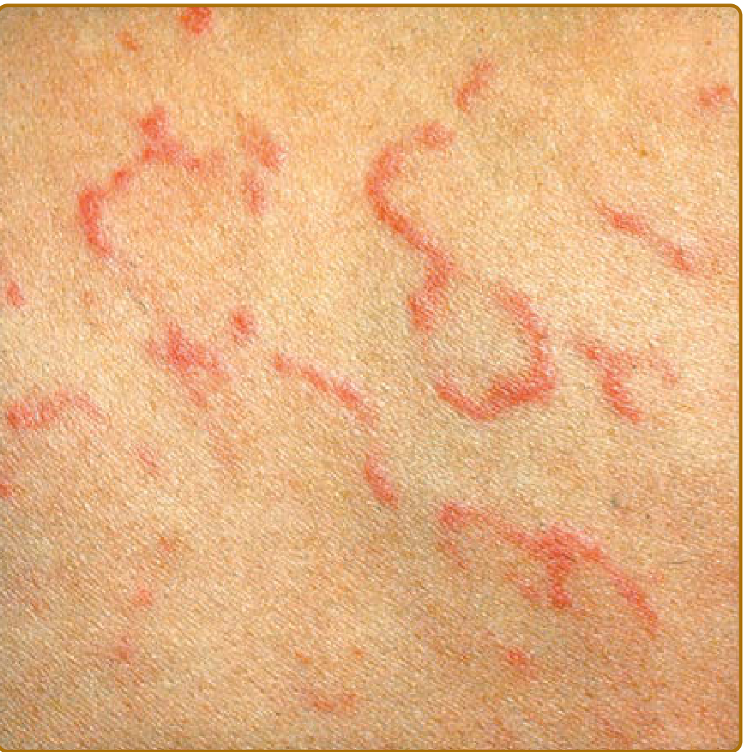
Erythema marginatum: expanding and shifting transient annular/polycyclic erythematous lesions - from Fitzpatrick's Dermatology
5. ⬜ SUBCUTANEOUS NODULES — <5% of cases
- Firm, painless, mobile, round nodules 0.5-2 cm
- Located over bony prominences and tendons: elbows, knees, wrists, occiput, spine
- Associated with severe carditis
- Resolve spontaneously in weeks
MINOR MANIFESTATIONS
| Criterion | Detail |
|---|---|
| Fever | ≥38.5°C (Low-risk populations); ≥38°C (High-risk populations) |
| Arthralgia | Joint pain WITHOUT objective arthritis (cannot use as minor if arthritis is a major criterion) |
| Elevated ESR | ≥60 mm/hr (low-risk); ≥30 mm/hr (high-risk populations) |
| Elevated CRP | ≥3.0 mg/dL |
| Prolonged PR interval | On ECG - indicates AV node involvement (cannot count if carditis is major criterion) |
5. DIAGNOSIS — 2015 REVISED JONES CRITERIA (AHA)
Diagnosis requires EVIDENCE OF PRECEDING GAS INFECTION PLUS:
Evidence of GAS Infection (MANDATORY - at least one):
- Positive throat culture for Group A Streptococcus
- Positive rapid antigen test (Rapid Strep Test)
- Elevated or rising ASO titre (Anti-Streptolysin O) - most commonly used
- Elevated Anti-DNase B antibodies
- Elevated Anti-hyaluronidase antibodies
Note: Absence of elevated ASO/antibody titer is strong evidence AGAINST rheumatic fever.
Diagnostic Rule:
| Scenario | Criteria Needed |
|---|---|
| Initial ARF | 2 Major OR 1 Major + 2 Minor criteria + evidence of GAS |
| Recurrent ARF | 2 Major OR 1 Major + 2 Minor OR 3 Minor criteria + evidence of GAS |
Risk Stratification (2015 Update):
| Population | Definition | Criteria Adjustment |
|---|---|---|
| Low-Risk | ARF incidence <2/100,000; RHD prevalence ≤1/1000 | Stricter (higher specificity) - only polyarthritis counts as major |
| Moderate-to-High Risk | Developing countries, indigenous populations, India | More sensitive - monoarthritis, polyarthralgia also count |
6. INVESTIGATIONS
| Test | Finding / Purpose |
|---|---|
| ASO Titre | Elevated (>200 Todd units in children); confirms recent GAS infection |
| Anti-DNase B | More sensitive than ASO; remains elevated longer |
| Throat culture | May be positive for GAS (often negative by the time RF develops) |
| Rapid Strep Test | Point-of-care antigen detection |
| CBC | Leukocytosis (neutrophilia); anemia in chronic carditis |
| ESR | Elevated (acute phase reactant) |
| CRP | Elevated |
| ECG | Prolonged PR interval (first-degree AV block) - most common ECG finding |
| Echocardiogram (2D + Doppler) | Gold standard for detecting subclinical carditis; valvular regurgitation; effusion |
| Chest X-ray | Cardiomegaly (if myocarditis/heart failure) |
| Synovial fluid | Sterile, inflammatory pattern (raised WBC, no organisms) |
7. TREATMENT
A. HOSPITALIZATION
All patients with ARF should be hospitalized for:
- Bed rest (especially if carditis present)
- Monitoring and management
- Initiation of prophylaxis
B. ERADICATION OF GAS (Treating the Trigger)
| Drug | Dose | Duration |
|---|---|---|
| Benzathine Penicillin G (IM) - PREFERRED | 1.2 million units IM (single dose) | Single injection |
| Oral Penicillin V | 500 mg PO 3x/day (250 mg in small children) | 10 days |
| Oral Amoxicillin | 500 mg PO twice daily | 10 days |
| Clarithromycin (Penicillin allergy) | 250 mg PO twice daily | 10 days |
C. TREATMENT OF ARTHRITIS AND FEVER
| Drug | Dose | Notes |
|---|---|---|
| Aspirin (Salicylate) - FIRST LINE | 80-100 mg/kg/day in 4 divided doses (max 4-8 g/day) | Dramatic response within 24-48 hrs; continue 1-2 weeks after ALL symptoms resolve |
| Naproxen | 10-20 mg/kg/day in 2 divided doses | Alternative NSAID; studied in small trials |
Important: NSAIDs/Aspirin treat arthritis and fever but do NOT prevent cardiac damage or alter the course of carditis.
D. TREATMENT OF CARDITIS
| Intervention | Indication |
|---|---|
| Bed rest | Mandatory during active carditis |
| Corticosteroids (Prednisolone 1-2 mg/kg/day, taper over 2-4 weeks) | Severe carditis with heart failure; evidence limited but used in practice |
| Diuretics (Furosemide) | Heart failure management |
| Angiotensin Receptor Blockers | Goal-directed heart failure therapy |
| Fluid restriction | Cardiac failure |
| Valve surgery | Rarely needed for acute RF; indicated for acute valve leaflet rupture |
E. TREATMENT OF SYDENHAM'S CHOREA
- Usually self-limiting - resolves in weeks to months without treatment
- Haloperidol or carbamazepine for severe/disabling chorea
- Valproate as alternative
- Protect child from self-injury during involuntary movements
8. SECONDARY PROPHYLAXIS (MOST CRITICAL PART)
The most important aspect of ARF management is preventing recurrence because each recurrence increases the risk of cumulative cardiac damage.
Drug of Choice: Benzathine Penicillin G (BPG) IM
- 1.2 million units IM every 4 weeks (every 3 weeks in high-risk regions)
| Duration of Prophylaxis | Indication |
|---|---|
| 10 years OR until age 21 (whichever is longer) | ARF without carditis |
| 10 years OR until age 25 | ARF with carditis, no residual valve disease |
| For life (until age 40 minimum) | ARF with severe RHD, valve surgery, or persisting carditis |
Alternatives (if Penicillin not possible):
- Oral Penicillin V 250 mg twice daily
- Oral Azithromycin 250 mg daily
- Oral Sulfadiazine (for penicillin-allergic patients)
9. COMPLICATIONS — RHEUMATIC HEART DISEASE (RHD)
- 35-70% of patients with ARF develop chronic RHD
- 18% have subclinical carditis on echocardiogram
| Valve | Involvement |
|---|---|
| Mitral valve | Nearly 100% of RHD cases; Mitral stenosis (most characteristic of chronic RHD) + Mitral regurgitation |
| Aortic valve | 20-30%; Aortic regurgitation |
| Tricuspid valve | Histologic changes; rarely clinical |
| Pulmonary valve | Rarely involved |
Mitral stenosis: The classic sequela - thickening, fusion of leaflets, narrowing of the mitral orifice → obstructed left atrial outflow → pulmonary hypertension → right heart failure.
10. NURSING CARE PLAN
Nursing Diagnosis 1: Acute Pain (Joint Pain)
| Interventions | Rationale |
|---|---|
| Assess pain on VAS/FLACC scale | Objective measurement |
| Administer Aspirin as prescribed | First-line anti-inflammatory |
| Gentle positioning; support painful joints with pillows | Reduces mechanical stress |
| Minimize movement during acute phase; bed rest | Prevents aggravation |
| Warm or cool compresses to affected joints | Reduces local inflammation |
Nursing Diagnosis 2: Decreased Cardiac Output (Carditis)
| Interventions | Rationale |
|---|---|
| Strict bed rest; elevate head of bed 30-45° | Reduces cardiac workload |
| Monitor heart rate, rhythm, BP every 4 hours | Detects dysrhythmias early |
| Continuous cardiac monitoring (ECG) | Detects PR prolongation, arrhythmias |
| Administer corticosteroids/diuretics as ordered | Manages cardiac inflammation and failure |
| Monitor fluid intake and output strictly | Prevents overload |
| Daily weight monitoring | Detects fluid retention |
| Restrict activity until ESR and CRP normalize | Prevents further cardiac strain |
Nursing Diagnosis 3: Hyperthermia
| Interventions | Rationale |
|---|---|
| Monitor temperature every 4 hours | Tracks fever trend |
| Administer Aspirin as prescribed (antipyretic + anti-inflammatory) | Dual therapeutic benefit |
| Tepid sponging; light clothing; adequate ventilation | Non-pharmacological cooling |
| Ensure adequate hydration | Prevents dehydration from sweating |
Nursing Diagnosis 4: Risk for Injury (Chorea)
| Interventions | Rationale |
|---|---|
| Padded bed rails | Prevents injury from involuntary movements |
| Calm, quiet environment | Reduces emotional triggers |
| Assist with feeding, ambulation | Prevents aspiration and falls |
| Administer sedatives/anticonvulsants as ordered | Controls severe chorea |
| Reassure child and family that chorea is temporary | Reduces anxiety |
Nursing Diagnosis 5: Parental/Child Anxiety and Knowledge Deficit
| Interventions | Rationale |
|---|---|
| Explain disease process, treatment, and prophylaxis in simple terms | Improves compliance |
| Emphasize lifelong prophylaxis importance | Prevents recurrence and RHD |
| Teach parents to report sore throats immediately for prompt treatment | Early GAS treatment prevents recurrence |
| Explain activity restrictions and gradual resumption | Prevents overexertion |
| Demonstrate IM injection technique if family to give home prophylaxis | Promotes adherence |
SUMMARY TABLE
| Feature | Key Point |
|---|---|
| Cause | Group A Strep pharyngitis → abnormal immune response |
| Onset | 2-3 weeks after throat infection |
| Age | 5-15 years |
| Diagnosis | 2015 Jones Criteria + evidence of GAS infection |
| Most common manifestation | Migratory polyarthritis (75%) |
| Most serious manifestation | Carditis (50-80%) → permanent valve damage |
| Pathognomonic sign | Aschoff bodies (histology); Erythema marginatum (clinical) |
| Lab test | Elevated ASO titre + ESR/CRP |
| ECG finding | Prolonged PR interval |
| Treatment of arthritis | Aspirin 80-100 mg/kg/day |
| Treatment of GAS | Benzathine Penicillin G IM single dose |
| Prophylaxis | Benzathine Penicillin G 1.2 MU IM every 4 weeks |
| Duration of prophylaxis | Minimum 10 years (lifelong if significant RHD) |
| Most important complication | Rheumatic Heart Disease (Mitral stenosis) |
| Key prevention message | "Treat every sore throat promptly with antibiotics" |
Sources: Fitzpatrick's Dermatology; Rosen's Emergency Medicine; Goldman-Cecil Medicine; Medical Microbiology 9e; AHA 2015 Revised Jones Criteria (Circulation 2015;131:1806-1818)
Nephtotic syndrome enlist
Nephrotic syndrome is characterized by the classic tetrad: massive proteinuria, hypoalbuminemia, edema, and hyperlipidemia/lipiduria. Most common cause in children is Minimal Change Disease.
Searching sources...
Finding Sources
Finding Sources
Reading File
Reading File
Finding Sources
Finding Sources
Reading File
Finding Sources
Here is the complete enlisted (comprehensive outline) answer on Nephrotic Syndrome:
NEPHROTIC SYNDROME — Complete Enlisted Overview
1. DEFINITION
Nephrotic syndrome is a clinical syndrome (not a disease in itself) defined by the classic tetrad:
| Feature | Diagnostic Threshold |
|---|---|
| Massive Proteinuria | >3.5 g/24 hours in adults; >40 mg/m²/hr or >50 mg/kg/24h in children |
| Hypoalbuminemia | Serum albumin <3 g/dL |
| Generalized Edema | Pitting edema, periorbital puffiness, ascites, pleural effusion, anasarca |
| Hyperlipidemia + Lipiduria | Serum cholesterol >300 mg/dL; oval fat bodies + fatty casts in urine |
Remember: "PHHE" — Proteinuria, Hypoalbuminemia, Hyperlipidemia, Edema
2. CAUSES / ETIOLOGY
A. PRIMARY (Idiopathic) — No identifiable cause
| Type | Age Group | Steroid Response | Notes |
|---|---|---|---|
| Minimal Change Disease (MCD/MCNS) | Children 2-6 yrs (most common in children) | ✅ 90% respond | "Nil disease" on biopsy; podocyte effacement on EM |
| Focal Segmental Glomerulosclerosis (FSGS) | All ages; higher in Blacks, Hispanics | ❌ <25% respond | Most common cause of SRNS in children |
| Membranous Nephropathy (MN) | Adults (most common adult primary) | ❌ Poor | Anti-PLA2R antibodies; secondary to Hep B, SLE, drugs |
| Mesangial Proliferation (MesPGN) | Children | Variable | |
| Membranoproliferative GN (MPGN) | Children, young adults | ❌ | Low complement |
B. SECONDARY — Identifiable underlying cause
| Category | Examples |
|---|---|
| Infections | Hepatitis B and C, HIV, Malaria (quartan malaria - P. malariae), Syphilis, Leprosy |
| Autoimmune/Systemic | Lupus nephritis (SLE) - most common secondary cause; Rheumatoid arthritis, Sjogren's |
| Metabolic/Endocrine | Diabetic nephropathy - most common cause of NS in adults worldwide |
| Drugs/Toxins | NSAIDs, Gold, Penicillamine, Mercury, Heroin, Captopril |
| Malignancy | Hodgkin's lymphoma (→ MCD), Solid tumors (→ MN), Leukemia |
| Allergic | Insect bites, pollens, immunizations |
| Hereditary | Congenital NS (Finnish type) - NPHS1 mutation; Alport syndrome |
| Miscellaneous | Amyloidosis, Pre-eclampsia |
C. CONGENITAL / INFANTILE
- Presents in first year of life
- Finnish type: NPHS1 (nephrin) gene mutation on chromosome 19
- Diffuse mesangial sclerosis
- Does NOT respond to steroids; requires bilateral nephrectomy + transplant
3. PATHOPHYSIOLOGY
Injury to Glomerular Filtration Barrier
(podocytes, glomerular basement membrane, slit diaphragm)
↓
LOSS OF NEGATIVE CHARGE on GBM
+ DISRUPTION OF SLIT DIAPHRAGM
↓
Massive proteinuria (especially albumin)
↓
┌─────────────────────────────────┐
↓ ↓
HYPOALBUMINEMIA HYPERLIPIDEMIA
(↓ oncotic pressure) (Liver overproduces lipoproteins
↓ to compensate ↓ oncotic pressure)
↓ Plasma oncotic + Lipiduria
pressure (lipoproteins lost in urine)
↓
Fluid shifts to interstitium
(UNDERFILL theory)
↓
EDEMA (periorbital,
dependent, anasarca)
↓
↓ Intravascular volume
↓
RAAS activation → Na+/H₂O retention
ADH release → Water retention
↓
Worsening EDEMA (OVERFILL)
4. CLINICAL FEATURES (ENLISTED)
A. Symptoms
- Periorbital puffiness - early morning, first noticed by parents; most common presenting complaint in children
- Pedal edema - bilateral pitting edema of feet and ankles
- Abdominal distension - due to ascites
- Frothy/foamy urine - due to heavy proteinuria (urine froths in toilet)
- Decreased urine output (oliguria) - due to volume depletion
- Anorexia, malaise, fatigue
- Breathlessness - pleural effusion (fluid in lungs)
- Scrotal/labial edema in severe cases
B. Signs on Examination
- Pitting edema - generalized; pits on finger pressure
- Periorbital puffiness - puffy eyelids, especially morning
- Ascites - shifting dullness, fluid thrill
- Pleural effusion - dullness at lung bases, reduced breath sounds
- Anasarca - total body edema
- Pallor - from protein loss and anemia
- Blood pressure - usually NORMAL (unlike nephritic syndrome); may be low in underfill
- Xanthelasma - in chronic cases (from hyperlipidemia)
- White bands on nails (Muehrcke lines) - from hypoalbuminemia
- Striae - stretch marks from rapid edema accumulation
5. INVESTIGATIONS (ENLISTED)
Urine
| Test | Finding |
|---|---|
| Urine dipstick | 3+ to 4+ protein |
| 24-hour urine protein | >3.5 g/24 hrs (adults); >40 mg/m²/hr (children) |
| Urine protein:creatinine ratio | >2.0 mg/mg (or >200 mg/mmol) |
| Urine microscopy | Oval fat bodies, fatty casts, lipid droplets (Maltese cross under polarized light) |
| Urine albumin:creatinine ratio | Markedly elevated |
Blood Tests
| Test | Finding |
|---|---|
| Serum Albumin | <3 g/dL (often <2 g/dL in severe cases) |
| Serum Cholesterol | >300 mg/dL (hypercholesterolemia) |
| Serum Triglycerides | Elevated |
| Total Protein | Low |
| Serum Creatinine/BUN | Usually normal initially; elevated if AKI |
| Serum Electrolytes | Hyponatremia (dilutional); hypokalemia |
| Serum Complement (C3, C4) | Low in MPGN, Lupus nephritis; NORMAL in MCD, MN |
| ANA, anti-dsDNA | Elevated in Lupus nephritis |
| HBsAg, HCV antibody | Rule out secondary causes |
| Serum Iron, TIBC | Low transferrin due to urinary loss |
| Coagulation profile (PT, aPTT) | May be deranged (hypercoagulable state) |
| CBC | Normochromic normocytic anemia |
Imaging
| Test | Findings |
|---|---|
| Ultrasound abdomen | Bilateral enlarged kidneys (in early NS); ascites; pleural effusion |
| Chest X-ray | Pleural effusion, cardiomegaly (if severe) |
| Echocardiogram | Pericardial effusion (if severe anasarca) |
Renal Biopsy (Indications)
- Age <1 year or >12 years (atypical age for MCD)
- Steroid resistance (no response in 8 weeks)
- Steroid dependence
- Macroscopic hematuria, persistent low complement
- Extrarenal features (rash, arthritis)
- Suspected secondary cause
| Histology | Light Microscopy | Immunofluorescence | Electron Microscopy |
|---|---|---|---|
| MCD | Normal ("nil disease") | Negative | Podocyte foot process effacement |
| FSGS | Focal (some glomeruli) + Segmental (part of tuft) sclerosis | IgM, C3 (non-specific) | Foot process effacement |
| Membranous | Thickened GBM, "spike and dome" | IgG, C3 granular (subepithelial) | Subepithelial deposits |
| MPGN | Lobular, "tram-track" GBM duplication | C3, IgG/IgM | Subendothelial deposits |
6. COMPLICATIONS (ENLISTED)
A. Infections (Most Common Cause of Death in Children)
- Spontaneous Bacterial Peritonitis (SBP) - 2-6% of cases; Streptococcus pneumoniae most common (because IgG and complement lost in urine → opsonic defect)
- Cellulitis - protein-rich edema fluid is a growth medium for bacteria
- Septicemia, Meningitis
- Pneumococcal infections - children especially vulnerable
- Gram-negative sepsis (E. coli, Klebsiella)
B. Thromboembolism (Most Serious Complication)
- Up to 20% of patients experience thrombotic events
- Renal vein thrombosis - flank pain, hematuria, acute deterioration of renal function; most common in membranous nephropathy
- Deep Vein Thrombosis (DVT)
- Pulmonary embolism
- Cerebral venous thrombosis
- Mechanism: Loss of antithrombotic proteins (Protein C, Protein S, Antithrombin III) in urine + elevated clotting factors (I, V, VIII, X) + hyperviscosity + platelet hyperaggregability
C. Metabolic Complications
- Protein malnutrition - muscle wasting, growth retardation in children
- Hypocalcemia - vitamin D-binding protein lost in urine → low 25-OH vitamin D → low Ca²⁺
- Osteoporosis/Rickets - from vitamin D deficiency + steroid therapy
- Iron deficiency anemia - transferrin lost in urine
- Zinc deficiency - impaired wound healing, growth failure
- Hypothyroidism - thyroid-binding globulin lost in urine
- Drug toxicity - drugs bound to albumin have increased free fraction (increased toxicity)
D. Renal Complications
- Acute Kidney Injury (AKI) - from volume depletion (underfill), sepsis, renal vein thrombosis, interstitial nephritis (from diuretics)
- Chronic Kidney Disease (CKD) - especially in FSGS and MPGN
- Progression to ESRD - particularly steroid-resistant forms
E. Cardiovascular
- Hypertension - from fluid overload
- Accelerated atherosclerosis - from chronic hyperlipidemia
- Pericardial effusion - in anasarca
7. TREATMENT (ENLISTED)
A. General / Supportive Measures
- Rest - during active edema phase
- Diet:
- Low sodium (1-2 g/day) - reduces edema
- Normal to high protein (1.5-2 g/kg/day) - to replace urinary losses
- Low fat - manage hyperlipidemia
- Adequate calories for growth (in children)
- Fluid restriction - only if severe hyponatremia
B. Treatment of Edema
| Drug | Dose | Notes |
|---|---|---|
| Furosemide (Loop diuretic) | 1-2 mg/kg/day | First-line for edema; hypoalbuminemia reduces response |
| Spironolactone | 2-3 mg/kg/day | Aldosterone antagonist; potassium-sparing; combine with furosemide |
| IV Albumin + Furosemide | 0.5-1 g/kg albumin, then furosemide | For resistant edema with severe hypoalbuminemia; not recommended routinely |
C. Specific Treatment — CORTICOSTEROIDS (Primary NS)
| Phase | Drug | Dose | Duration |
|---|---|---|---|
| Initial treatment | Prednisolone | 60 mg/m²/day or 2 mg/kg/day (max 60 mg) | 4-6 weeks (until urine protein-free) |
| Maintenance | Prednisolone | Alternate day, then taper | Over 6 weeks |
Definitions of Response:
- Remission: Urine protein negative/trace for 3 consecutive days
- Relapse: Urine protein 3+ for 3 consecutive days after remission
- Steroid Sensitive NS (SSNS): Remission within 8 weeks (90% of MCD)
- Steroid Resistant NS (SRNS): No remission after 8 weeks of full-dose steroids
- Frequent Relapsing NS (FRNS): ≥2 relapses in 6 months or ≥4 in 12 months
- Steroid Dependent NS (SDNS): Relapses during steroid taper or within 2 weeks of stopping
D. Steroid-Sparing Agents (for FRNS/SDNS/SRNS)
| Drug | Use |
|---|---|
| Cyclophosphamide | Oral; 2-3 mg/kg/day for 8-12 weeks; FRNS/SDNS |
| Mycophenolate Mofetil (MMF) | FRNS, SDNS; fewer side effects |
| Calcineurin Inhibitors (Tacrolimus, Cyclosporine) | SRNS, SDNS; risk of nephrotoxicity |
| Rituximab (anti-CD20) | Severe FRNS/SDNS; biological agent |
| Levamisole | Immunomodulator; used in developing countries |
E. Treatment of Hyperlipidemia
- Statins (Atorvastatin, Rosuvastatin) for persistent hyperlipidemia or high cardiovascular risk
- Dietary fat restriction
F. Prevention / Treatment of Thrombosis
- Low molecular weight heparin (LMWH) - if albumin <2 g/dL and additional risk factors
- Warfarin - for established thrombosis (DVT, pulmonary embolism, renal vein thrombosis)
- Maintain adequate hydration
G. Prevention / Treatment of Infections
- Pneumococcal vaccine - strongly recommended in all children with NS
- Varicella vaccine - before steroid therapy if possible
- Penicillin prophylaxis - during active nephrotic state in some centers
- Prompt antibiotic treatment for infections (cefotaxime for peritonitis)
H. Treatment of Specific Causes
- Membranous (secondary): ACE inhibitor/ARB + immunosuppression
- Lupus nephritis: Hydroxychloroquine + steroids + MMF or cyclophosphamide
- Diabetic nephropathy: Tight glucose control + ACE inhibitor/ARB (renoprotective)
- FSGS: High-dose steroids; CNIs if resistant
8. NURSING CARE PLAN (ENLISTED)
| Nursing Diagnosis | Key Interventions |
|---|---|
| Excess Fluid Volume (edema, ascites) | Daily weight; strict I/O; low sodium diet; diuretics as ordered; elevate edematous limbs; skin care for stretched skin |
| Imbalanced Nutrition (protein loss) | High-protein diet; dietitian referral; monitor serum albumin; supplement vitamins |
| Risk for Infection | Hand hygiene; isolate from infectious contacts; monitor temperature; avoid IM injections in edematous areas; vaccinations |
| Risk for Impaired Skin Integrity (edema, steroids) | 2-hourly position change; pressure-relieving mattress; avoid trauma to edematous skin; careful NG/catheter care |
| Risk for Thrombosis | Adequate hydration; passive exercises; compression stockings if appropriate; monitor for DVT signs |
| Body Image Disturbance (moon face, weight gain from steroids, edema) | Reassure child and family; explain steroid effects are reversible; support groups |
| Parental/Child Knowledge Deficit | Teach urine dipstick testing at home; teach low-sodium diet; teach signs of relapse; explain prophylaxis importance |
| Activity Intolerance (edema, fatigue) | Graduated activity; bed rest during acute phase; play therapy to maintain development |
SUMMARY — "NEPHROTIC" Mnemonic
| Letter | Feature |
|---|---|
| N | eNormous proteinuria (>3.5 g/day) |
| E | Edema (periorbital, ascites, anasarca) |
| P | Proteinuria (massive, selective for albumin) |
| H | Hypoalbuminemia (<3 g/dL) |
| R | RAAS activation (renin-angiotensin) |
| O | Oval fat bodies in urine (lipiduria) |
| T | Thromboembolism risk (renal vein, DVT, PE) |
| I | Infections (pneumococcal, peritonitis) |
| C | Cholesterol elevated (hyperlipidemia) |
Sources: Campbell-Walsh-Wein Urology; Brenner & Rector's The Kidney; Comprehensive Clinical Nephrology 7e; Goldman-Cecil Medicine; Smith & Tanagho's General Urology
This is a shared conversation. Sign in to Orris to start your own chat.