
Course of femoral nerve
femoral nerve course anatomy diagram
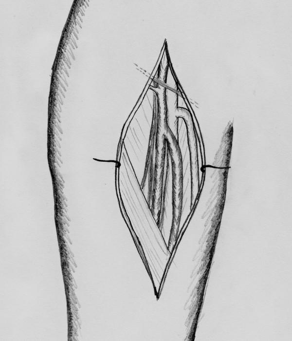
This anatomical diagram is a grayscale illustration depicting the surgical exposure of the superficial femoral artery (SFA) in the thigh, prepared for harvesting as a vascular graft. The drawing shows a vertical, spindle-shaped skin incision held open by retractors to reveal the neurovascular bundle. Centrally located within the surgical field is the superficial femoral artery, positioned between the femoral vein and the associated femoral nerve. The illustration highlights the relationship between these parallel longitudinal structures and includes fine lines suggesting surgical sutures or vessel loops used for vessel isolation. This diagram serves an educational purpose in vascular and oncological surgery, specifically illustrating the donor site preparation for carotid artery reconstruction using an autologous femoral artery transplant. Key concepts include surgical anatomy of the femoral triangle, vessel harvesting techniques, and the orientation of the femoral neurovascular sheath.
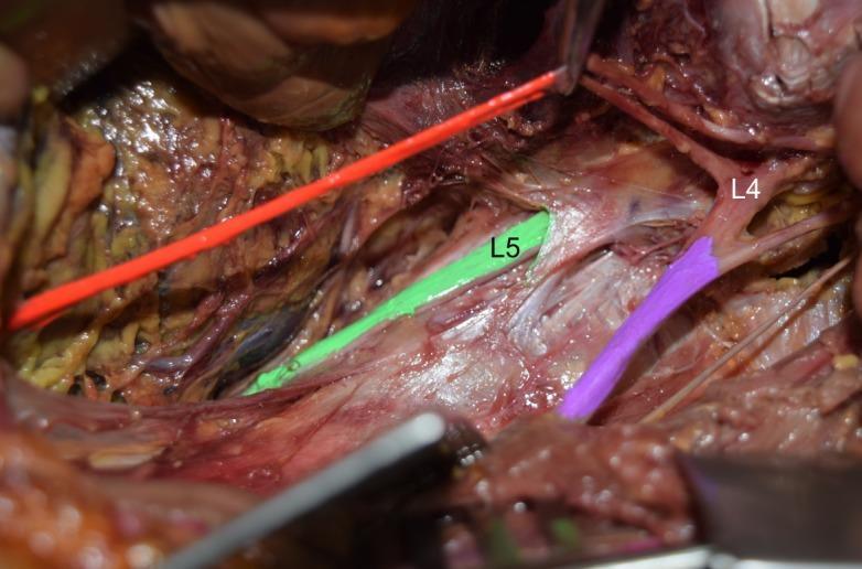
Anatomical Diagram/Clinical Photograph: This image depicts a cadaveric dissection of the left posterior abdominal wall focusing on the lumbar plexus. The psoas major muscle has been removed to expose the neural structures. Key anatomical landmarks are color-coded for clarity: the L4 ventral ramus and femoral nerve are highlighted in purple, the L5 ventral ramus is highlighted in green, and the obturator nerve is shown in orange/red. The primary educational focus is an anatomical variation where the lumbosacral trunk is absent. Visually, there is a clear separation between the L4 and L5 ventral rami, with no evidence of the standard fusion that typically forms the lumbosacral trunk. The femoral nerve (purple) is seen emerging from the L4 level, running lateral and parallel to the L5 ramus (green). This clinical photograph serves as an educational tool for gross anatomy and neurosurgery, illustrating variations in lumbosacral plexus connectivity and its implications for regional anesthesia or surgical approaches to the pelvic and lumbar regions.
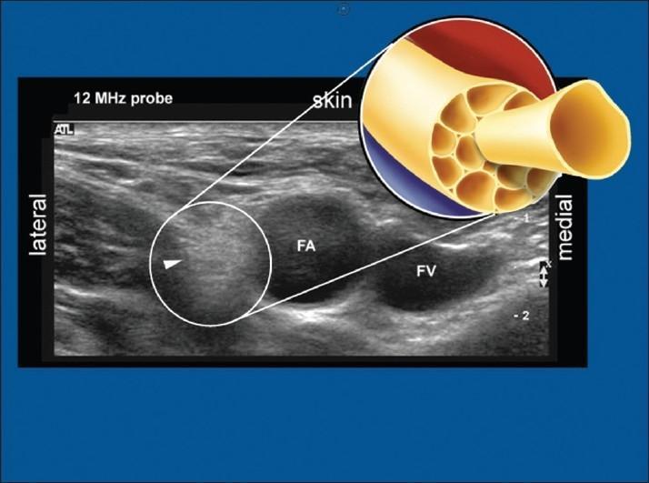
This composite educational image features a diagnostic ultrasound scan paired with an anatomical diagram, focusing on the femoral neurovascular bundle. The ultrasound, captured with a 12 MHz high-frequency linear probe, shows the femoral nerve in a transverse cross-section. The nerve is characterized as a distinct hyperechoic structure (brighter than surrounding tissue) located lateral to the pulsatile femoral artery (FA) and the compressible femoral vein (FV). An arrowhead points to the nerve, which exhibits a characteristic 'honeycomb' echotexture. A circular call-out diagram provides a histological illustration of the nerve's internal morphology, detailing numerous nerve fascicles bundled together by a dense perineurium and surrounded by epineurium. This visual comparison highlights the correlation between peripheral nerve microanatomy (fascicular arrangement) and its sonographic appearance. This material is essential for regional anesthesia training and ultrasound-guided femoral nerve block procedures within the fields of anesthesiology, radiology, and emergency medicine.
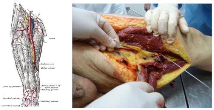
This dual-panel image provides a comparison between normal anatomy and traumatic vascular pathology of the lower limb. The left panel is a classical anatomical diagram of the right thigh, illustrating the femoral artery, its major branches, and associated nerves such as the saphenous nerve and genicular arteries. The right panel is a clinical photograph from a forensic or surgical setting showing a deep, penetrating injury to the left medial thigh. The photograph demonstrates a large, gaping wound with clear stratification of tissues: the pale yellow subcutaneous adipose tissue, the dark red skeletal muscle fibers of the adductor or quadriceps groups, and a centrally located, severed femoral artery. A metal surgical probe or instrument is inserted to demonstrate the depth of the track and the complete transection of the vascular bundle. Hemorrhagic staining and tissue maceration are visible throughout the wound bed. This image serves as an educational tool for identifying vascular trauma and understanding the surgical and forensic anatomy of femoral artery injuries resulting from high-energy mechanisms, such as gunshot wounds or sharp force trauma.
Femoral Nerve - Complete Course
Origin
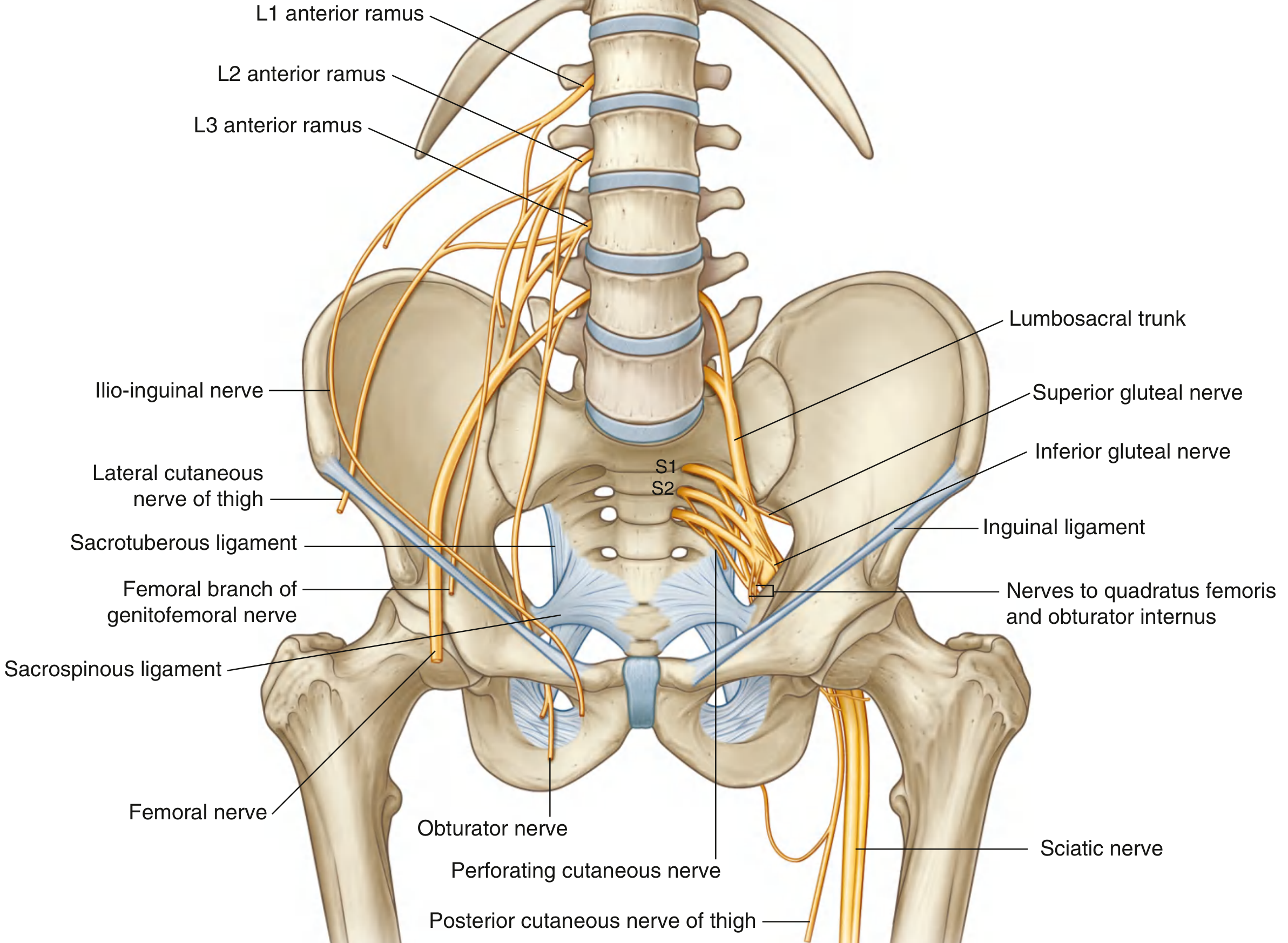
- Arises from the posterior divisions of the ventral rami of L2, L3, and L4 - making it the largest branch of the lumbar plexus.
- Forms within the substance of the psoas major muscle, in the posterior abdominal wall.
Abdominal (Pelvic) Segment
- Descends through psoas major, emerging from its lateral border.
- Runs in the groove between psoas major (medially) and iliacus (laterally) - the iliac fossa.
- Here it lies within the iliac fascia (iliacus compartment).
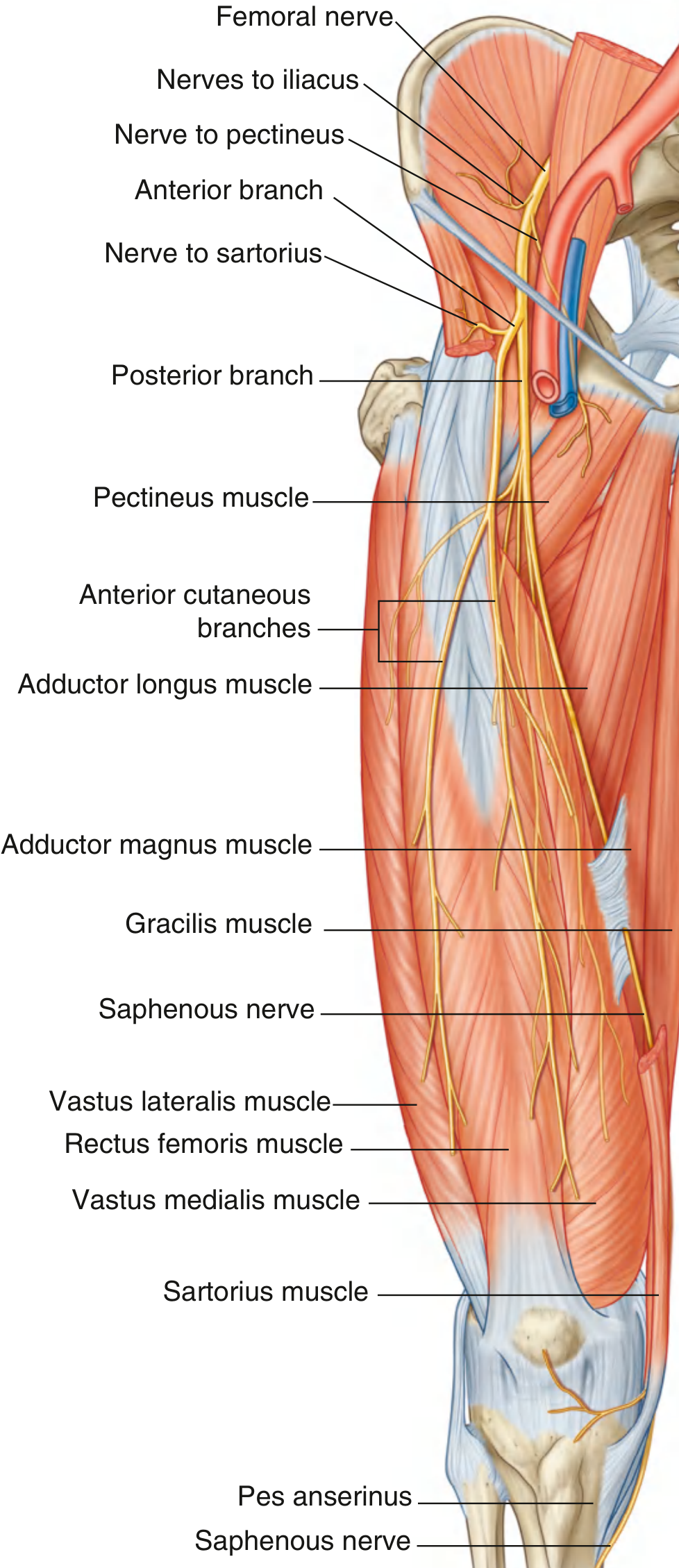
- While still above the inguinal ligament, it gives off branches to:
- Iliacus muscle (motor branch arising ~4-5 cm proximal to the inguinal ligament)
- Pectineus muscle
At the Inguinal Ligament
- Passes under the inguinal ligament through the muscular lacuna (lateral compartment), together with iliacus, to enter the femoral triangle.
- It lies lateral to the femoral artery and femoral sheath.
- Importantly, the femoral nerve is outside the femoral sheath (which surrounds only the femoral artery, vein, and canal).
- Relationship at the inguinal ligament (lateral to medial): NAVY - Nerve, Artery, Vein, Y-fronts (lymphatics).
In the Femoral Triangle
- The femoral triangle is bounded by the inguinal ligament (above), sartorius (laterally), and adductor longus (medially).
- Immediately after entering the femoral triangle, the nerve divides into anterior and posterior branches.
- Medial cutaneous nerve of thigh (anterior cutaneous branches) - skin over anterior and medial thigh
- Nerve to sartorius
- Nerves to rectus femoris, vastus lateralis, vastus intermedius, vastus medialis
- The saphenous nerve - the longest branch
The Saphenous Nerve (terminal sensory branch)
- Accompanies the femoral artery into and through the adductor canal (Hunter's canal).
- Unlike the femoral artery, the saphenous nerve does not pass through the adductor hiatus. Instead it exits the canal by piercing through connective tissue near its distal end, emerging between sartorius and gracilis on the medial side of the knee.
- Descends the medial side of the leg alongside the great saphenous vein.
- Supplies skin on the medial side of the knee, leg, and foot (as far as the medial side of the big toe).
Summary of Distribution
| Component | Structures |
|---|---|
| Motor | Iliacus, pectineus, sartorius, quadriceps femoris (rectus femoris, vastus lateralis, vastus medialis, vastus intermedius) |
| Sensory | Anterior thigh, anteromedial knee, medial leg, medial foot (via saphenous nerve) |
| Reflex | Knee jerk (L3, L4) |
Diagrams
Clinical Relevance
- Femoral nerve block targets the nerve just lateral to the femoral artery at the inguinal ligament.
- Femoral nerve injury (most commonly iatrogenic) causes: weakness of knee extension, absent knee jerk, and sensory loss over the anterior thigh and medial leg. Hip flexion is weak if the lesion is above the inguinal ligament (iliacus denervation).
- Common causes: retroperitoneal hematoma (after anticoagulation or femoral catheterization), self-retaining retractors during pelvic surgery, lithotomy positioning, total hip replacement.