
Daigrams for relations of lungs on all sides
Reading File
Now let me search the medical library for lung relations diagrams and anatomy content in parallel.
Finding Sources
Finding Sources
Finding Sources
Finding Sources
Good - there are anatomy books available. Let me search specifically in anatomy books for lung relations.
Finding Sources
Finding Sources
Finding Sources
Excellent! Found the key sections. Let me read all three relevant sections in parallel - "Right lung", "Left lung", "Surfaces of the Lung", and the Color Atlas section.
Reading File
Reading File
I have rich content. Now let me fetch all the key diagrams in parallel.
Loading Image
Loading Image
Reading File
Now let me fetch the left lung relations diagram and the Color Atlas surface images.
Reading File
Reading File
Loading Image
Loading Image
Loading Image
I now have all the key diagrams and textual content. Let me compile the comprehensive answer.
Here are the complete anatomical diagrams and descriptions for the relations of the lungs on all sides, from Gray's Anatomy for Students and the Color Atlas of Human Anatomy Vol. 2.
Overview: Surfaces and Borders of Both Lungs
Each lung has the shape of a half cone. Its surfaces conform to surrounding structures.
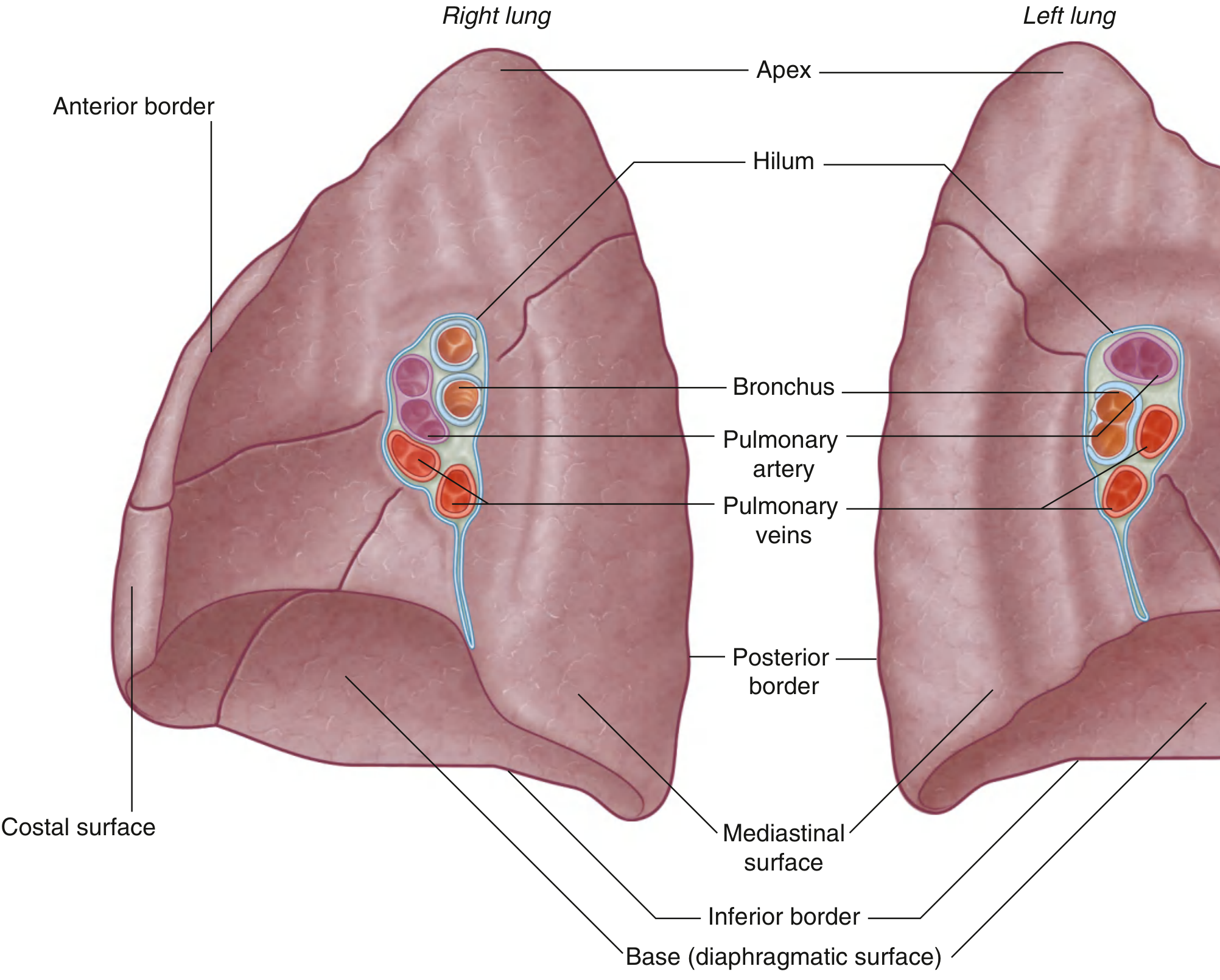
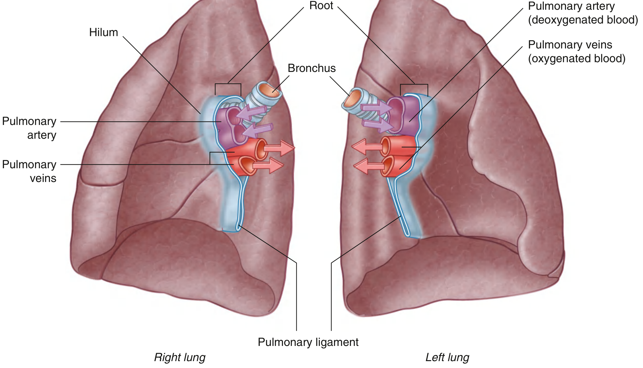
Roots and Hila
Four Surfaces of Each Lung (Color Atlas)
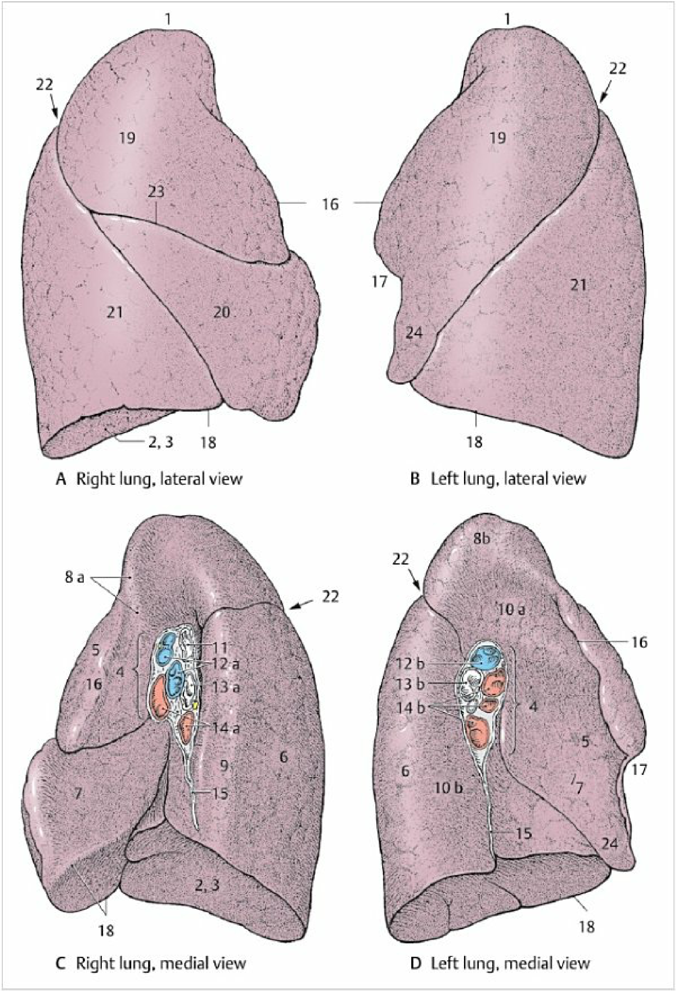
The four main surfaces and their relations:
| Surface | Relations |
|---|---|
| Costal (outer/lateral) | Ribs and intercostal muscles - convex |
| Mediastinal (medial) | Heart, great vessels, esophagus, bronchi |
| Diaphragmatic (base) | Diaphragm - concave |
| Apex | Projects 2-3 cm above clavicle into root of neck; crossed by subclavian vessels |
Right Lung - Lobes, Fissures & Mediastinal Relations
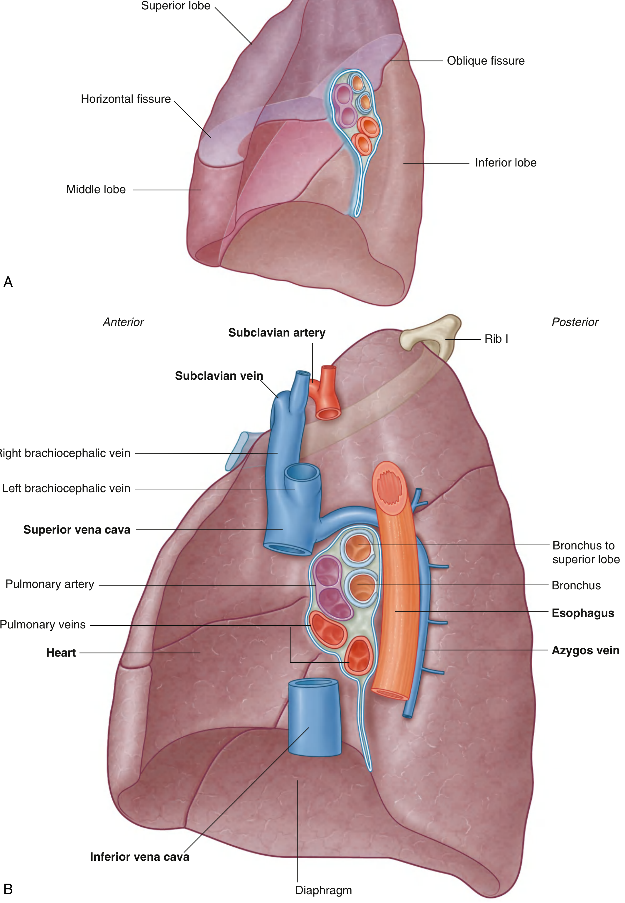
Mediastinal (medial surface) relations of the RIGHT lung:
- Heart (cardiac impression)
- Superior vena cava (anterior/superior)
- Inferior vena cava (inferior)
- Azygos vein (arches over the hilum posteriorly)
- Esophagus (posterior)
- Right subclavian artery and vein (arch over the apex / superior lobe)
Left Lung - Lobes, Fissures & Mediastinal Relations
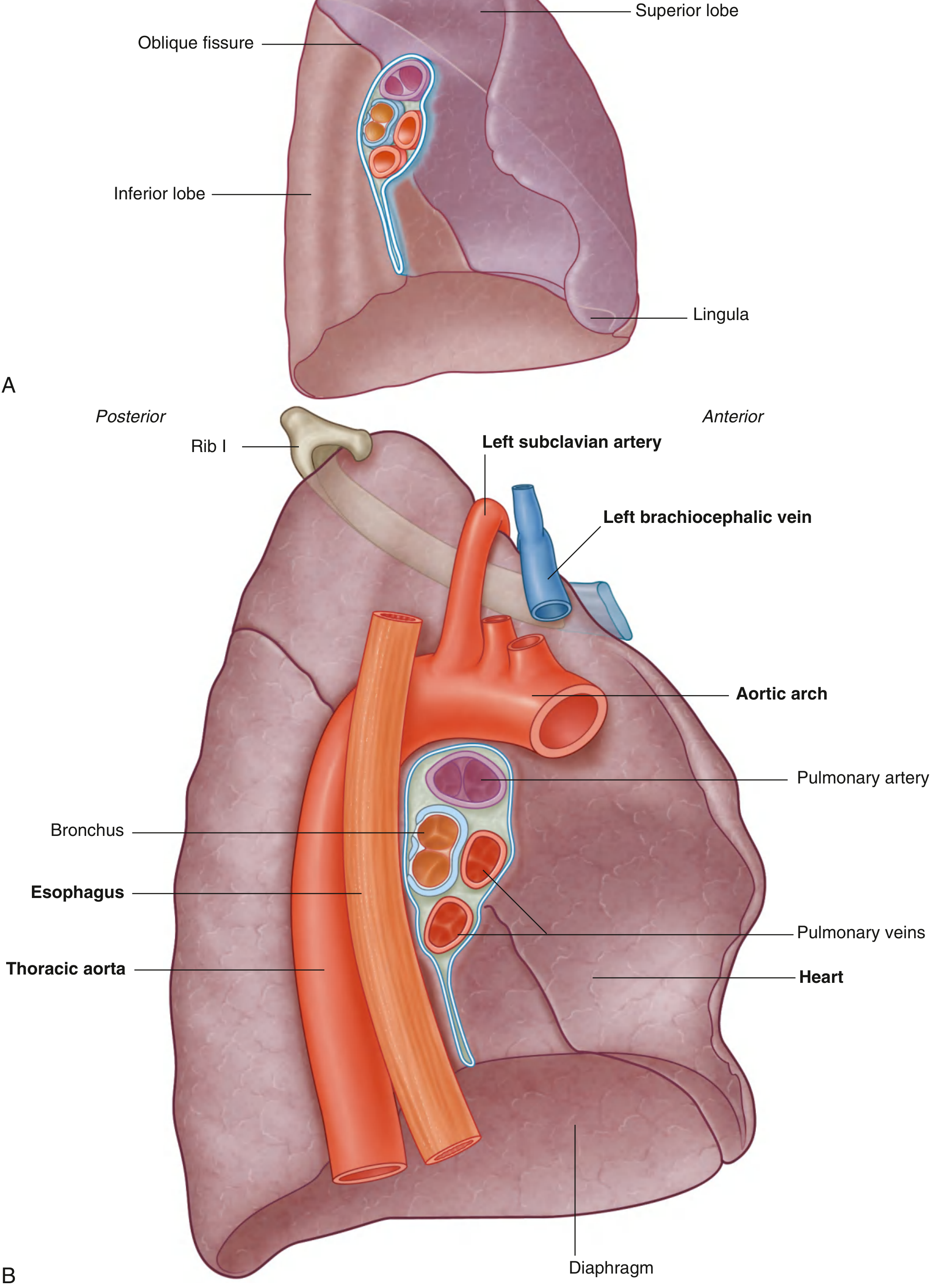
Mediastinal (medial surface) relations of the LEFT lung:
- Heart (larger cardiac impression - heart projects more into left pleural cavity)
- Aortic arch (grooves the superior aspect of the mediastinal surface)
- Thoracic aorta (posterior groove, runs along the entire posterior mediastinal surface)
- Esophagus (posterior, inferior)
- Left subclavian artery (groove at apex)
- Left brachiocephalic vein (superior)
- Pulmonary artery (at hilum)
Summary Table: Relations on All Sides
| Side | Right Lung | Left Lung |
|---|---|---|
| Costal (lateral) | Ribs 1-10, intercostal spaces | Ribs 1-10, intercostal spaces |
| Mediastinal (medial) | Heart, SVC, IVC, azygos vein, esophagus | Heart (larger impression), aortic arch, thoracic aorta, esophagus |
| Diaphragmatic (base/inferior) | Right dome of diaphragm (higher, over liver) | Left dome of diaphragm (lower) |
| Apex (superior) | Right subclavian artery & vein, cervical pleura, 1st rib | Left subclavian artery & vein, aortic arch, cervical pleura |
| Posterior (vertebral) | Vertebral column (T1-T10), sympathetic chain | Vertebral column, descending thoracic aorta |
| Anterior border | Meets opposite lung behind sternum | Has cardiac notch (heart bulges left) + lingula |
Key differences between the two lungs:
- The right lung is shorter (pushed up by right dome of diaphragm / liver) and wider, has 3 lobes (superior, middle, inferior) and 2 fissures (oblique + horizontal)
- The left lung is narrower and longer, has 2 lobes (superior, inferior) and 1 fissure (oblique); its medial surface has a prominent cardiac notch and lingula
- On the right, the azygos vein arches over the hilum - a key posterior relation unique to the right side
- On the left, the aortic arch and thoracic aorta are the dominant posterior/medial relations unique to the left side
Sources: Gray's Anatomy for Students, pp. 205-209 | Color Atlas of Human Anatomy Vol. 2 (Internal Organs), pp. 211-212
Full structure of the relation needed
Reading File
Finding Sources
Reading File
Finding Sources
Reading File
Finding Sources
Finding Sources
Finding Sources
There's a surgery textbook with a dedicated mediastinal surface diagram. Let me fetch that image and also read the detailed content from Fischer's Surgery.
Reading File
Finding Sources
Reading File
Loading Image
This is an excellent, highly detailed diagram. Now let me fetch the remaining key diagrams from the previous session and get one more detailed diagram showing the costal/posterior relations.
Loading Image
Loading Image
Loading Image
Loading Image
Loading Image
This is a shared conversation. Sign in to Orris to start your own chat.