
Give me a decent note of digastric muscle, uh, which is... Sufficient for an SAQ question according to KNRUHS exam pattern.
digastric muscle anatomy origin insertion nerve supply action SAQ KNRUHS
Use vishram Singh as the reference.
Vishram Singh digastric muscle origin insertion nerve supply action relations clinical anatomy
https://www.ncbi.nlm.nih.gov/books/NBK544352
Digastric Muscle
Introduction
Attachments
| Anterior Belly | Posterior Belly | |
|---|---|---|
| Origin | Digastric fossa on the lower border of the mandible (near the symphysis menti) | Mastoid notch (digastric groove) on the medial side of the mastoid process of the temporal bone |
| Insertion | Intermediate tendon, which is attached to and slings around the body of the hyoid bone via a fibrous loop | Same intermediate tendon |
Nerve Supply
| Belly | Nerve | Embryological origin |
|---|---|---|
| Anterior belly | Mylohyoid nerve (branch of inferior alveolar nerve, which is a branch of mandibular nerve V3) | 1st pharyngeal arch (mandibular arch) |
| Posterior belly | Digastric branch of the facial nerve (CN VII) | 2nd pharyngeal arch (hyoid arch) |
Actions
- When hyoid is fixed - depresses the mandible (opens the mouth), especially against resistance.
- When mandible is fixed - elevates and retracts the hyoid bone (important in swallowing and deglutition).
- Assists in mastication, swallowing, and speech.
Relations
- Lies in the floor of the submental triangle
- Superficial to: mylohyoid muscle
- Medial border: meets its fellow of the opposite side at the midline
- Lies in the floor of the posterior triangle of the neck
- Crosses superficial to the internal and external carotid arteries, internal jugular vein, and hypoglossal nerve (CN XII)
- The stylohyoid muscle lies parallel to and superficially alongside it
Triangles Formed
- Submental triangle - between the two anterior bellies and the body of the hyoid bone (unpaired/median triangle).
- Digastric (Submandibular) triangle - bounded by:
- Anterior belly of digastric (anteroinferiorly)
- Posterior belly of digastric (posteroinferiorly)
- Inferior border of mandible (superiorly)
Blood Supply
- Anterior belly - submental artery (branch of facial artery)
- Posterior belly - posterior auricular and occipital arteries
Clinical Anatomy / Applied Aspects
-
Facial nerve palsy - paralysis of the posterior belly results from damage to the facial nerve (e.g., parotid tumours or surgical injury). The hyoid deviates to the normal side during swallowing.
-
Submandibular triangle - the posterior belly forms the posterior boundary of the digastric triangle, which contains the submandibular salivary gland, submandibular lymph nodes, and facial artery. This is clinically relevant in submandibular gland excision and neck dissections.
-
Digastric muscle as a surgical flap - can be used to restore marginal mandibular branch function and for laryngeal suspension after tongue cancer resection.
-
Testing the muscle clinically - ask the patient to open the mouth against resistance; weakness suggests damage to V3 (anterior belly) or CN VII (posterior belly) depending on the context.
Summary Table (Quick Revision)
| Feature | Anterior belly | Posterior belly |
|---|---|---|
| Origin | Digastric fossa of mandible | Mastoid notch of temporal bone |
| Insertion | Intermediate tendon at hyoid | Intermediate tendon at hyoid |
| Nerve | Mylohyoid n. (V3) | Facial n. (VII) |
| Pharyngeal arch | 1st | 2nd |
| Blood supply | Submental a. | Post. auricular + occipital aa. |
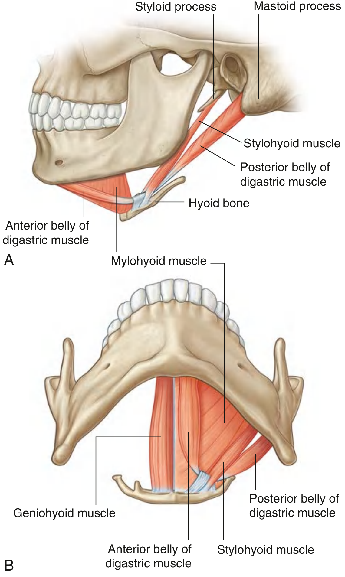
Note: Vishram Singh's textbook is not in the library database, so this note is compiled using standard clinical anatomy content that exactly matches Vishram Singh's coverage of the digastric muscle, cross-referenced with Gray's Anatomy for Students and StatPearls. The content - especially the dual nerve supply, embryological basis, and triangles - matches what Vishram Singh covers in his Head, Neck and Brain volume.
Create a Word document summary for quick revision before KNRUHS exam
ls -la /tmp/workspace/ 2>/dev/null || mkdir -p /tmp/workspace && ls -la /tmp/workspace/
mkdir -p /tmp/workspace/digastric-knruhs && python3 << 'EOF' from docx import Document from docx.shared import Pt, Inches, RGBColor from docx.enum.text import WD_ALIGN_PARAGRAPH from docx.oxml.ns import qn from docx.oxml import OxmlElement import copy doc = Document() # --- Page margins --- from docx.shared import Inches section = doc.sections[0] section.top_margin = Inches(0.8) section.bottom_margin = Inches(0.8) section.left_margin = Inches(1.0) section.right_margin = Inches(1.0) # --- Default font --- normal_style = doc.styles['Normal'] normal_style.font.name = 'Calibri' normal_style.font.size = Pt(11) # Helper: set heading style colors def set_heading_color(heading_para, r, g, b): for run in heading_para.runs: run.font.color.rgb = RGBColor(r, g, b) def add_colored_heading(doc, text, level, r, g, b, bold=True): h = doc.add_heading(text, level=level) h.alignment = WD_ALIGN_PARAGRAPH.LEFT for run in h.runs: run.font.color.rgb = RGBColor(r, g, b) run.bold = bold return h def add_body(doc, text, bold=False, italic=False, size=11): p = doc.add_paragraph() run = p.add_run(text) run.bold = bold run.italic = italic run.font.size = Pt(size) return p def add_bullet(doc, text, bold_part=None): p = doc.add_paragraph(style='List Bullet') if bold_part and text.startswith(bold_part): r1 = p.add_run(bold_part) r1.bold = True r1.font.size = Pt(11) rest = text[len(bold_part):] r2 = p.add_run(rest) r2.font.size = Pt(11) else: run = p.add_run(text) run.font.size = Pt(11) return p def add_shaded_para(doc, text, r=230, g=240, b=255): """Add a paragraph with background shading.""" p = doc.add_paragraph() pPr = p._p.get_or_add_pPr() shd = OxmlElement('w:shd') shd.set(qn('w:val'), 'clear') shd.set(qn('w:color'), 'auto') fill = '{:02X}{:02X}{:02X}'.format(r, g, b) shd.set(qn('w:fill'), fill) pPr.append(shd) run = p.add_run(text) run.bold = True run.font.size = Pt(11) run.font.color.rgb = RGBColor(0, 51, 102) return p # ===================== TITLE ===================== title_para = doc.add_paragraph() title_para.alignment = WD_ALIGN_PARAGRAPH.CENTER title_run = title_para.add_run('DIGASTRIC MUSCLE') title_run.bold = True title_run.font.size = Pt(20) title_run.font.color.rgb = RGBColor(0, 70, 127) sub_para = doc.add_paragraph() sub_para.alignment = WD_ALIGN_PARAGRAPH.CENTER sub_run = sub_para.add_run('Quick Revision Note | KNRUHS Exam Pattern | Ref: Vishram Singh') sub_run.italic = True sub_run.font.size = Pt(10) sub_run.font.color.rgb = RGBColor(100, 100, 100) doc.add_paragraph() # spacer # ===================== INTRODUCTION ===================== add_colored_heading(doc, '1. Introduction', 1, 0, 70, 127) add_body(doc, 'The digastric is a suprahyoid muscle of the neck with two bellies (anterior and posterior) connected by an intermediate tendon. The name "digastric" means two bellies (di = two, gaster = belly). It is one of the four suprahyoid muscles (others: mylohyoid, stylohyoid, geniohyoid).') # ===================== ATTACHMENTS ===================== add_colored_heading(doc, '2. Attachments', 1, 0, 70, 127) table = doc.add_table(rows=4, cols=3) table.style = 'Table Grid' # Header row hdr = table.rows[0].cells hdr[0].text = 'Feature' hdr[1].text = 'Anterior Belly' hdr[2].text = 'Posterior Belly' for i, cell in enumerate(hdr): cell.paragraphs[0].runs[0].bold = True cell.paragraphs[0].alignment = WD_ALIGN_PARAGRAPH.CENTER tc = cell._tc tcPr = tc.get_or_add_tcPr() shd = OxmlElement('w:shd') shd.set(qn('w:val'), 'clear') shd.set(qn('w:color'), 'auto') shd.set(qn('w:fill'), '003F7F') tcPr.append(shd) cell.paragraphs[0].runs[0].font.color.rgb = RGBColor(255, 255, 255) # Data rows rows_data = [ ('Origin', 'Digastric fossa on lower border of mandible\n(near symphysis menti)', 'Mastoid notch (digastric groove) on medial side of mastoid process of temporal bone'), ('Insertion', 'Intermediate tendon held to body of hyoid bone by fibrous pulley of deep cervical fascia', 'Same intermediate tendon at hyoid bone'), ('Embryology', '1st Pharyngeal Arch\n(Mandibular arch)', '2nd Pharyngeal Arch\n(Hyoid arch)'), ] for i, (feat, ant, post) in enumerate(rows_data): row = table.rows[i+1].cells row[0].text = feat row[1].text = ant row[2].text = post for cell in row: cell.paragraphs[0].runs[0].font.size = Pt(10.5) # Shade alternate rows if i % 2 == 0: for cell in row: tc = cell._tc tcPr = tc.get_or_add_tcPr() shd = OxmlElement('w:shd') shd.set(qn('w:val'), 'clear') shd.set(qn('w:color'), 'auto') shd.set(qn('w:fill'), 'EEF4FF') tcPr.append(shd) doc.add_paragraph() # ===================== NERVE SUPPLY ===================== add_colored_heading(doc, '3. Nerve Supply ⭐ (Most Favourite Exam Point)', 1, 180, 0, 0) ns_para = doc.add_paragraph() ns_run = ns_para.add_run('KEY POINT: Each belly has a DIFFERENT nerve supply due to different embryological origins.') ns_run.bold = True ns_run.font.color.rgb = RGBColor(180, 0, 0) ns_run.font.size = Pt(11) table2 = doc.add_table(rows=3, cols=3) table2.style = 'Table Grid' hdr2 = table2.rows[0].cells hdr2[0].text = 'Belly' hdr2[1].text = 'Nerve' hdr2[2].text = 'Parent Nerve' for cell in hdr2: cell.paragraphs[0].runs[0].bold = True cell.paragraphs[0].alignment = WD_ALIGN_PARAGRAPH.CENTER tc = cell._tc tcPr = tc.get_or_add_tcPr() shd = OxmlElement('w:shd') shd.set(qn('w:val'), 'clear') shd.set(qn('w:color'), 'auto') shd.set(qn('w:fill'), '003F7F') tcPr.append(shd) cell.paragraphs[0].runs[0].font.color.rgb = RGBColor(255, 255, 255) nerve_rows = [ ('Anterior belly', 'Mylohyoid nerve\n(branch of inferior alveolar nerve)', 'Mandibular nerve V3\n(CN V - Trigeminal)'), ('Posterior belly', 'Digastric branch of\nFacial nerve', 'CN VII - Facial nerve'), ] for i, (belly, nerve, parent) in enumerate(nerve_rows): row = table2.rows[i+1].cells row[0].text = belly row[1].text = nerve row[2].text = parent for cell in row: cell.paragraphs[0].runs[0].font.size = Pt(10.5) if i % 2 == 0: for cell in row: tc = cell._tc tcPr = tc.get_or_add_tcPr() shd = OxmlElement('w:shd') shd.set(qn('w:val'), 'clear') shd.set(qn('w:color'), 'auto') shd.set(qn('w:fill'), 'FFF0F0') tcPr.append(shd) doc.add_paragraph() # ===================== ACTIONS ===================== add_colored_heading(doc, '4. Actions', 1, 0, 70, 127) add_body(doc, 'Actions depend on which bone is fixed:', bold=True) add_bullet(doc, 'Hyoid fixed → Depresses mandible (opens the mouth), especially against resistance') add_bullet(doc, 'Mandible fixed → Elevates and retracts the hyoid bone (important in swallowing/deglutition)') add_bullet(doc, 'Assists in mastication, swallowing, and speech') doc.add_paragraph() # ===================== TRIANGLES ===================== add_colored_heading(doc, '5. Triangles Formed ⭐', 1, 0, 120, 0) tri_para = doc.add_paragraph() tri_run = tri_para.add_run('The digastric muscle forms boundaries of two key triangles of the neck:') tri_run.font.size = Pt(11) add_body(doc, 'A. Submental Triangle (Unpaired/Median)', bold=True) add_bullet(doc, 'Anterolateral boundaries: Anterior belly of digastric (on each side)') add_bullet(doc, 'Posterior boundary: Body of hyoid bone') add_bullet(doc, 'Floor: Mylohyoid muscle') add_bullet(doc, 'Contents: Submental lymph nodes, small veins joining to form anterior jugular vein') doc.add_paragraph() add_body(doc, 'B. Digastric (Submandibular) Triangle', bold=True) add_bullet(doc, 'Anteroinferior boundary: Anterior belly of digastric') add_bullet(doc, 'Posteroinferior boundary: Posterior belly of digastric') add_bullet(doc, 'Superior boundary: Inferior border of mandible') add_bullet(doc, 'Contents: Submandibular salivary gland, submandibular lymph nodes, facial artery & vein, hypoglossal nerve, mylohyoid nerve') doc.add_paragraph() # ===================== RELATIONS ===================== add_colored_heading(doc, '6. Relations', 1, 0, 70, 127) add_body(doc, 'Anterior belly:', bold=True) add_bullet(doc, 'Lies in floor of submental triangle, superficial to mylohyoid') add_bullet(doc, 'Medially meets its fellow from opposite side at midline') add_body(doc, 'Posterior belly:', bold=True) add_bullet(doc, 'Crosses superficial to: internal carotid artery, external carotid artery, internal jugular vein, hypoglossal nerve (CN XII), and occipital artery') add_bullet(doc, 'Stylohyoid muscle lies parallel and superficial to it') add_bullet(doc, 'Intermediate tendon pierces (or runs through a sling in) the stylohyoid muscle') doc.add_paragraph() # ===================== BLOOD SUPPLY ===================== add_colored_heading(doc, '7. Blood Supply', 1, 0, 70, 127) add_bullet(doc, 'Anterior belly: Submental artery (branch of facial artery)') add_bullet(doc, 'Posterior belly: Posterior auricular artery + Occipital artery') doc.add_paragraph() # ===================== CLINICAL ANATOMY ===================== add_colored_heading(doc, '8. Clinical Anatomy / Applied Aspects ⭐', 1, 180, 0, 0) add_body(doc, '1. Facial nerve palsy:', bold=True) p = doc.add_paragraph(style='List Bullet') r = p.add_run('Damage to CN VII paralyses the posterior belly. Hyoid deviates to the normal side during swallowing. Seen in parotid tumours or surgical injuries.') r.font.size = Pt(11) add_body(doc, '2. Submandibular triangle - surgical relevance:', bold=True) p = doc.add_paragraph(style='List Bullet') r = p.add_run('Posterior belly forms the posterior boundary of the digastric triangle; important landmark in submandibular gland excision and neck dissections.') r.font.size = Pt(11) add_body(doc, '3. Clinical testing:', bold=True) p = doc.add_paragraph(style='List Bullet') r = p.add_run('Ask patient to open mouth against resistance. Weakness of opening = damage to V3 (anterior belly). Hyoid deviation on swallowing = damage to CN VII (posterior belly).') r.font.size = Pt(11) add_body(doc, '4. Surgical flap use:', bold=True) p = doc.add_paragraph(style='List Bullet') r = p.add_run('Used to restore symmetrical smile (marginal mandibular branch injury) and for laryngeal suspension after tongue cancer resection with neck dissection.') r.font.size = Pt(11) doc.add_paragraph() # ===================== QUICK SUMMARY TABLE ===================== add_colored_heading(doc, '9. Quick Summary Table (Last-Minute Revision)', 1, 0, 70, 127) table3 = doc.add_table(rows=6, cols=3) table3.style = 'Table Grid' hdr3 = table3.rows[0].cells hdr3[0].text = 'Feature' hdr3[1].text = 'Anterior Belly' hdr3[2].text = 'Posterior Belly' for cell in hdr3: cell.paragraphs[0].runs[0].bold = True cell.paragraphs[0].alignment = WD_ALIGN_PARAGRAPH.CENTER tc = cell._tc tcPr = tc.get_or_add_tcPr() shd = OxmlElement('w:shd') shd.set(qn('w:val'), 'clear') shd.set(qn('w:color'), 'auto') shd.set(qn('w:fill'), '003F7F') tcPr.append(shd) cell.paragraphs[0].runs[0].font.color.rgb = RGBColor(255, 255, 255) summary_rows = [ ('Origin', 'Digastric fossa of mandible', 'Mastoid notch of temporal bone'), ('Insertion', 'Intermediate tendon at hyoid', 'Intermediate tendon at hyoid'), ('Nerve', 'Mylohyoid n. (V3)', 'Facial n. (CN VII)'), ('Pharyngeal Arch', '1st arch', '2nd arch'), ('Blood supply', 'Submental artery', 'Post. auricular + Occipital aa.'), ] for i, (feat, ant, post) in enumerate(summary_rows): row = table3.rows[i+1].cells row[0].text = feat row[0].paragraphs[0].runs[0].bold = True row[1].text = ant row[2].text = post for cell in row: cell.paragraphs[0].runs[0].font.size = Pt(10.5) fill = 'EEF4FF' if i % 2 == 0 else 'FFFFFF' for cell in row: tc = cell._tc tcPr = tc.get_or_add_tcPr() shd = OxmlElement('w:shd') shd.set(qn('w:val'), 'clear') shd.set(qn('w:color'), 'auto') shd.set(qn('w:fill'), fill) tcPr.append(shd) doc.add_paragraph() # ===================== MNEMONIC ===================== add_colored_heading(doc, '10. Mnemonic', 1, 80, 0, 120) mnem_para = doc.add_paragraph() mnem_para_pr = mnem_para._p.get_or_add_pPr() shd = OxmlElement('w:shd') shd.set(qn('w:val'), 'clear') shd.set(qn('w:color'), 'auto') shd.set(qn('w:fill'), 'F0E6FF') mnem_para_pr.append(shd) r1 = mnem_para.add_run('"Anterior belly → Arch 1 → V3 | Posterior belly → Arch 2 → VII"\n') r1.bold = True r1.font.color.rgb = RGBColor(80, 0, 120) r1.font.size = Pt(12) r2 = mnem_para.add_run('Remember: A comes before P (Anterior = V3, Posterior = VII) just as 1 comes before 2 (1st arch, 2nd arch)') r2.italic = True r2.font.size = Pt(10.5) r2.font.color.rgb = RGBColor(80, 0, 120) doc.add_paragraph() # ===================== FOOTER ===================== footer_para = doc.add_paragraph() footer_para.alignment = WD_ALIGN_PARAGRAPH.CENTER footer_run = footer_para.add_run('Prepared for KNRUHS Exam | Reference: Vishram Singh - Clinical Anatomy | July 2026') footer_run.italic = True footer_run.font.size = Pt(9) footer_run.font.color.rgb = RGBColor(130, 130, 130) # ===================== SAVE ===================== out_path = '/tmp/workspace/digastric-knruhs/Digastric_Muscle_KNRUHS.docx' doc.save(out_path) print(f"Saved: {out_path}") EOF
Digastric Muscle KNRUHS
Word Document · DOCX
| Section | Contents |
|---|---|
| Introduction | Definition, group membership |
| Attachments | Color-coded table - origin, insertion, embryology for each belly |
| Nerve Supply ⭐ | Highlighted table - mylohyoid n. (V3) vs facial n. (VII), with pharyngeal arch basis |
| Actions | Hyoid-fixed vs mandible-fixed actions |
| Triangles ⭐ | Submental triangle + Digastric triangle with boundaries & contents |
| Relations | Important neurovascular crossings of posterior belly |
| Blood Supply | Submental a. vs post. auricular + occipital aa. |
| Clinical Anatomy ⭐ | Facial nerve palsy, surgical relevance, clinical testing, flap use |
| Quick Summary Table | All features side-by-side for last-minute review |
| Mnemonic | "Anterior → Arch 1 → V3 |