
What is poixh of doglus
The "pouch of Douglas" (also written as Pouch of Douglas) is a medical/anatomical term.
Searching sources...
Reading File
Finding Sources
Searching the Web
pouch of Douglas anatomy function clinical significance
Reading File
Loading Image
Pouch of Douglas (Rectouterine Pouch)
You're asking about the Pouch of Douglas - a key anatomical structure in the female pelvis. Here is a complete overview:
What It Is
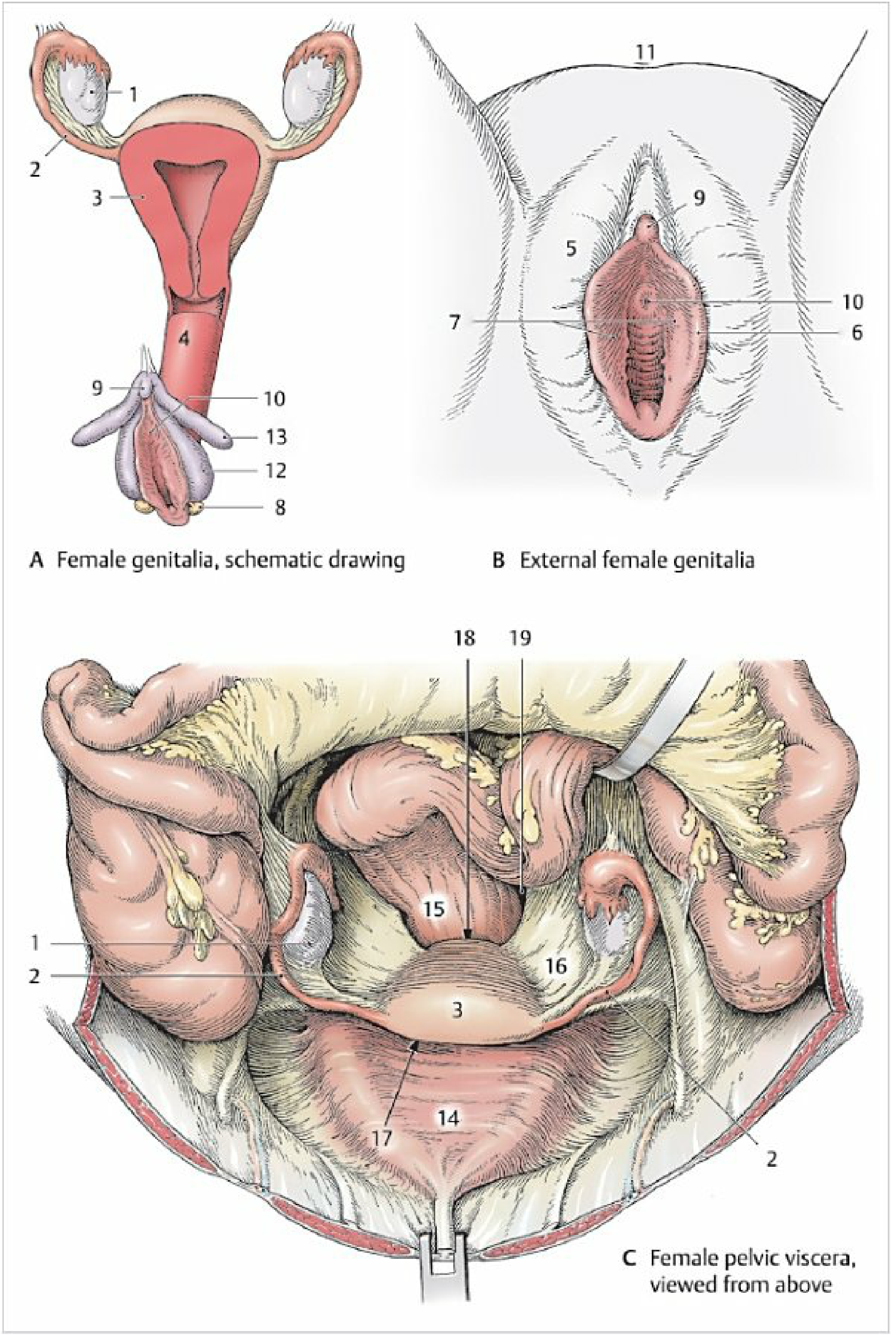
The Pouch of Douglas (formally called the rectouterine pouch) is a peritoneal pocket - a bag-shaped extension of the peritoneum - located between the posterior wall of the uterus and the front of the rectum in females. It is named after the Scottish anatomist James Douglas (1675-1742).
It represents the deepest point of the peritoneal (abdominal) cavity in women, making it anatomically and clinically very important.
Anatomy & Boundaries
- Anterior: posterior surface of the uterus and the posterior fornix of the vagina
- Posterior: anterior wall of the rectum
- Lateral: rectouterine folds (C19), which contain the sacrouterine ligaments and the inferior hypogastric nerve plexus
Because it is the most dependent (lowest) part of the peritoneal cavity, fluid naturally drains and pools here by gravity.
- Color Atlas of Human Anatomy, Vol. 2, p. 443
Diagram
Clinical Significance
Because it is the lowest point of the abdominal cavity, it is clinically important in several ways:
| Condition | How the Pouch Is Involved |
|---|---|
| Ascites | Free fluid collects here first |
| Pelvic abscess | Pus from abdominal diseases (e.g., appendicitis, diverticulitis, Crohn disease) pools here, causing swinging fever, rectal mucous discharge, and urinary frequency |
| Ectopic pregnancy rupture | Blood pools in the pouch |
| Endometriosis | Fibrotic plaques form and can obliterate the pouch, sometimes involving the rectosigmoid colon |
| Drop metastases | Cancer cells from ovarian, colonic, or other pelvic tumors seed here |
| Peritoneal dialysis | Catheter tip is often placed here for fluid drainage |
- Pye's Surgical Handicraft, 22nd ed.
- Grainger & Allison's Diagnostic Radiology
Clinical Procedures
- Culdocentesis - needle aspiration of fluid through the posterior vaginal fornix into the pouch; used to diagnose ruptured ectopic pregnancy or pelvic abscess
- Culdoscopy - endoscopic examination via the same route
- Digital rectal examination - the pouch can be palpated through the rectum; tenderness suggests pelvic peritonitis or a collection
- Surgical entry - in colorectal and gynecological surgery (e.g., the Altemeier perineal proctosigmoidectomy), the surgeon deliberately enters the pouch anteriorly to begin pelvic dissection
In Males
The equivalent space in males is called the rectovesical pouch - located between the bladder and the rectum. It has less clinical prominence but also acts as a dependent collection space. The two layers of Denonvilliers' fascia (behind the prostate) are embryologic remnants of the pouch of Douglas that extended down to the urogenital diaphragm.
- Smith and Tanagho's General Urology, 19th ed.
In short, the Pouch of Douglas is a gravity-dependent peritoneal recess that acts as a natural sump for the pelvis - making it a critical landmark in gynecology, general surgery, and clinical examination.
This is a shared conversation. Sign in to Orris to start your own chat.