
I am 2nd year medical student i want you to make notes for reproductive system histology
Which reproductive system do you want covered? · What format works best for your notes?
seminiferous tubule spermatogenesis histology
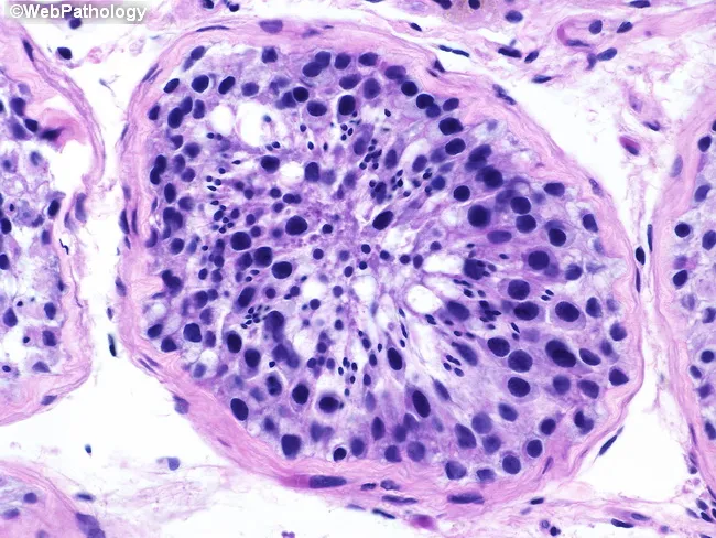
Histology, Testis, Seminiferous tubule, Hematoxylin and Eosin stain, Brightfield microscopy; Cross-sectional view of a seminiferous tubule demonstrates orderly spermatogenesis with a basal germ cell compartment containing spermatogonia, progressing through primary and secondary spermatocytes, round and elongated spermatids, to mature spermatozoa in the tubule lumen. The germinal epithelium exhibits a concentric arrangement along the basement membrane with Sertoli cells interspersed, providing nourishment and structural support. Notable features include intact basement membrane, high cellular density, and a full spectrum of germ cell stages, indicating active spermatogenic activity and normal testicular histology. Pathology domain features: tissue architecture; cellular morphology; luminal maturation; presence of multiple germ cell types at defined maturation stages. Diagnostic significance lies in confirming fertility potential and serving as a baseline for infertility workups or post-therapy monitoring. Differential considerations include maturation arrest and Sertoli cell-only patterns if germ cells are absent or arrested. Clinically relevant for urology, andrology, reproductive endocrinology, and pathology education; suitable for radiology-pathology correlation, histology textbooks, and medical education.
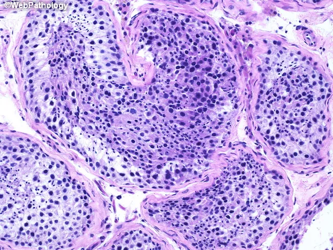
This histopathology image depicts a testicular biopsy prepared for light microscopy. Using Hematoxylin and Eosin (H&E) staining, the tissue reveals multiple cross-sections of seminiferous tubules with preserved architecture and active germ cell maturation. The tubules show intact basal membranes and orderly germinal epithelium containing all stages of spermatogenesis. Within several tubules, spermatogonia lie adjacent to the basement membrane, followed by primary spermatocytes in prophase I, round and elongated spermatids, and mature spermatozoa occupying tubule lumina in places, consistent with complete spermatogenic effort. Sertoli cells provide supportive scaffolding, appearing columnar with prominent nuclei. Interstitium between tubules contains connective tissue and rare Leydig cells without overt inflammation or fibrosis. Overall, there is no evidence of tubular sclerosis, hyalinization, maturation arrest, or neoplastic processes. The image emphasizes normal histology with germ cells at multiple maturation stages and intact tubule integrity. Clinically, such a biopsy supports normal spermatogenic function and fertility potential, assuming no systemic hormonal disturbances. The finding argues against major gonadal causes of infertility such as Sertoli cell-only syndrome or spermatogenic failure. In practice, results correlate with normal semen parameters and typical serum testosterone, FSH, and LH profiles, guiding management toward non-testicular etiologies if infertility persists. This description highlights the diagnostic significance of preserved spermatogenesis in evaluating male fertility and testicular pathology.
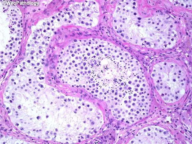
This histopathology image is a Hematoxylin and Eosin (H&E) stained light microscopy section of testicular tissue, illustrating intratubular germ cell neoplasia in situ (GCNIS) alongside a morphologically normal seminiferous tubule showing active spermatogenesis. The specimen is testicular parenchyma, prepared as a paraffin-embedded section, with transverse sections through multiple tubules. The central tubule demonstrates ongoing germ cell maturation and spermatogenesis, characterized by orderly germ cells and prominent spermatogenic cords. Surrounding tubules, however, contain neoplastic germ cells confined to the tubule basement membranes, consistent with GCNIS; these cells typically exhibit enlarged, relatively uniform nuclei, increased nuclear-to-cytoplasmic ratio, and conspicuous nucleoli, with disruption of normal maturation. Notably, there is no invasion into the interstitium or vasculature evident in this field. Clinically, GCNIS is a premalignant lesion with a high propensity to progress to invasive testicular germ cell tumors, especially when bilateral involvement is present. Management varies: unilateral GCNIS commonly necessitates orchiectomy, whereas bilateral disease may be treated with low-dose external beam radiotherapy to preserve testicular tissue and hormonal function. The image underscores the importance of correlating histology with clinical strategy, surveillance, and fertility considerations in germ cell neoplasia. Further histologic context includes assessment for invasion and ancillary markers as clinically indicated.
ovarian follicle development histology graafian corpus luteum
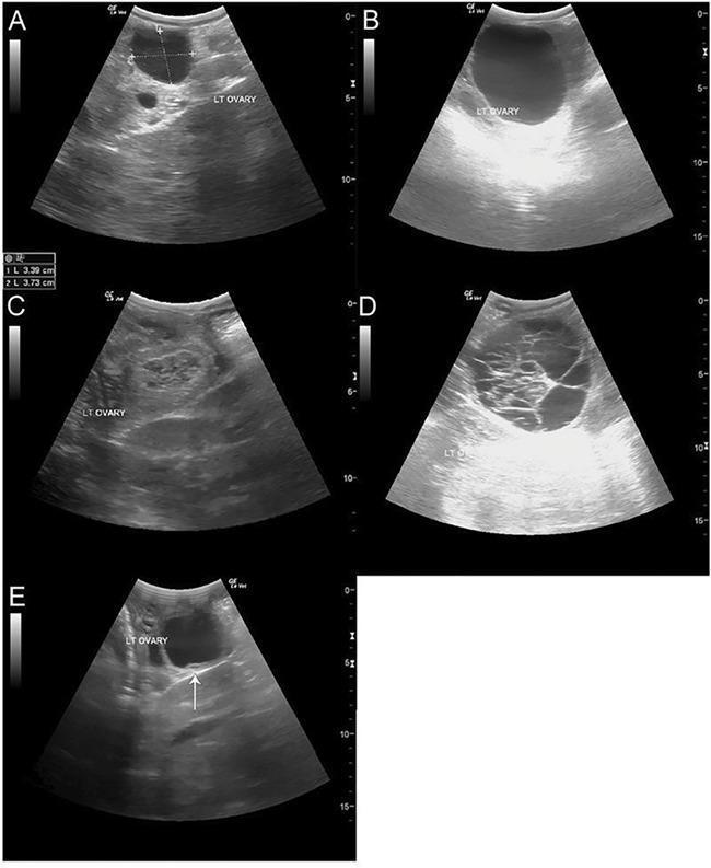
A series of five transabdominal ultrasound images (A-E) illustrating various stages and pathologies of ovarian follicular development. (A) Shows a dominant, pre-ovulatory follicle on the left ovary, measuring approximately 3.39 x 3.73 cm, displaying a characteristic hypoechoic, fluid-filled center. (B) Displays a significantly enlarged dominant follicle (approximately 60 mm) that regressed without ovulating. (C) Depicts a corpus luteum (CL) characterized by a heterogeneous, more echogenic, and solid-appearing internal texture compared to fluid-filled follicles. (D) Illustrates a large hemorrhagic anovulatory follicle (HAF) with a complex internal architecture containing multiple hyperechoic fibrous or echogenic bands and septations. (E) Shows an unstimulated atretic follicle; a white arrow highlights a distinctive ultrasound sign of atresia where the granulosa cell layer has separated or lifted from the follicle wall. These images provide diagnostic criteria for monitoring ovulation induction, distinguishing between functional luteal tissue and anovulatory structures.
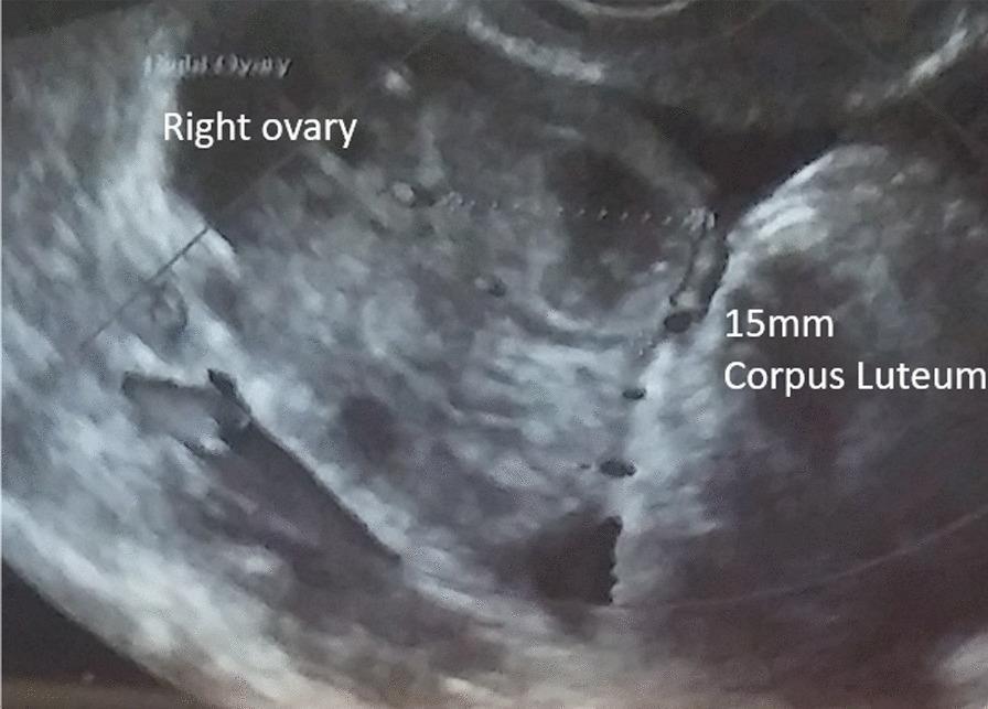
This diagnostic image is a transvaginal ultrasound of the right ovary, demonstrating post-ovulatory changes consistent with follicle rupture. The central finding is a 15mm corpus luteum, which appears as a hypoechoic (darker) cystic structure with irregular, somewhat thick walls and internal echoes, located within the heterogeneous ovarian stroma. The surrounding ovarian tissue exhibits a characteristic grainy echotexture. This imaging modality is used in reproductive medicine and gynecology for follicle tracking during ovulation induction or IVF/ICSI cycles. The visual transition from a clear, fluid-filled mature follicle to this collapsed, irregularly shaped corpus luteum confirms that ovulation has occurred. The image is annotated with labels identifying the 'Right ovary' and the '15mm Corpus Luteum,' making it an essential educational resource for understanding sonographic signs of the menstrual cycle's luteal phase.
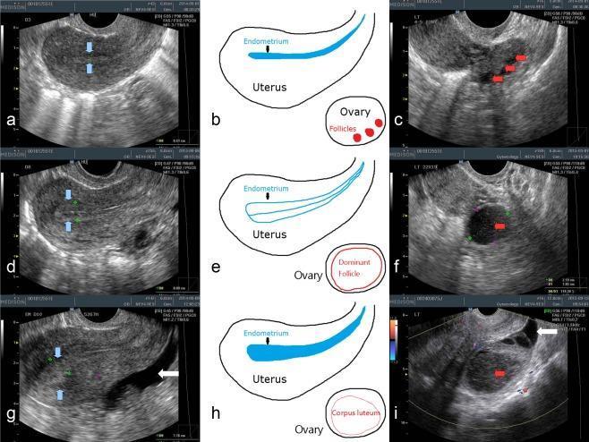
This composite figure illustrates transvaginal ultrasonography (TVUS) findings of the uterus and ovaries throughout the menstrual cycle, organized into three phases: early follicular (a-c), late follicular (d-f), and post-ovulatory luteal (g-i). In the early follicular phase, the endometrium is thin and homogenous (blue arrows), with multiple small antral follicles (red arrows) visible in the ovary. The late follicular phase displays a thickened endometrium with a characteristic 'triple-line' pattern (d) and a large, hypoechoic dominant follicle approximately 2 cm in diameter (f). The luteal phase shows a 'luteinized' endometrium with increased echogenicity (g) and the transformation of the dominant follicle into a corpus luteum (i), characterized by an inhomogenous internal structure and filament-like contents. Anechoic free fluid in the pelvis, indicative of recent ovulation, is highlighted by white arrows in the late follicular and luteal phase images. Schematic diagrams (b, e, h) provide anatomical context for the sonographic changes in endometrial morphology and ovarian follicle development.
uterus endometrium histology menstrual cycle phases
This composite figure illustrates transvaginal ultrasonography (TVUS) findings of the uterus and ovaries throughout the menstrual cycle, organized into three phases: early follicular (a-c), late follicular (d-f), and post-ovulatory luteal (g-i). In the early follicular phase, the endometrium is thin and homogenous (blue arrows), with multiple small antral follicles (red arrows) visible in the ovary. The late follicular phase displays a thickened endometrium with a characteristic 'triple-line' pattern (d) and a large, hypoechoic dominant follicle approximately 2 cm in diameter (f). The luteal phase shows a 'luteinized' endometrium with increased echogenicity (g) and the transformation of the dominant follicle into a corpus luteum (i), characterized by an inhomogenous internal structure and filament-like contents. Anechoic free fluid in the pelvis, indicative of recent ovulation, is highlighted by white arrows in the late follicular and luteal phase images. Schematic diagrams (b, e, h) provide anatomical context for the sonographic changes in endometrial morphology and ovarian follicle development.
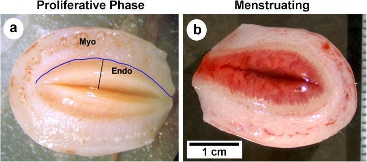
A side-by-side comparison of two macroscopic cross-sections of a rhesus macaque uterus, demonstrating physiological changes during the menstrual cycle. Image (a) represents the proliferative phase (day 14), showing a thickened endometrium (Endo) with a pale tan-to-pink hue. A clear blue line highlights the smooth, well-defined endometrial-myometrial border, with the underlying myometrium (Myo) clearly visible. Image (b) represents the second day of the menstrual phase, where the endometrium appears significantly thinner due to tissue sloughing and breakdown. In this phase, the upper third of the endometrium (functionalis zone) is visibly engorged with blood, exhibiting a deep red, hemorrhagic appearance, while the basalis layer remains intact. The endometrial-myometrial junction in the menstruating specimen is less distinct compared to the proliferative phase. A 1 cm scale bar is provided for reference. This comparison illustrates the cyclic changes in endometrial thickness, vascularity, and tissue integrity characteristic of menstruating primates.
prostate gland histology corpora amylacea
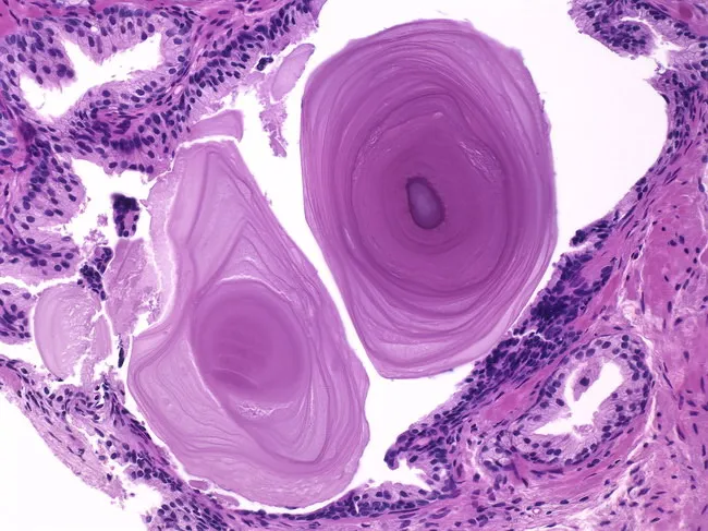
Imaging modality: Light microscopy of Hematoxylin and Eosin stained prostatic tissue section from a biopsy or surgical specimen. The slide shows corpora amylacea: round to oval, lamellated, laminated bodies within the luminal spaces of prostatic glands. These acellular, eosinophilic concentric rings encircle a central core. They vary in size and color, appearing pink to purple to golden brown depending on staining and illumination. Originates from layers of inspissated secretions deposited around shed acinar epithelial cells. In the surrounding stroma, prostatic glands show benign features with intact architecture; no infiltration or atypia is observed. The corpora amylacea are typically incidental and benign; they may be seen in aging prostates and with chronic prostatitis; their presence does not imply malignancy. Histologically, corpora amylacea can form within lumens of acini and ducts, representing late secretory products that calcify over time. The diagnostic significance is mainly incidental; the differential includes calcifications and other laminated concretions; correlation with clinical context and age is recommended. This image is relevant for teaching prostatic histology, aging changes, benign prostatic pathology, and differentiation from neoplastic processes in the prostate. Potential clinical uses include educational case discussions, pathology lab QA, and reference for identifying corpora amylacea in prostatic biopsies.
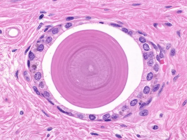
This brightfield light microscopy image presents a histologic cross-section through prostatic glandular tissue, highlighting a prominent corpora amylacea in the central acinar lumen. The central portion exhibits radiating, laminated rims that extend outward, producing concentric bands with a bull’s-eye appearance. Surrounding the laminated core are circular to oval prostatic acini lined by benign cuboidal epithelium and supported by fibromuscular stroma. The corpora amylacea are eosinophilic and often PAS-positive; they may undergo calcification to form gritty calculi that can be palpated in radical prostatectomy specimens. The adjacent stroma and epithelial components show no nuclear atypia, mitotic activity, or infiltrative growth, consistent with benign prostatic glands. This structure is not a neoplasm; corpora amylacea are age-related and occur more commonly in benign glands than in carcinoma, though their presence can occasionally complicate interpretation of small foci of atypia. Morphologically, the laminated bodies illustrate chronic, degenerative changes and may serve as incidental histologic markers of aging prostate tissue. Clinically, recognizing corpora amylacea helps avoid misdiagnosing benign calcified bodies as malignant processes and supports age-appropriate differential diagnoses in radical prostatectomy evaluation. Correlation with patient history, PSA values, and imaging findings can further refine the clinical interpretation and minimize unnecessary treatment in routine practice today.
🔬 Reproductive System Histology Notes
PART 1: MALE REPRODUCTIVE SYSTEM
1. Overview
- Testes - spermatogenesis + steroidogenesis
- Excurrent ducts - tubuli recti → rete testis → efferent ductules → epididymis → ductus (vas) deferens → ejaculatory duct → urethra
- Accessory sex glands - seminal vesicles, prostate, bulbourethral glands
- External genitalia - penis, scrotum
2. Testis
Gross/Microscopic Structure
- Covered by tunica albuginea - thick dense irregular connective tissue capsule
- Inner layer = tunica vasculosa - loose CT with blood/lymph vessels
- Posterior thickening = mediastinum testis - where vessels and ducts enter/exit
- Septa divide testis into ~250 lobules, each containing 1-4 seminiferous tubules
- Between tubules = interstitium containing Leydig cells, macrophages, lymphatics
Cells of the Testis - Quick Table
| Cell | Location | Key Features | Function |
|---|---|---|---|
| Sertoli cell | Seminiferous tubule, resting on basement membrane | Elongated pale nucleus + prominent nucleolus; cytoplasm extends to lumen | Support, nourish germ cells; form blood-testis barrier; phagocytose residual bodies |
| Leydig cell | Interstitium (in clusters) | Small round nucleus; eosinophilic cytoplasm; may have Reinke crystals | Produce testosterone |
| Myoid cells | Peritubular (lamina propria) | Smooth muscle-like; form peritubular investment | Peristaltic contraction of tubules |
Key histology point: Sertoli cells are identified by their pale-staining, elongated nuclei with a prominent nucleolus, unlike the round, dark-staining nuclei of spermatogonia.
3. Spermatogenesis
Phases of Spermatogenesis
A. Spermatogonial Phase (Mitotic)
| Cell | Ploidy | Location | Notes |
|---|---|---|---|
| Type A dark spermatogonia | 2n | Periphery | Stem cells; only cells with complete cytoplasmic division |
| Type A pale spermatogonia | 2n | Periphery | Replenish stem cell pool |
| Type B spermatogonia | 2n | Periphery | Last step before entering meiosis |
- All daughter cells (except type A dark) remain connected by intercellular bridges
B. Spermatocyte Phase (Meiosis)
- Primary spermatocytes: 2n, 4d (DNA replicated before meiosis I)
- Prophase I lasts up to 22 days - the longest phase, why most cells seen in tubules are primary spermatocytes
- Crossing-over occurs here → genetic diversity
- Secondary spermatocytes: 1n, 2d (after meiosis I) - short-lived, rarely seen
- Spermatids: 1n, 1d (after meiosis II) - round cells near lumen
C. Spermatid Phase = Spermiogenesis
- Acrosome formation - from Golgi apparatus; contains hydrolytic enzymes (hyaluronidase, acrosin)
- Flagellum development - from centrioles
- Nuclear condensation - chromatin compaction, nucleus elongates
- Cytoplasm loss - residual body phagocytosed by Sertoli cell
- Manchette - microtubule cylinder that shapes the nucleus
Mature Spermatozoon Structure
Head → Neck → Middle piece → Principal piece → End piece
| Part | Contents |
|---|---|
| Head | Nucleus (haploid, 1n) + acrosome cap |
| Neck | Connecting piece, proximal centriole |
| Middle piece | Mitochondrial sheath (energy); axoneme (9+2 microtubules) |
| Principal piece | Fibrous sheath; no mitochondria |
| End piece | Axoneme only; no sheath |
4. Blood-Testis Barrier (BTB)
- Formed by tight junctions between adjacent Sertoli cells
- Divides seminiferous tubule into:
- Basal compartment - spermatogonia; in contact with interstitium
- Adluminal compartment - spermatocytes onward; immunologically privileged
- Prevents immune attack on haploid germ cells (which express novel antigens)
5. Excurrent Duct System
Comparison Table
| Duct | Epithelium | Special Features |
|---|---|---|
| Tubuli recti | Simple cuboidal/Sertoli cells only | Short connecting tubules from lobules |
| Rete testis | Simple cuboidal with single cilium | Network in mediastinum testis |
| Efferent ductules (10-15) | Pseudostratified - alternating tall ciliated + short non-ciliated cells → "scalloped/festooned" appearance | Absorb most testicular fluid |
| Epididymis | Pseudostratified columnar with stereocilia (non-motile; actually microvilli) | Long stereocilia; principal cells + basal cells; sperm maturation/storage |
| Ductus deferens | Pseudostratified columnar with stereocilia | Thick muscularis = 3 layers (inner longitudinal, middle circular, outer longitudinal); peristalsis during ejaculation |
Exam Tip - Efferent ductules vs Epididymis: Efferent ductules = scalloped/uneven luminal border (alternating tall + short cells) Epididymis = even/smooth luminal border (uniform height with long stereocilia)
6. Accessory Sex Glands
Seminal Vesicles
- Paired glands, highly tortuous/folded mucosa with complex papillary projections
- Epithelium: pseudostratified columnar (or simple columnar)
- Secretion: fructose-rich, alkaline fluid (60-70% of semen volume)
- Contains fructose (energy for sperm), prostaglandins, fibrinogen, vitamin C
Prostate Gland
- Surrounds the urethra; ~30 tubuloalveolar glands embedded in fibromuscular stroma
- Epithelium: simple to pseudostratified columnar
- Characteristic feature: Corpora amylacea - concentrically laminated, eosinophilic calcified concretions in glandular lumens (increase with age; benign)
- 3 zones: periurethral (central/transition) → inner mucosal → outer main glands
- BPH = enlargement of transition zone; carcinoma most common in peripheral zone
Bulbourethral Glands (Cowper's Glands)
- Paired; in urogenital diaphragm
- Epithelium: simple columnar mucus-secreting
- Pre-ejaculatory secretion: lubricates urethra; neutralizes acid urine
7. Penis
- 3 erectile bodies: 2 corpora cavernosa (paired) + 1 corpus spongiosum (surrounds urethra)
- Each corpus = cavernous spaces lined by endothelium surrounded by smooth muscle and CT
- Tunica albuginea covers each corpus cavernosum (dense CT; limits expansion)
- Mechanism of erection: NO (nitric oxide) → cGMP → smooth muscle relaxation → blood fills lacunae
PART 2: FEMALE REPRODUCTIVE SYSTEM
8. Ovary
General Structure
- Outer cortex: germinal epithelium (simple cuboidal/columnar) + tunica albuginea beneath → cortical stroma with follicles
- Inner medulla: loose CT, blood vessels, lymphatics
Follicular Development - Summary Table
| Follicle Type | Oocyte Stage | Granulosa Cells | Special Features |
|---|---|---|---|
| Primordial | Primary oocyte (prophase I arrested) | Single layer flattened (squamous) cells | Most abundant; quiescent |
| Primary (unilaminar) | Primary oocyte | Single layer cuboidal cells | FSH-independent growth |
| Primary (multilaminar) | Primary oocyte | Multiple layers; zona pellucida appears | ZP = 3 glycoproteins (ZP1, ZP2, ZP3) |
| Secondary/Antral | Primary oocyte | Multiple layers; antrum forming (follicular fluid) | Theca interna + theca externa develop |
| Graafian (mature) | Primary oocyte (meiosis I not yet complete) | Large antrum; cumulus oophorus + corona radiata | Dominant follicle; ready to ovulate |
Note on naming: "Secondary follicle" in some texts = antral follicle - still contains a primary oocyte (meiosis I not yet complete)
Theca Layers
| Layer | Cells | Function |
|---|---|---|
| Theca interna | Vascularized; secretory cells | Produce androgens (androstenedione) → converted to estrogen by granulosa cells |
| Theca externa | Fibromuscular; no vessels in inner layer | Structural support |
Two-Cell Theory of Estrogen Production
- LH → Theca interna → androstenedione
- FSH → Granulosa cells → aromatase → estradiol
9. Ovulation and Corpus Luteum
Ovulation
- LH surge → dominant Graafian follicle ruptures at stigma
- Released: secondary oocyte (arrested at metaphase II) + corona radiata
- Meiosis II only completes at fertilization
Corpus Luteum
| Feature | Detail |
|---|---|
| Origin | Remnant granulosa + theca cells after follicle rupture |
| Cell types | Granulosa lutein cells (80%; large, pale; from granulosa; secrete progesterone & estrogen) + Theca lutein cells (20%; smaller, darker; from theca interna) |
| Histology | Large folded gland; cells have abundant smooth ER + lipid droplets (steroidogenesis) |
| Fate (no pregnancy) | Degenerates 8-10 days after ovulation → corpus albicans (hyaline scar) |
| Fate (pregnancy) | Maintained by hCG from trophoblast until 3rd month |
Atresia
- Most follicles degenerate before reaching maturity
- Atretic follicles: degenerating oocyte + granulosa cells + glassy membrane persists → eventually replaced by stroma
10. Uterine Tubes (Fallopian Tubes / Oviducts)
Regions (lateral to medial)
Histology
| Layer | Features |
|---|---|
| Mucosa | Highly folded (especially in ampulla - most folds); simple columnar epithelium with ciliated cells (beat toward uterus) + secretory (peg) cells |
| Muscularis | Inner circular + outer longitudinal smooth muscle; main driver of embryo transport via peristalsis |
| Serosa | Peritoneum |
- Fertilization normally occurs in the ampulla
- Ectopic pregnancy most common in ampulla
11. Uterus
Layers
| Layer | Structure |
|---|---|
| Endometrium (mucosa) | Simple columnar epithelium + uterine glands + CT stroma; two zones: basal (permanent) + functional (shed monthly) |
| Myometrium | Thick smooth muscle; 3 poorly defined layers; contains arcuate arteries |
| Perimetrium | Serosa (peritoneum) anteriorly = adventitia |
Endometrium - Two Zones
| Zone | Blood Supply | Changes with Cycle |
|---|---|---|
| Basal layer | Straight arteries (from arcuate aa.) | Stable; not shed; regenerates functional layer |
| Functional layer | Spiral arteries (coiled; progesterone-sensitive) | Proliferates → secretes → sheds |
Key histology: Spiral arteries = hallmark of functional layer; sensitive to progesterone withdrawal → ischemia → menstruation
Menstrual Cycle - Endometrial Changes
| Phase | Days | Dominant Hormone | Endometrial Histology |
|---|---|---|---|
| Menstrual | 1-4 | Progesterone withdrawal | Sloughing of functional layer; hemorrhage |
| Proliferative | 5-14 | Estrogen | Straight/tubular glands; mitoses present; thin compact stroma |
| Secretory | 15-28 | Progesterone | Coiled/tortuous glands; subnuclear glycogen vacuoles (day 16-17); sawtooth gland pattern; decidual change in stroma |
Classic exam Q: Subnuclear glycogen vacuoles = earliest histologic sign of secretory phase (day 16-17)
12. Cervix
- Narrow inferior uterine segment
- Endocervix (canal): simple columnar mucus-secreting epithelium + large branched cervical glands (Nabothian cysts when occluded)
- Ectocervix (portio vaginalis): nonkeratinized stratified squamous epithelium
- Transformation zone (squamocolumnar junction): junction of two epithelia; site of highest risk for cervical carcinoma (HPV infection)
- Cervical mucosa does NOT undergo cyclic shedding (unlike endometrium)
- Cycle changes: mucus composition changes (thin/watery mid-cycle; thick at luteal phase)
13. Vagina
- Wall: nonkeratinized stratified squamous epithelium (rich in glycogen)
- No glands - lubrication from cervical mucus + transudate
- Muscularis: inner circular + outer longitudinal smooth muscle
- Adventitia: dense fibrous CT with elastic fibers
- Doderlein bacilli (Lactobacillus) ferment glycogen → lactic acid → acidic pH → protection
14. Summary Quick-Reference: Female Reproductive Epithelium
| Structure | Epithelium Type |
|---|---|
| Ovarian surface | Simple cuboidal (germinal epithelium) |
| Fallopian tube | Simple columnar (ciliated + secretory) |
| Uterus (endometrium) | Simple columnar |
| Cervix (endocervix) | Simple columnar mucus-secreting |
| Cervix (ectocervix) | Nonkeratinized stratified squamous |
| Vagina | Nonkeratinized stratified squamous |
HIGH-YIELD EXAM POINTS
- Sertoli cells = pale elongated nucleus + prominent nucleolus; form BTB via tight junctions
- Leydig cells = eosinophilic cytoplasm; interstitium; testosterone production
- Efferent ductules = scalloped lumen (uneven height); epididymis = smooth lumen (stereocilia)
- Prostate hallmark = corpora amylacea (laminated concretions)
- Middle piece of sperm = mitochondria (energy); axoneme = 9+2 microtubule pattern
- Ductus deferens = thickest muscular wall (3 layers)
Provide histlogical slides
testis seminiferous tubule Sertoli Leydig cells H&E histology slide
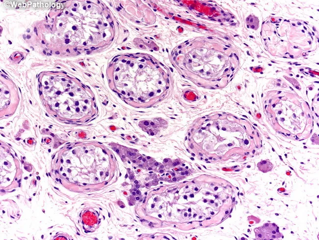
Imaging modality and technique: Light microscopy histology of a cryptorchid testis with splenogonadal fusion. Specimen is gonadal tissue from a testes showing ectopic splenic tissue attachment. The section is a transverse histologic plane stained with Hematoxylin and Eosin (H&E). In this field, seminiferous tubules appear atrophic, with thickened basement membranes and Sertoli cells lining the tubular walls. Germinal epithelium is sparse, and round to elongated nuclei are variably present. Interstitial spaces show clusters of Leydig cells amid fibrous stroma. Overall architecture demonstrates disruption of normal seminiferous tubule integrity, reduced germ cell lineage, and a general paucity of actively proliferating germ cells consistent with cryptorchidism. Accompanying ectopic splenic tissue, a hallmark of splenogonadal fusion, is not readily apparent within the tubules but may be apposed to the gonadal parenchyma in adjacent sections. The pathogenesis involves an early embryologic mis-migration of spleen anlage between week 5 and 8, with later gonadal descent carrying splenic tissue into the testis. Clinically, this histology supports a congenital non-neoplastic anomaly, not a primary neoplasm, and explains potential infertility risk in unilateral or bilateral cryptorchidism. Differential considerations include splenosis, accessory spleen, or heterotopic splenic tissue accompanying the gonad. The image underpins diagnosis, pathophysiology, and surgical decision-making oftentimes.
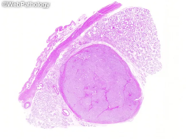
Imaging modality and technique: Hematoxylin and eosin (H&E) stained histopathology slide of testicular tissue examined under light microscopy at low power. The specimen shows a small, well-circumscribed intratesticular mass within the testicular parenchyma. Tunica albuginea is identifiable at the upper left border, establishing an intraparenchymal location. The lesion displays cords and tubule-like structures lined by tall, uniform Sertoli cells with oval nuclei and scant cytoplasm, set in a fibrous to densely collagenous stroma. The tumor margin appears sharp, with preservation of adjacent seminiferous tubule architecture and only mild adjacent tubulopathy. No overt necrosis, hemorrhage, or significant cytologic atypia is seen in this field. These features are characteristic of a Sertoli cell tumor, a sex cord-stromal neoplasm of the testis that is typically small and well circumscribed. Clinically, Sertoli cell tumors may present as a testicular mass and are often benign but can occasionally show malignant potential; differential diagnosis includes germ cell tumors such as seminoma, as well as Leydig cell tumor and fibroma. Immunohistochemistry (if performed) may show Sertoli cell markers (e.g., cytokeratin, inhibin) supporting the diagnosis. This image is valuable for educational and diagnostic context, aiding tumor characterization and surgical planning. Useful for radiology-pathology correlation and tumor typing education.
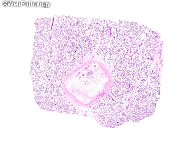
Digital whole-slide histology image of a testicular parenchymal section stained with Hematoxylin and Eosin (H&E), captured as a high-resolution slide to illustrate a partially regressed Leydig cell tumor. The lesion is well-circumscribed and hyalinized, located in the interstitial compartment adjacent to normal seminiferous tubules. The tumor area is largely supplanted by vascularized fibroconnective tissue, producing a prominent fibrous core with few residual clusters of Leydig tumor cells, which exhibit eosinophilic cytoplasm and occasional nuclear atypia, set within a fibrotic stroma. The surrounding tubules appear morphologically normal, with preserved spermatogenic epithelium at the periphery. The histologic pattern is consistent with sex cord-stromal neoplasm of Leydig cell lineage, but with marked regression; immunohistochemical confirmation (inhibin-α, calretinin, Melan-A) would support Sertoli-Leydig differentiation and help exclude germ cell tumors such as seminoma. The diagnostic significance lies in recognizing regression/regenerative changes in Leydig cell tumors, which may mimic fibrous scar or other testicular neoplasms on gross exam and cytology. Differential considerations include germ cell tumors (seminoma, embryonal carcinoma) and Sertoli cell tumors; clinical correlation with hormonal profile and imaging aids management decisions. This image is valuable for education, pathology review, and research on gonadal tumor regression and stromal remodeling.
epididymis histology pseudostratified stereocilia slide
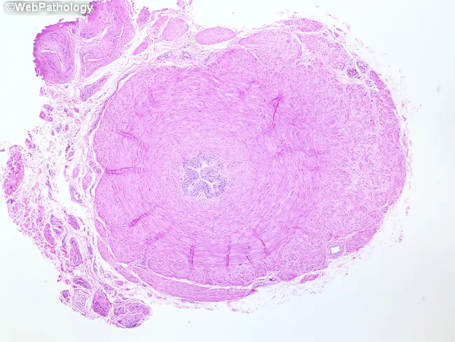
Comprehensive description (approx 200 words): This image depicts a transverse histologic cross-section of the ductus deferens (vas deferens) from the human male reproductive tract, prepared on a hematoxylin and eosin stained slide and viewed with microscopy. The structure is a single, rounded duct with a conspicuously thick muscular wall reflecting the muscularis externa, which in this region comprises inner longitudinal, middle circular, and outer longitudinal muscle layers. The mucosal surface lines a narrow lumen; in classic vas deferens histology, the mucosa is lined by tall pseudostratified columnar epithelium with stereocilia, though the epithelial detail may be partially compressed in this section and not readily visualized. The surrounding stroma shows dense connective tissue and occasional smooth muscle fibers; a well-developed periductal vascular network may be present in the resolved plane. This duct arises embryologically in the male, transports spermatozoa from the epididymis to the ejaculatory duct, and measures roughly 30–40 cm in vivo; histology here emphasizes architecture over content. Clinically, the duct is relevant to fertility and vasectomy planning, since obstruction or scarring can impair sperm transport. This image serves as a reference for normal ductal anatomy, layering, and epithelial morphology, and is useful in educational settings (anatomy, histology, pathology) for learning identification and differential diagnosis within the male reproductive tract.
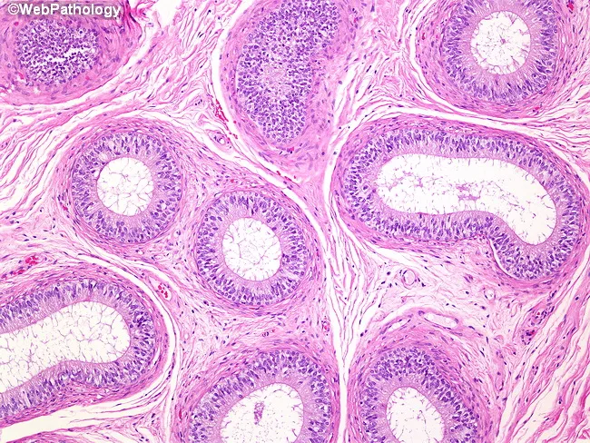
Imaging modality: Light microscopy of a formalin-fixed epididymal tissue section stained with Hematoxylin and Eosin (H&E). Anatomical context: Epididymis of the male reproductive tract, examining tubules that represent the head, body, and tail regions. Core histology: Tubules are lined by tall, pseudostratified columnar epithelium with apical cilia; the epithelial cells include dark-staining columnar cells and basal cells, with occasional clear cells. The lumina are widened and irregular, containing little luminal debris. The tubules sit within a thick tunica muscularis externa composed of multiple concentric smooth muscle layers, which supports peristaltic movement of sperm. Surrounding stroma is relatively loose with fibroelastic connective tissue. The mucosae display intact polarity, with nuclei oriented basally and apical margins bearing dense cilia. This image captures typical epididymal architecture: a specialized tall epithelium, narrow lumens, and a prominent muscular coat essential for sperm maturation, concentration, and propulsion. Pathologic deviations to this pattern would include epithelial desquamation, loss of ciliation, basement membrane disruption, or inflammatory infiltrates. Clinical relevance: normal epididymal histology demonstrates coordinated epithelial activity and smooth muscle contractility; findings aid education in male reproductive pathology, histology teaching, and differential diagnosis of scrotal pathology. In educational databases, this image enhances recognition of epididymal histology.
ovary follicle primordial primary secondary graafian histology H&E
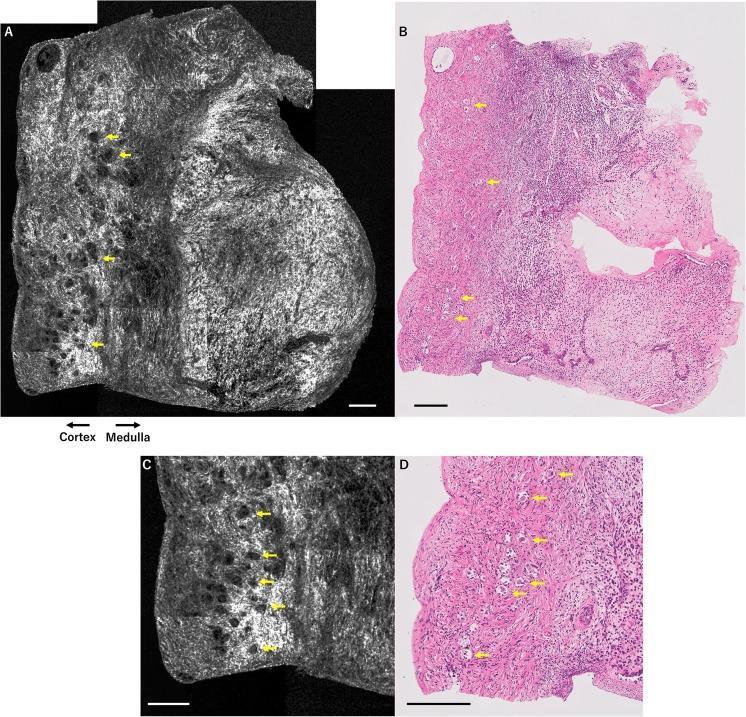
A comparison of human ovarian tissue imaging using two different modalities: Optical Coherence Tomography (OCT) and Hematoxylin and Eosin (H&E) histology. Panel A (OCT) and Panel B (H&E) show low-magnification cross-sections of the ovary, clearly demarcating the outer cortex and inner medulla. In the OCT images (A, C), primordial and primary follicles appear as small, dark, rounded signal-void structures embedded within the hyper-reflective, fibrous ovarian stroma. Panels C and D provide magnified views of these structures. Yellow arrows highlight the direct correlation between the dark circular features in the grayscale OCT images and the corresponding microscopic follicles in the pink/purple H&E-stained sections. This comparison illustrates OCT's utility in non-invasive, real-time assessment of ovarian reserve by visualizing follicle distribution and density within the cortical layer. The scale bars represent 200 micrometers. This material is targeted toward intermediate to advanced medical education in gynecology, reproductive endocrinology, and diagnostic imaging.
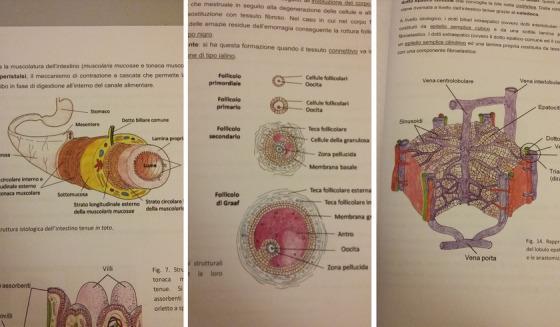
This composite educational resource consists of three histology diagrams. The first diagram illustrates the cross-sectional anatomy of the small intestine, highlighting the concentric layers: mucosa (including villi and lamina propria), submucosa, and the muscularis externa with its circular and longitudinal muscle layers. The second diagram depicts the chronological progression of oogenesis and follicular development in the ovary, identifying the primordial follicle, primary follicle, secondary follicle, and mature Graafian follicle. Key labels include the oocyte, zona pellucida, follicular cells (granulosa cells), theca layers (interna and externa), and the fluid-filled antrum. The third diagram shows a three-dimensional representation of a liver lobule, emphasizing the structural organization of hepatocytes and the microvasculature. It demonstrates the blood flow from the portal triad (interlobular veins and hepatic arteries) through the sinusoids toward the central vein (vena centrolobulare). These illustrations are designed for histology education, focusing on tissue microarchitecture and functional anatomical relationships in the gastrointestinal, reproductive, and hepatobiliary systems.
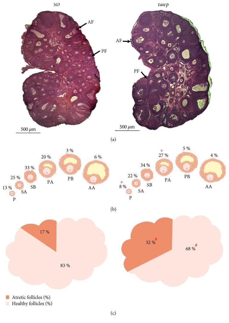
This composite educational graphic compares ovarian follicle populations between Sprague-Dawley (SD) and taiep rat models at the juvenile stage. (a) Histological sections stained with hematoxylin and eosin (H&E) demonstrate the ovarian cortex and medulla. Labels identify antral follicles (AF) with distinct fluid-filled cavities and preantral follicles (PF). The SD rat ovary shows a higher density of primordial and developing follicles compared to the taiep rat. (b) A series of schematic diagrams illustrates the percentages of follicles across development stages: primary (P), secondary (SA, SB), preantral (PA, PB), and antral (AA). Notably, the taiep rat shows a significant decrease in primary follicles (8% vs. 13%) and an increase in preantral A follicles (27% vs. 20%). (c) Comparative pie charts represent follicle health. The SD rat ovary contains 83% healthy and 17% atretic follicles. In contrast, the taiep rat ovary exhibits a significantly higher proportion of atretic follicles (32%) and a reduced percentage of healthy follicles (68%), indicating accelerated follicular atresia in the mutant model.
corpus luteum histology granulosa lutein cells H&E slide
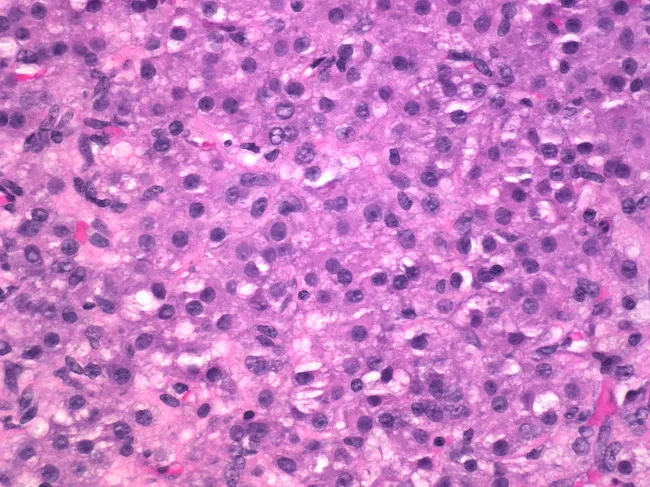
This is a light microscopy image of ovarian tissue, prepared as an H&E-stained section, viewed at high magnification (approximately 400x). The field demonstrates a population of theca-lutein–type cells contiguous within stromal background. The cells are polygonal with abundant eosinophilic cytoplasm and centrally located, round to oval nuclei, with conspicuous nucleoli. Nuclear features include mild pleomorphism and occasional mitotic figures, consistent with steroid-secreting histology rather than a high-grade carcinoma. The cells are arranged in cords and small nests within stromal background; there is no overt necrosis or extensive hemorrhage evident in this field. Background appears relatively uniform with small, occasional intercellular lipid vacuoles not clearly defined. The histologic pattern is compatible with a theca-lutein phenotype commonly seen in luteinized granulosa/theca cell populations; in isolation these features may reflect luteinization associated with corpus luteum formation or hormonal stimulation. Differential diagnoses include thecoma, granulosa cell tumor, steroid cell tumor, Sertoli-Leydig cell tumor, or fibroma-thecoma continuum. Clinically, this morphology is relevant for estrogenic activity or androgenic effects depending on tumor burden and endocrine status. Correlation with patient age, clinical presentation, serum hormone levels, and imaging is advised to establish a precise diagnosis and guide management.
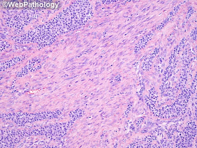
Histopathology, hematoxylin and eosin (H&E) stained slide of ovarian tumor showing fibrothecomatous stroma interposed between cords and sheets of granulosa cells. The tumor displays two components: prominent fibromatous (fibrothecoma-like) stroma and areas of classic granulosa cell proliferation arranged in small solid cords and ill-defined trabeculae. Granulosa cells characteristically exhibit scant cytoplasm and angulated, coffee-bean nuclei with nuclear grooves; occasional Call-Exner bodies (rosette-like microfollicular structures with eosinophilic fluid surrounds) may be present, though in the fibrous zones such features are reduced. The fibrous stroma is variably cellular, with spindle-shaped fibroblasts and collagen deposition; peripherally the diagnostic granulosa cell tumor areas are often evident when the fibrothecomatous component is prominent. The overall pattern is consistent with an adult-type granulosa cell tumor with fibromatous change; this histology can mimic pure fibromas or thecomas if granulosa components are sparse. Immunohistochemical staining typically shows inhibin and calretinin positivity; FOXL2 p.C134W mutation is characteristic but not visible here. Diagnostic significance lies in recognizing the mixed stromal pattern and the granulosa cell cords that support a diagnosis of GCT, which has implications for hormonal activity and potential malignant behavior. Relevance extends to differential diagnosis, surgical planning, and surveillance. This image aids education, pattern recognition, and diagnostic literacy.
uterus endometrium proliferative secretory phase histology glands
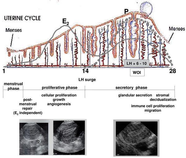
This composite educational graphic illustrates the morphological and hormonal changes of the human endometrium across a standard 28-day uterine cycle. The top section features a pathophysiology diagram showing fluctuations in estradiol (E2) and progesterone (P). E2 peaks during the proliferative phase, correlating with endometrial growth, while P dominates the secretory phase, driving glandular coiling and the 'window of implantation' (WOI). Below the hormone curves, a cross-sectional illustration depicts the histological evolution of endometrial glands and spiral arteries. It transitions from a thin, shedding layer in the menstrual phase to elongated glands during the proliferative phase, and finally to highly coiled, secretory glands with increased angiogenesis in the secretory phase. The bottom section displays three diagnostic ultrasound images of the uterus, providing clinical correlation of endometrial thickness: 2 mm (early proliferative), 11 mm (late proliferative), and 14 mm (mid-secretory). Key physiological processes such as post-menstrual repair, cellular proliferation, angiogenesis, and stromal decidualization are annotated to link hormonal signaling with macroscopic and microscopic changes.
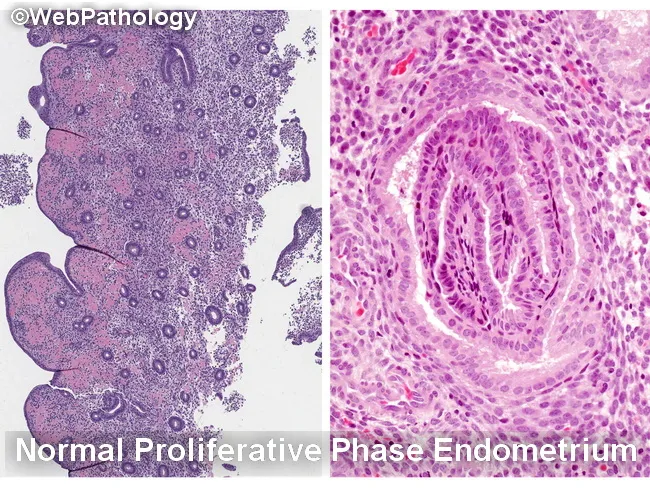
Histology image illustrating the endometrium in two complementary views. The left panel depicts normal proliferative phase endometrium with relatively sparse glands embedded in a cellular stroma, producing a low gland-to-stroma ratio and open gland spacing. The right panel shows characteristic epithelial morphology from proliferative endometrium with elongated, back-to-back glands and prominent, basally oriented nuclei at high power. The specimen emphasizes normal architectural patterns and avoids overcalling hyperplasia when glands are well separated or when fragmentation and telescoping create artifactual crowding. Key differential considerations include endometrial hyperplasia (EH) with or without atypia, disordered proliferative endometrium, reparative or regenerative changes, and endocervical/metaplastic proliferations. Within EH, nonatypical EH must be distinguished from atypical endometrial hyperplasia/endometrioid intraepithelial neoplasia (AEH/EIN), and AEH/EIN must be differentiated from endometrioid carcinoma and from endocervical lesions. Normal proliferative glands show minimal cytologic atypia, whereas AEH/EIN demonstrates architectural complexity (crumpled, crowded glands) and cytologic atypia. Artifacts such as glandular fragmentation, fragmentation- or telescoping-related crowding, and sample artifact can mimic AEH/EIN if the specimen is not intact. This image supports educational differentiation for gynecologic pathology, endometrial biopsy interpretation, and clinical correlation with bleeding patterns and imaging findings. Keywords: endometrium, proliferative phase, hyperplasia, AEH, EIN, histology, biopsy, gland-stroma ratio, artifacts, differential diagnosis teaching.
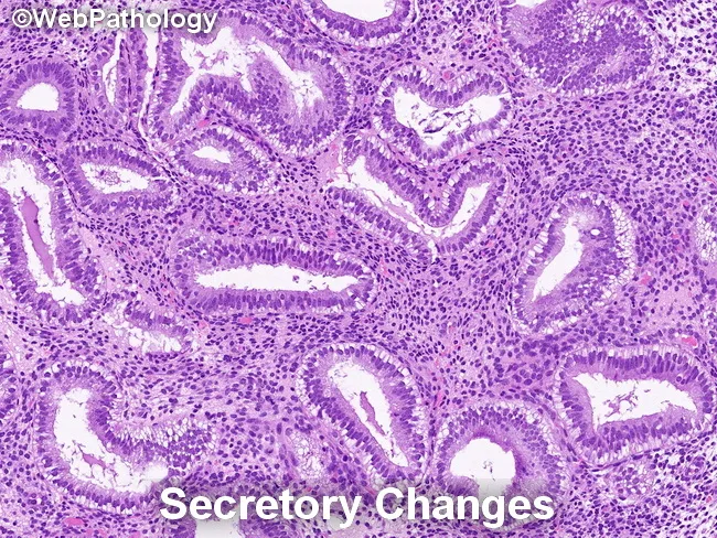
Endometrial histology in secretory phase demonstrated by light microscopy on a hematoxylin and eosin stained endometrial biopsy specimen. Glandular architecture shows irregular serration with gland crowding in focal areas, while glands are lined by tall, columnar secretory epithelium. Nuclei are enlarged and may display mild pleomorphism and loss of polarity, consistent with secretory transformation. The background stroma is edematous and commonly predecidualized, with cytoplasmic vacuolization of stromal cells. Together, these findings reflect physiologic secretory changes rather than overt endometrial hyperplasia. Notably, secretory endometrium can mimic endometrial hyperplasia/endometrioid intraepithelial neoplasia (EIN) in some cases, but the absence of marked architectural crowding, complex glandular patterns, or cytologic atypia argues against a neoplastic process. Differential considerations include secretory change, simple endometrial hyperplasia without atypia, and EIN in a background of secretory endometrium; careful architectural assessment and clinical correlation are essential. Diagnostic significance lies in distinguishing benign secretory transformation from premalignant or malignant entities to avoid overtreatment. This image is educational for pathology training, differential diagnosis exercises, and research on endometrial cycle pathology. Correlate with patient menstrual history, pregnancy status, and cycle timing; repeat sampling or adjunct studies may be warranted when discordance is suspected. Interpret interpretation should integrate histology with clinical data and follow-up.
prostate gland tubuloalveolar histology H&E normal slide
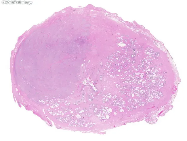
Prostate gland histopathology slide, whole-mount preparation, hematoxylin and eosin (H&E) stained. The primary subject is prostatic adenocarcinoma with a dominant left-sided nodule, in which the normal prostatic architecture is completely effaced by tumor tissue. The right hemiprostate shows relatively preserved benign acini, highlighting focal contrast between malignant and benign glands. The lesion displays infiltrative, irregular glandular structures with neoplastic epithelial cells, showing cytologic atypia and nuclear enlargement in a cribriform to poorly formed gland pattern. The tumor nests invade the stroma and disrupt normal glandular contours, with loss of basal cell layer typical of invasive carcinoma; perineural invasion may be present but not definitively demonstrable at this low magnification. Overall, this image illustrates classic focal prostatic adenocarcinoma within a radical prostatectomy whole-mount, emphasizing potential multifocal disease while presenting a single dominant nodule. The left-sided dominance underscores intra-glandular heterogeneity common in prostate cancer. This histology snapshot is relevant for Gleason grading, risk stratification, and surgical planning, as well as for educational discussion of tumor biology, clonal variation, and the pitfall of conflating malignant foci with benign hyperplasia in limited sampling. Clinically, such patterns influence prognosis and management, including potential focal therapy versus radical intervention.
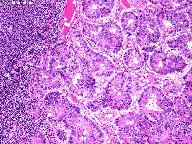
This image depicts a high-magnification bright-field histology slide of a pelvic lymph node effaced by metastatic prostatic adenocarcinoma. The specimen is a paraffin-embedded lymph node section stained with Hematoxylin and Eosin (H&E). Gland-forming malignant epithelial cells infiltrate the nodal parenchyma against a backdrop of residual lymphoid tissue. Tumor glands appear irregular, cribriform or fused, with back-to-back arrangements and desmoplastic stroma. Individual tumor cells show enlarged, hyperchromatic nuclei with conspicuous nucleoli and moderate cytoplasm; nuclear atypia is evident; mitotic activity may be present but not overtly prominent at this field. Lymphoid tissue on the left side remains densely packed with small round cells (reactive lymphocytes), highlighting contrast between neoplastic glands and normal nodes. The architectural invasion into the lymph node supports metastatic spread rather than benign inclusions. Although immunohistochemistry is not shown, prostatic origin is suggested by glandular morphology and is typically confirmed with PSA, PSAP, and NKX3-1 staining. Clinically, nodal metastasis from prostate cancer corresponds to stage N1 and has implications for prognosis and systemic therapy. This image is valuable for education in surgical pathology, oncology, and urology, illustrating the histopathologic hallmarks of metastatic adenocarcinoma in lymphatic tissue and the need for ancillary immunostaining for definitive origin.
fallopian tube oviduct histology ciliated cells ampulla
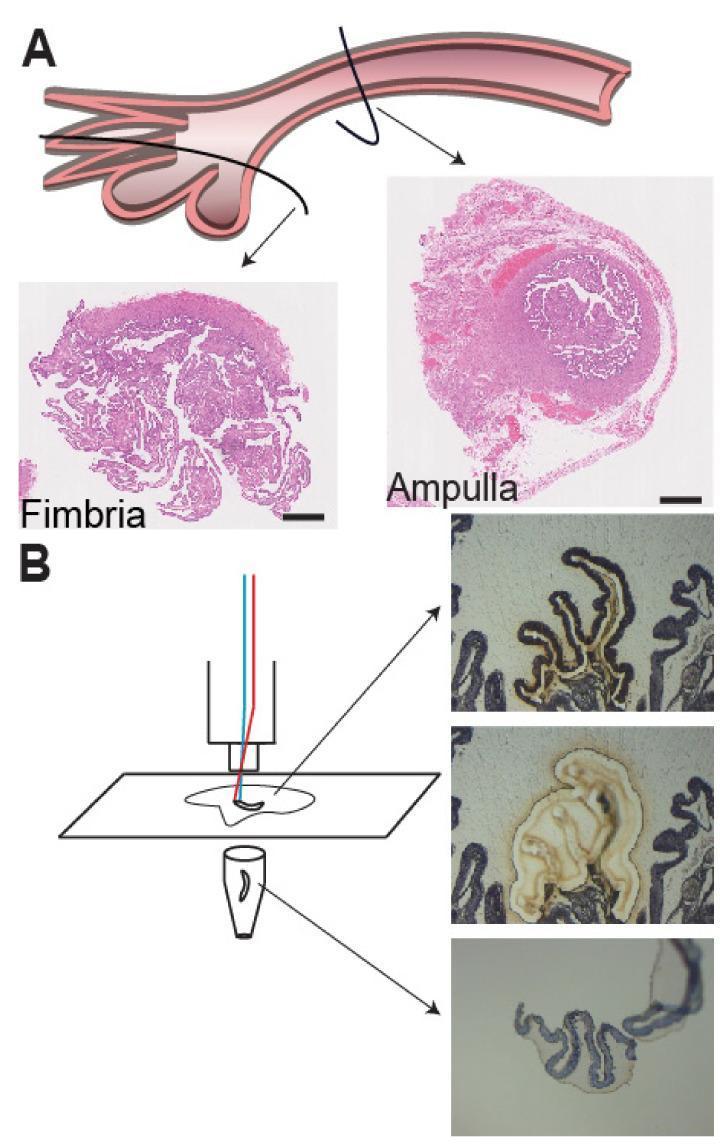
Educational composite image detailing fallopian tube histology and tissue processing. Panel A presents an anatomical diagram of the fallopian tube with arrows indicating two distinct regions: the distal fimbria and the more proximal ampulla. Corresponding Hematoxylin and Eosin (H&E) stained cross-sections are shown for each. The fimbria micrograph displays a highly complex, ruffled architecture with numerous branching papillary projections. In contrast, the ampulla micrograph shows a more defined circular profile with a central lumen and more consolidated wall structure. Panel B illustrates the Laser-Capture Microdissection (LCM) workflow used for RNA isolation. A schematic shows a laser targeting a tissue section on a PEN membrane slide, collecting the sample into a microcentrifuge tube. A series of three sequential images demonstrate the technique: the first shows the initial epithelial fold selected (marked by a dark outline), the second shows the tissue after the laser cut (appearing lighter), and the final image shows the isolated epithelium captured in the collection tube, leaving a void in the original tissue section. Scale bar: 5 mm.
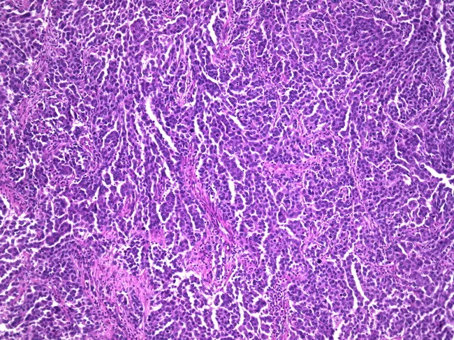
Histopathology examination of fallopian tube tissue demonstrating serous carcinoma. Imaging modality: Histopathology with light microscopy, hematoxylin and eosin (H&E) stained section. Primary site: fallopian tube (oviduct); tubal mucosa involved with neoplastic epithelium. Anatomical context: female reproductive tract, adnexal region; left or right not specified. Visual features: densely packed neoplastic cells forming papillary, cribriform, and glandular patterns with complex architecture; high-grade nuclear atypia, marked pleomorphism, conspicuous nucleoli; abundant mitotic figures; invasion into the underlying stroma; rare psammoma bodies may be observed. Diagnostic categorization: high-grade serous carcinoma of tubal origin (serous carcinoma); consistent with 70% of tubal malignancies. Immunophenotype often shows WT1 positivity in routine practice (not visible in image). Clinical significance: tubal origin serous carcinomas are associated with concurrent ovarian and endometrial malignancies, BRCA mutations, and heightened risk for breast cancer. Differential considerations include metastatic serous carcinoma from ovary or peritoneum; tubal intraepithelial carcinoma (STIC) precursor; endometrioid carcinoma in related contexts. Clinical relevance: histology supports staging and management decisions, including surgical cytoreduction and adjuvant therapy; informs genetic counseling and surveillance for patients and at-risk relatives. Use cases: educational histology references; diagnostic atlases; research on tubal carcinogenesis and BRCA-associated cancers.
cervix squamocolumnar junction transformation zone histology
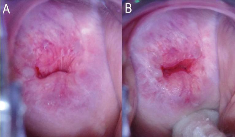
Two side-by-side colposcopic clinical photographs (A and B) demonstrating the assessment of the Transformation Zone (TZ) and Squamocolumnar Junction (SCJ) of the human cervix. In Image A, the cervix shows a pinkish, smooth ectocervix with a partially obscured SCJ, where the upper limit of the junction disappears into the endocervical canal without manipulation. This is categorized as TZ Type 3A because the entire circumference of the SCJ is not visible. In Image B, the same cervix is shown while a cotton-tipped applicator is used to exert pressure on the posterior fornix, successfully exposing the endocervical canal. This maneuver reveals the entire circumference of the SCJ, which is found to extend less than 5 mm into the canal, reclassifying the finding as TZ Type 2A. These images illustrate the importance of clinical manipulation techniques in cervical cancer screening to differentiate between patients eligible for ablative therapy (TZ 1 and 2) versus those requiring excisional procedures (TZ 3).
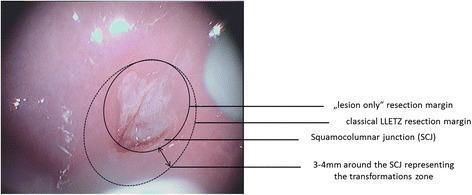
A colposcopic clinical photograph of a cervix showing a comparison of two surgical resection methods for cervical intraepithelial neoplasia (CIN). The image displays an acetowhite dysplastic lesion centered on the squamocolumnar junction (SCJ). Two concentric circular overlays illustrate different surgical margins: a solid line indicates the 'lesion only' resection margin, which closely contours the visible pathology; a larger dashed line represents the 'classical LLETZ' (Large Loop Excision of the Transformation Zone) resection margin, which encompasses the entire transformation zone. The transformation zone is visually defined as extending 3 to 4 mm peripheral to the SCJ. Labels and arrows specify the SCJ, the transformation zone dimensions, and the comparative extent of both excision techniques. This comparison chart serves as an educational tool for gynecological oncology and dysplasia management, highlighting the difference between targeted lesion excision and traditional excision of the whole transformation zone.
🔬 Reproductive System - Histological Slides
MALE REPRODUCTIVE SYSTEM
1. Testis & Epididymis - Gross Histology (Low Power)
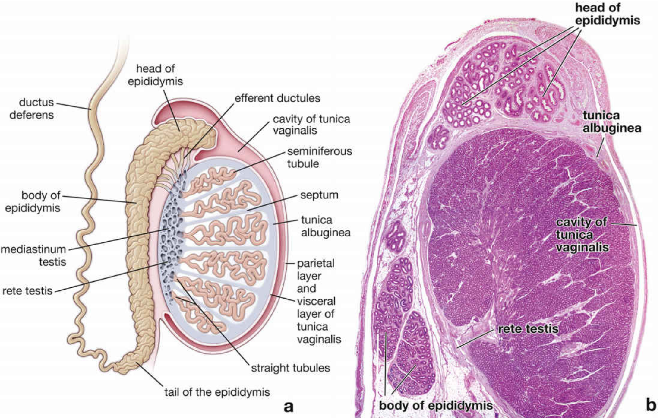
- Tunica albuginea - thick white capsule around the testis
- Seminiferous tubules - coiled tubes packed inside lobules
- Rete testis - network at mediastinum testis (posterior)
- Efferent ductules - upper head of epididymis (small coiled ducts)
- Head, body, tail of epididymis - single coiled duct system along the side
2. Seminiferous Tubules - Active Spermatogenesis (H&E)
- Spermatogonia - small dark cells on the basement membrane (outermost)
- Primary spermatocytes - largest cells, large round nucleus, mid-zone (most numerous - longest phase)
- Spermatids - small cells near the lumen
- Sertoli cells - tall cells with pale elongated nucleus + prominent nucleolus; span the full tubule height
- Leydig cells - clusters in the interstitium between tubules; eosinophilic cytoplasm, round nucleus
- Lamina propria/myoid cells - thin layer around each tubule
3. Epididymis (H&E)
- Pseudostratified columnar epithelium - two rows of nuclei (basal cells below, principal cells above)
- Stereocilia - long non-motile microvilli projecting from principal cells into lumen (very distinctive!)
- Smooth lumen (uniform height unlike efferent ductules)
- Thick muscularis - concentric smooth muscle layers
- Lumen may contain spermatozoa
Exam tip: Efferent ductules have a SCALLOPED/uneven luminal border (alternating tall ciliated + short non-ciliated cells). Epididymis has a SMOOTH even border with long stereocilia.
4. Ductus Deferens (H&E)

- Very thick muscularis - 3 layers: inner longitudinal / middle circular / outer longitudinal
- Narrow lumen - small relative to wall thickness
- Pseudostratified columnar epithelium with stereocilia lining the lumen
- This is the thickest-walled duct in the male reproductive system
5. Prostate Gland - Corpora Amylacea (H&E)
- Tubuloalveolar glands - irregular folded glands within fibromuscular stroma
- Simple to pseudostratified columnar epithelium lining the glands
- Corpora amylacea - concentrically laminated (onion-ring), eosinophilic, calcified concretions inside gland lumens. Hallmark of the prostate - increase with age, benign finding
- Fibromuscular stroma - dense stroma surrounding glands (50% smooth muscle, 50% fibroelastic CT)
FEMALE REPRODUCTIVE SYSTEM
6. Antral (Secondary) Follicle - H&E
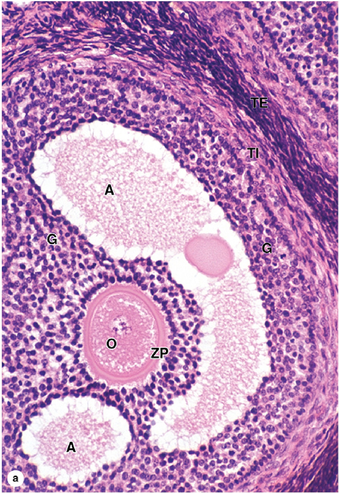
- O = Oocyte (large, pale cell in center)
- ZP = Zona pellucida - pink homogeneous ring around oocyte
- G = Granulosa cells - multiple layers of small dark cells surrounding the oocyte
- A = Antrum - fluid-filled space(s) forming within the granulosa layer
- TI = Theca interna - vascularized, secretory cells immediately outside the follicle
- TE = Theca externa - fibromuscular outer layer
7. Mature (Graafian / Preovulatory) Follicle - H&E
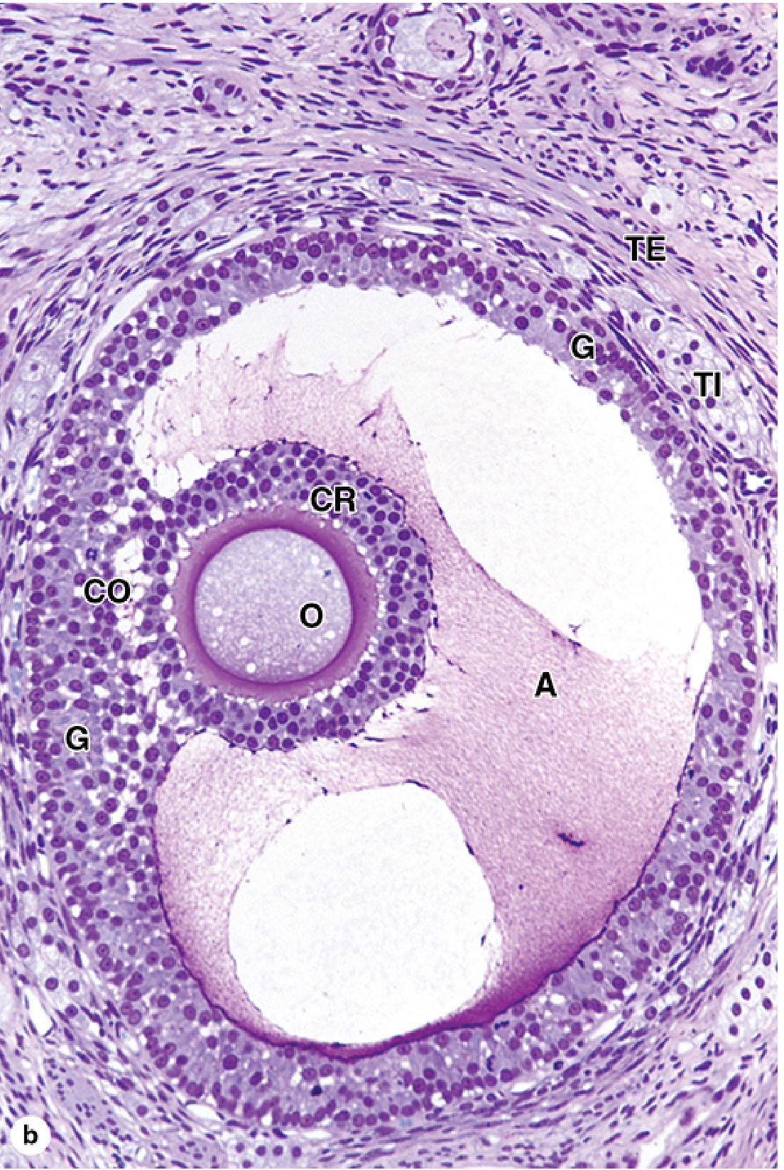
- A = Large single antrum - dominant, fluid-filled; follicle has "one big antrum"
- O = Oocyte - now projects into the antrum, eccentrically placed
- CR = Corona radiata - granulosa cells tightly adherent around the oocyte
- CO = Cumulus oophorus - stalk of granulosa cells connecting oocyte to the follicle wall
- G = Granulosa layer lining the antrum
- TI/TE = Theca interna + externa as before
8. Endometrium - Arterial Supply Diagram (Junqueira's)
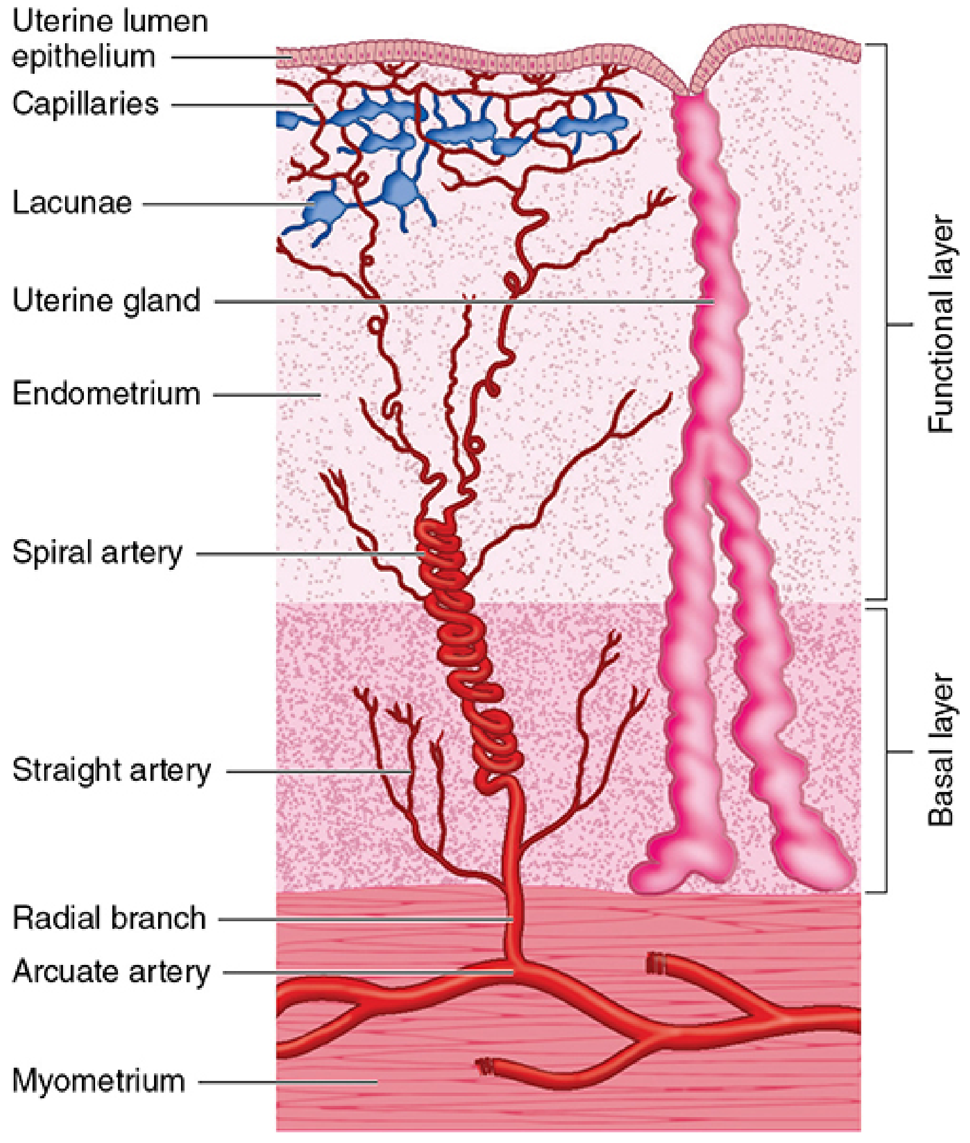
- Myometrium at the base
- Arcuate artery → Radial branches → splits into:
- Straight arteries → supply basal layer only (NOT hormone-sensitive)
- Spiral arteries → coil through functional layer → supply capillaries + lacunae
- Uterine glands (pink, tortuous) extend through both layers
- Functional layer = shed during menstruation (when spiral arteries constrict)
- Basal layer = regenerates the functional layer every cycle
9. Endometrium - Proliferative Phase (H&E)
- Tubular, straight glands (not tortuous yet)
- Pseudo-stratified columnar epithelium with basally located nuclei
- Mitotic figures in gland cells (estrogen-driven proliferation)
- Dense cellular stroma with low gland-to-stroma ratio
- Thin endometrium overall
10. Endometrium - Secretory Phase (H&E)
- Tortuous/coiled glands with a "sawtooth" or "corkscrew" profile
- Subnuclear glycogen vacuoles (early secretory, days 16-17) - pale vacuoles between nucleus and basement membrane
- Intraluminal secretion - pink material in gland lumens (late secretory)
- Edematous stroma - loose, watery stroma (progesterone effect)
- Predecidual stromal change - stromal cells become large and polygonal near spiral arteries
11. Fallopian Tube - Ampulla (H&E)
- Highly complex folded mucosa - finger-like papillary folds projecting into lumen (most elaborate in ampulla)
- Simple columnar epithelium - two cell types:
- Ciliated cells - have cilia (beat toward uterus - transport ovum)
- Peg cells (secretory cells) - no cilia; secrete nutritive fluid
- Thin muscularis compared to ductus deferens
- The fimbriae (infundibulum) have the most complex folds; isthmus has the least
Quick Visual Comparison - Exam Mnemonics
| Slide | Key Visual Landmark |
|---|---|
| Seminiferous tubule | Concentric layers: Spermatogonia → Spermatocytes → Spermatids toward lumen |
| Efferent ductules | Scalloped/uneven lumen (tall ciliated + short non-ciliated cells alternate) |
| Epididymis | Smooth lumen + long stereocilia |
| Ductus deferens | Incredibly thick muscular wall, narrow lumen |
| Prostate | Corpora amylacea (laminated concretions) in gland lumens |
| Primordial follicle | Single layer flat cells around oocyte |
| Primary follicle | Single layer cuboidal cells + ZP appears |
| Secondary follicle | Multiple layers + antrum forming |
| Graafian follicle | Single large antrum + cumulus oophorus + corona radiata |
| Corpus luteum | Large folded gland; pale large granulosa lutein cells (progesterone) |
| Proliferative endometrium | Straight glands, mitoses, compact stroma |
| Secretory endometrium | Tortuous sawtooth glands + subnuclear vacuoles + edematous stroma |
| Fallopian tube | Highly folded mucosa, ciliated cells |