
About back pain and its types from textbook, essentials of orthopaedic and Campbell orthopaedic textbook, 15th edition
Reading File
Now let me search both textbooks for back pain content in parallel.
Finding Sources
Finding Sources
Now let me search specifically in Campbell's and Miller's (Essentials of Orthopaedics) for back pain content:
Finding Sources
Finding Sources
Reading File
Reading File
Reading File
Good - I found a helpful categories table from Miller's. Now let me search for more detailed back pain content in Campbell's (block 20 has selective indications, and I need the main spine chapter):
Finding Sources
Reading File
Now let me get the main back pain chapter from Campbell's (block 20 - this is Chapter 46 on lumbar spine) and more from Miller's:
Reading File
Finding Sources
Excellent - I'm getting rich content from Campbell's block 20 on back pain. Let me continue reading more of that section and also get the discogenic/classification content:
Reading File
Finding Sources
Reading File
I have rich content. Let me also get the Miller's discogenic back pain section and the postoperative back pain section from Campbell's:
Reading File
Reading File
Now let me get more of the Campbell's Chapter 46 main section covering classification and types of back pain, and also fetch the relevant spinal anatomy diagram:
Reading File
Finding Sources
I now have substantial content from both textbooks. Let me also fetch the spine diagram image and get information about specific types of back pain (facet, radicular, stenotic):
Reading File
I now have extensive content from both textbooks. Let me fetch the spine innervation diagram and the MRI disc image to include visually:
Loading Image
Loading Image
I now have comprehensive content from both textbooks with images. Let me compile the full detailed response.
Back Pain and Its Types
Sources: Campbell's Operative Orthopaedics, 15th Edition (2026) & Miller's Review of Orthopaedics, 9th Edition
1. Introduction and Epidemiology
Back pain - specifically axial spine pain - is the most frequent musculoskeletal complaint worldwide. As stated in Campbell's Operative Orthopaedics, 15th Ed (Ch. 46):
- Approximately 80% of individuals are affected by back pain at some point in their lives.
- Impairments of the back and spine are the most frequent cause of activity limitation in individuals younger than 45 years (National Center for Health Statistics).
- The total cost of low back pain in the United States exceeds $100 billion per year - one-third from direct care costs, two-thirds from lost productivity and absenteeism.
- About 90% of patients return to work by 3 months, with most returning within 1 month. Patients off work for 6 months have only a 50% probability of ever returning; at 1 year this drops to 25%.
Axial spine pain - whether cervical, thoracic, or lumbar - is often attributed to disc degeneration, but this degenerative process does not always cause pain. It can lead to several distinct pathologic entities: internal disc derangement, disc herniation, facet arthrosis, degenerative spondylolisthesis, and stenosis.
2. Innervation - Understanding Why the Back Hurts
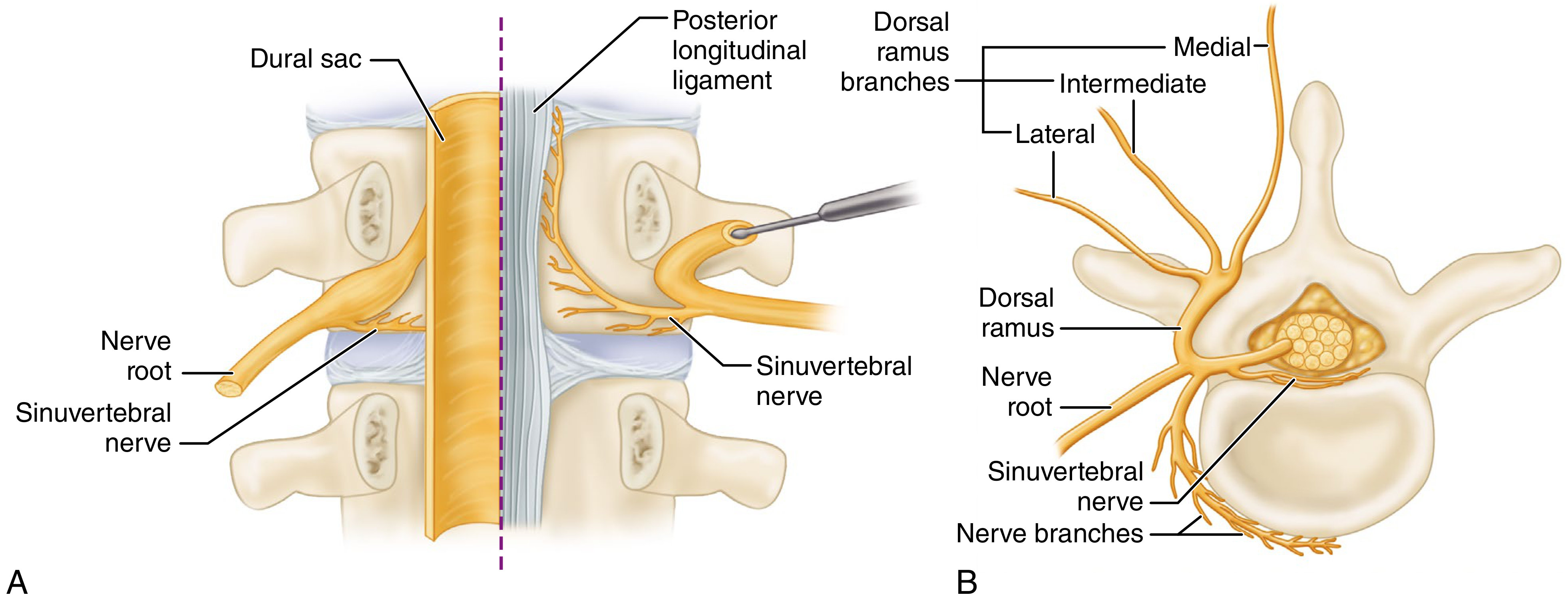
At the intervertebral foramen, three branches arise from the spinal nerve (Campbell's Ch. 46):
- Ventral ramus - supplies all ventral structures
- Sinuvertebral nerve - a recurrent filamentous branch that innervates the posterior disc, vertebral bodies, and posterior longitudinal ligament
- Dorsal ramus - three sub-branches: lateral and intermediate (posterior musculature/skin), and medial branch (innervates facet joints at that level and adjacent levels above and below)
The disc has multilevel, nonsegmental innervation from the index level and two additional superior levels through the sinuvertebral nerves, plus further innervation through the sympathetic trunk. The vertebral endplates are innervated through the basivertebral nerve (a branch of the sinuvertebral nerve) at a density similar to the outer annulus - making endplates as important to pain generation as the annulus itself.
3. Natural History of Disc Degeneration (Three Stages)
Campbell's describes disc degeneration in three stages with distinct findings:
| Stage | Age Group | Features |
|---|---|---|
| Dysfunction | 15-45 years | Circumferential and radial tears in annulus; localized synovitis of facet joints |
| Instability | 35-70 years | Internal disc disruption, progressive disc resorption, facet degeneration with capsular laxity, subluxation, and joint erosion |
| Stabilization | >60 years | Progressive hypertrophic bone around disc and facets leads to segmental stiffening or frank ankylosis |
- Disc herniation is a complication of disc degeneration occurring in the dysfunction and instability stages.
- Spinal stenosis from degenerative arthritis is a complication of bony overgrowth in the late instability and early stabilization stages.
4. Classification of Back Pain by Etiology
From Miller's Review of Orthopaedics, 9th Ed (Table 3.10) - Pediatric/General Framework:
| Category | Specific Conditions |
|---|---|
| Musculoskeletal | Nonspecific back pain/sprain-strain, spondylolysis/spondylolisthesis, fracture, intervertebral disc degeneration or herniation, scoliosis, Scheuermann kyphosis |
| Infectious | Discitis, vertebral osteomyelitis, epidural abscess, sacroiliac septic arthritis, paraspinal abscess, nonspinal infection (pneumonia, pyelonephritis, appendicitis) |
| Inflammatory | Ankylosing spondylitis, psoriatic arthritis, reactive arthritis |
| Neoplastic | Osteoid osteoma/osteoblastoma, aneurysmal bone cyst, neurofibroma, eosinophilic granuloma, leukemia/lymphoma, solid malignancy (Ewing sarcoma, osteosarcoma), primary spinal cord tumors, metastatic disease |
| Miscellaneous | Sickle cell crisis, syringomyelia/tethered cord, idiopathic juvenile osteoporosis, chronic multifocal recurrent osteomyelitis, conversion disorder |
Note: Up to 75-80% of children with low back pain have no identifiable diagnosis.
5. Specific Types of Back Pain (Campbell's & Miller's)
A. Nonspecific / Axial Lumbar Pain
- The most common type - occurs at some point in most people's lives
- Nonspecific axial pain = no identifiable structural pathology; accounts for the majority of presentations
- Management: 1-3 days relative rest, anti-inflammatory regimen, rapid progression to active exercise, expected return to full activity in 4-8 weeks
- Psychosocial factors (depression, job dissatisfaction, anxiety, marital status) are intimately intertwined with complaints and disability
- Risk factors: genetic factors, age, gender, smoking, increased intraabdominal fat, metabolic syndrome
B. Discogenic Back Pain (Miller's Review, Ch. 8)
Pain secondary to intervertebral disc degeneration without other pathologic entities (no instability, fractures, dislocations, or neural compression).
Diagnosis:
- Paucity of physical findings
- Back pain greater than leg pain
- No radiculopathy - absence of tension signs
- Radiographs: may show disc space narrowing or spondylosis but negative for instability
- MRI: decreased signal intensity on T2-weighted images ("dark disc"), with or without annular tear or high-intensity zone (HIZ)
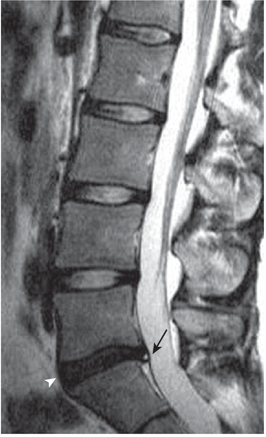
- Discography (controversial): needle placed into disc, concordant pain elicited upon injection confirms that disc is a pain generator; however, evidence suggests annular tears from the needle may accelerate degeneration, so this is falling out of favor
Treatment:
- Almost always conservative
- More than half recover within 1 week; 90% within 1-3 months
- NSAIDs, physical therapy, conditioning
- Patient education about self-limiting nature
C. Disc Herniation / Radicular Pain
- A complication of disc degeneration (dysfunction and instability stages)
- Pain radiates to extremity following dermatomal pattern
- In the lumbar spine, an L4-5 posterolateral disc herniation typically causes L5 nerve root symptoms (because the named root exits below the named pedicle)
- Favorable outcome in most patients on natural history
- Primary surgical benefit noted in first year (SPORT trial), with maintained benefit at 8-year follow-up
- Non-progressive neurologic deficits can be treated nonoperatively; surgery can usually be delayed 6-12 weeks
D. Facet-Mediated (Zygapophyseal Joint) Pain
- Mediated through the medial branch of the dorsal ramus (from Campbell's innervation anatomy)
- Associated with facet arthrosis arising from disc degeneration and secondary loading of facet joints
- Spondylotic changes in foramina result from subsequent facet joint loading (Miller's)
- Can cause segmental instability (spondylolisthesis) from disc collapse and facet incompetence
- Treatment: medial branch blocks and radiofrequency ablation if confirmed
E. Spinal Stenosis
- A complication of bony overgrowth (hypertrophic bone around disc and facet joints) in the late instability and early stabilization stages
- Compromises neural tissue (central canal or foramen)
- Presents as neurogenic claudication - pain with walking, relieved by flexion (sitting/leaning forward)
F. Mechanical Back Pain (Spondylolysis / Spondylolisthesis)
From Campbell's Ch. on Spondylolisthesis: Patients present with symptoms of mechanical low back pain aggravated by high activity levels or competitive sports. Pain is diminished with activity restriction and rest.
G. Postoperative Back Pain
From Campbell's Ch. 3 (Imaging):
In a patient with persistent postoperative back pain, the following must be considered:
- Residual disc material
- Epidural hematoma or abscess
- Discitis
MRI with gadolinium is the key diagnostic tool:
- Scar/fibrosis enhances on T1 post-gadolinium images
- Disc material (in the absence of infection) does not enhance beyond the immediate postoperative period
- The triad of vertebral body endplate enhancement + disc space enhancement + posterior longitudinal ligament enhancement is highly suggestive of postoperative discitis
6. Red Flags for Radiography in Acute Low Back Pain
From Campbell's Box 46.1, imaging is selectively indicated when:
- Age >50 years
- Significant trauma
- Neuromuscular deficits
- Unexplained weight loss (>10 lb in 6 months)
- Suspicion of ankylosing spondylitis
- Drug or alcohol abuse
- History of cancer
- Use of corticosteroids
- Temperature ≥37.8°C (≥100°F)
- Recent visit (≤1 month) for same problem with no improvement
- Patient seeking compensation for back pain
7. Genetic and Risk Factors
Campbell's highlights that the genetic influence on disc degeneration involves multiple gene loci, including variants in:
- Aggrecan gene
- Metalloproteinase-3 gene
- Collagen type IX and alpha 2 and 3 gene forms
Identified risk factors for radiographically apparent disc disorders include: genetic factors, age, gender, smoking, increased intraabdominal fat, and metabolic syndrome. Notably, socioeconomic factors (job dissatisfaction, physically strenuous work, psychologically stressful work, low educational attainment, workers' compensation insurance) play a large role in disability even if not in the disc disorder itself.
Sources:
- Campbell's Operative Orthopaedics, 15th Ed 2026 - Chapters 3 (Imaging), 46 (Axial Lumbar Pain/Disc Degeneration)
- Miller's Review of Orthopaedics, 9th Ed - Chapter 8 (Discogenic Back Pain), Chapter 3 (Pediatric Orthopaedics - Low Back Pain Table 3.10)
This is a shared conversation. Sign in to Orris to start your own chat.