
Placenta formation in slides show in this chat
Reading File
Now let me search the medical library for placenta formation content and images simultaneously.
Finding Sources
Finding Sources
Good, I found key sources. Let me now read the main placenta formation sections from the textbooks, including image-rich content.
Reading File
Reading File
Reading File
Reading File
Excellent! I now have rich content and image URLs. Let me fetch all the key diagrams simultaneously.
Loading Image
Loading Image
Loading Image
Reading File
Reading File
Finding Sources
Reading File
Reading File
I now have all the content I need. Let me fetch the remaining key images.
Loading Image
Loading Image
I now have all the content and images needed for a comprehensive slide-by-slide presentation on placenta formation. Here it is:
🧬 Placenta Formation - Visual Slide Show
Slide 1: Overview - What is the Placenta?
The placenta is a transient, disc-shaped organ that connects the developing fetus to the uterine wall. It serves three core functions throughout pregnancy:
- Nutrition & Gas Exchange - transfers O₂, glucose, amino acids, fatty acids to the fetus
- Endocrine Function - secretes hCG, hPL, progesterone, estrogens
- Immunologic Barrier - protects the semi-allogeneic fetus from maternal rejection
It has both fetal components (trophoblast, chorionic villi) and a maternal component (decidua basalis).
Slide 2: Starting Point - Blastocyst to Trophoblast (Days 4.5-6)
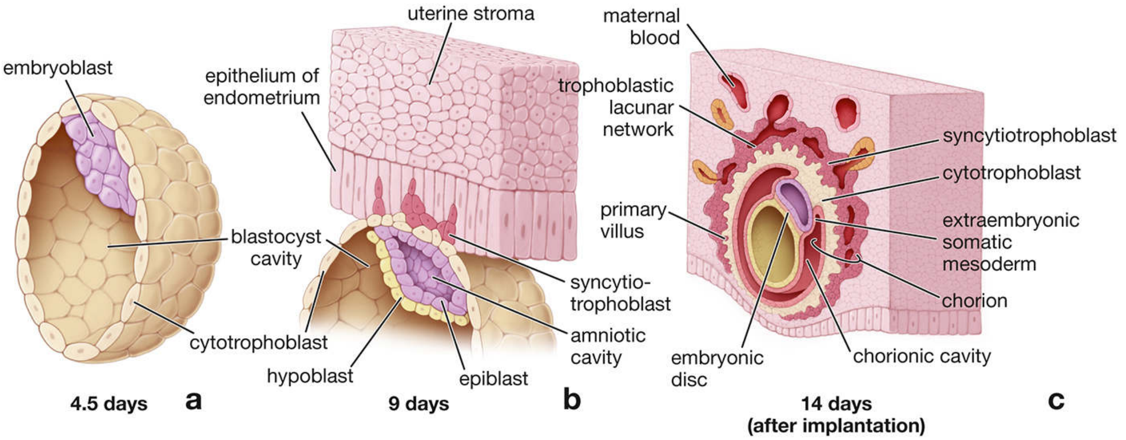
Key events:
- After fertilization, the zygote undergoes cleavage to form a morula (12-16 cells)
- The morula develops a fluid-filled cavity to become the blastocyst
- The blastocyst has two cell populations:
- Inner cell mass (embryoblast) → forms the embryo
- Outer cell mass (trophoblast) → forms the placenta
- The zona pellucida dissolves, freeing the blastocyst for implantation
Source: Histology - A Text and Atlas (Pawlina), p.2251; The Developing Human (Moore)
Slide 3: Implantation - The Window of Opportunity (Days 6-10)
The Implantation Window opens on day 6 after the LH surge and closes by day 10, driven by progesterone and estrogen acting on the endometrium.
Key molecular events at attachment:
- Endometrial epithelium expresses HB-EGF (heparin-binding EGF) in pinopodia
- Polar trophectoderm of blastocyst expresses ErbB1/ErbB4 receptors for HB-EGF
- HB-EGF binding = critical first step of attachment
- Other factors: LIF (leukemia inhibitory factor), HOXA10/HOXA11, WNT signaling
Once attached: trophoblast cells rapidly proliferate and invade the endometrium → the cells differentiate into syncytiotrophoblast (outer) and cytotrophoblast (inner).
Source: Creasy & Resnik's Maternal-Fetal Medicine, p.73
Slide 4: Trophoblast Differentiation Pathway
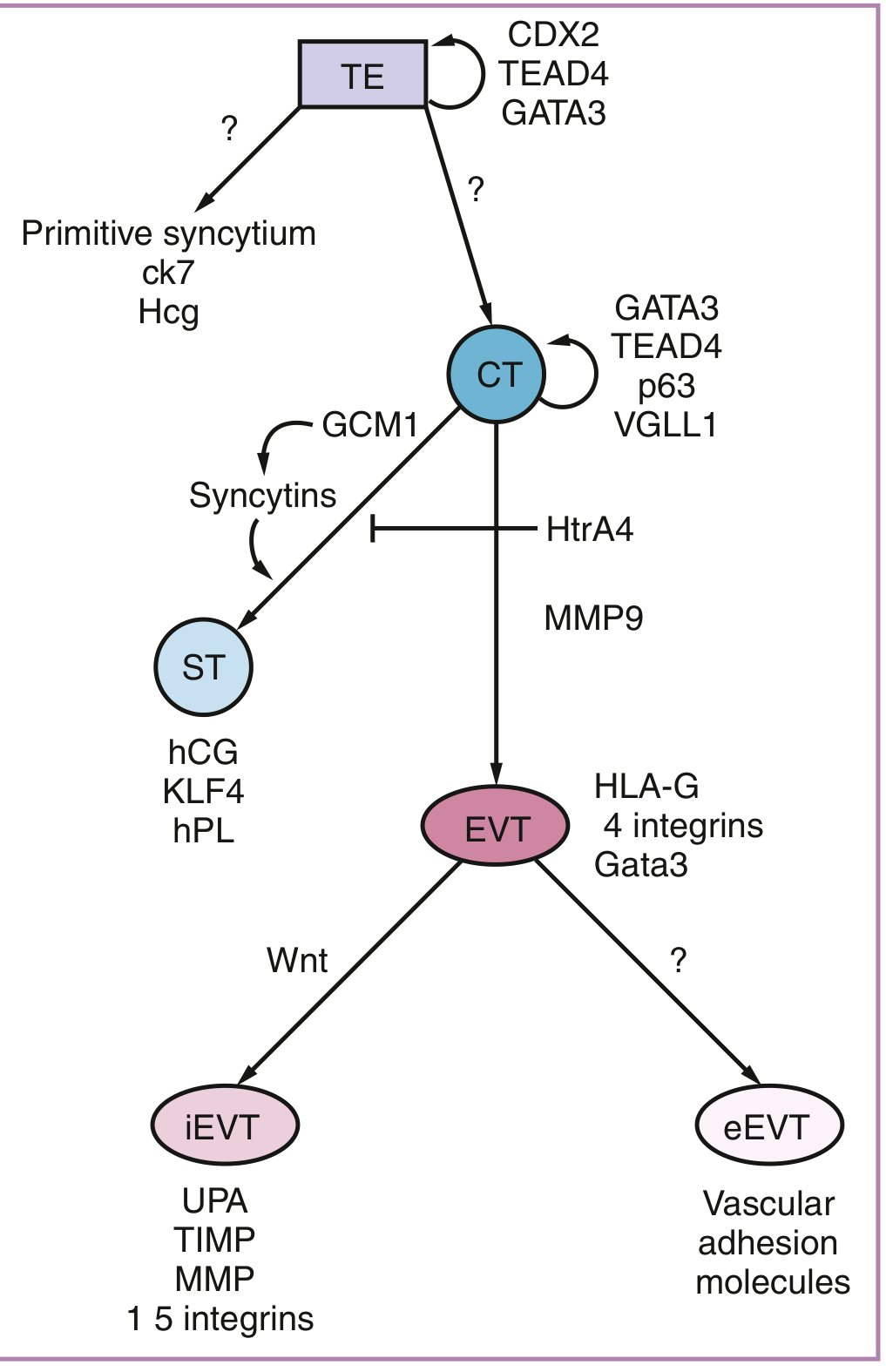
The trophectoderm (TE) gives rise to two main lineages via the cytotrophoblast (CT) progenitor:
| Cell Type | Location | Function | Key Markers |
|---|---|---|---|
| Cytotrophoblast (CT) | Inner villous layer | Progenitor/stem cell | p63, VGLL1, GATA3 |
| Syncytiotrophoblast (STB) | Outer villous surface | Transport, endocrine | hCG, hPL, KLF4 |
| EVT - Invasive (iEVT) | Decidua/myometrium | Anchoring | UPA, TIMP, MMP, integrins |
| EVT - Endovascular (eEVT) | Maternal spiral arteries | Vascular remodeling | Vascular adhesion molecules, HLA-G |
The transition from CT to STB is driven by GCM1 transcription factor, which induces syncytin proteins (captured retroviral envelope proteins) that mediate cell fusion.
Source: Creasy & Resnik's Maternal-Fetal Medicine, p.75-76
Slide 5: Formation of the Intervillous Space (Days 9-13)
At day 9, the syncytiotrophoblast (STB) secretes enzymes that break down surrounding decidual cells and matrix, forming fluid-filled spaces called lacunae. These lacunae:
- Coalesce to form the intervillous space (trophoblastic lacunar network - see diagram above at 9 days)
- Are initially filled with endometrial glandular secretions
- Provide nutrition via histiotrophic nutrition before maternal blood flow begins
The primitive syncytium taps directly into endometrial glands, providing the embryo's nutritional support during weeks 3-10.
Slide 6: Chorionic Villi Development - Three Stages
The chorionic villi are the functional units of the placenta. They develop in three successive stages:
Primary Chorionic Villi (Days 11-13)
- Cytotrophoblast cells form finger-like projections into the trophoblastic lacunae
- Composed only of: outer syncytiotrophoblast + inner cytotrophoblast core
- No mesenchyme, no blood vessels
Secondary Chorionic Villi (Day 16 onward)
- Extraembryonic mesenchyme grows into the primary villi core
- Structure: syncytiotrophoblast → cytotrophoblast → mesenchymal core
- Cover the entire surface of the chorionic sac
Tertiary Chorionic Villi (End of Week 3)
- Mesenchymal cells within the core differentiate into capillaries and blood cells (vasculogenesis)
- Blood vessels connect to the embryonic circulation
- Functional gas/nutrient exchange can now occur
Source: The Developing Human (Moore), p.219; Histology - A Text and Atlas (Pawlina), p.2263
Slide 7: Vascular Remodeling - Spiral Artery Invasion
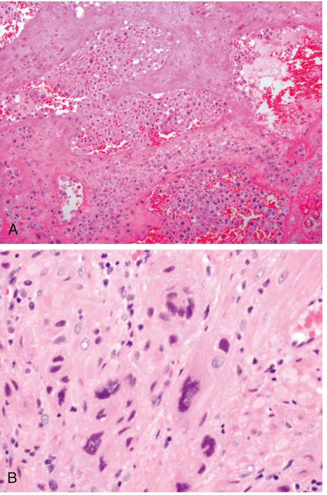
Extravillous trophoblast (EVT) has two key jobs:
-
Interstitial EVT (iEVT): Invade the decidua and superficial one-third of the myometrium to anchor the placenta. They secrete MMPs and plasminogen activators to degrade extracellular matrix.
-
Endovascular EVT (eEVT): Enter maternal spiral arteries, replace the endothelium, and transform muscular high-resistance vessels into wide, low-resistance vessels. Initially they plug these arteries, maintaining low O₂ until ~10-12 weeks.
Why low oxygen early? Hypoxia-inducible factor (HIF) drives EVT differentiation. This ensures proper placental development before high-flow maternal blood arrives.
Immunologic tolerance: EVT express non-classical HLA-G, -C, -E (no classical HLA-A or -B), enabling them to evade maternal NK cell killing.
Source: Creasy & Resnik's Maternal-Fetal Medicine, p.76-77
Slide 8: Histology of the Mature Placenta
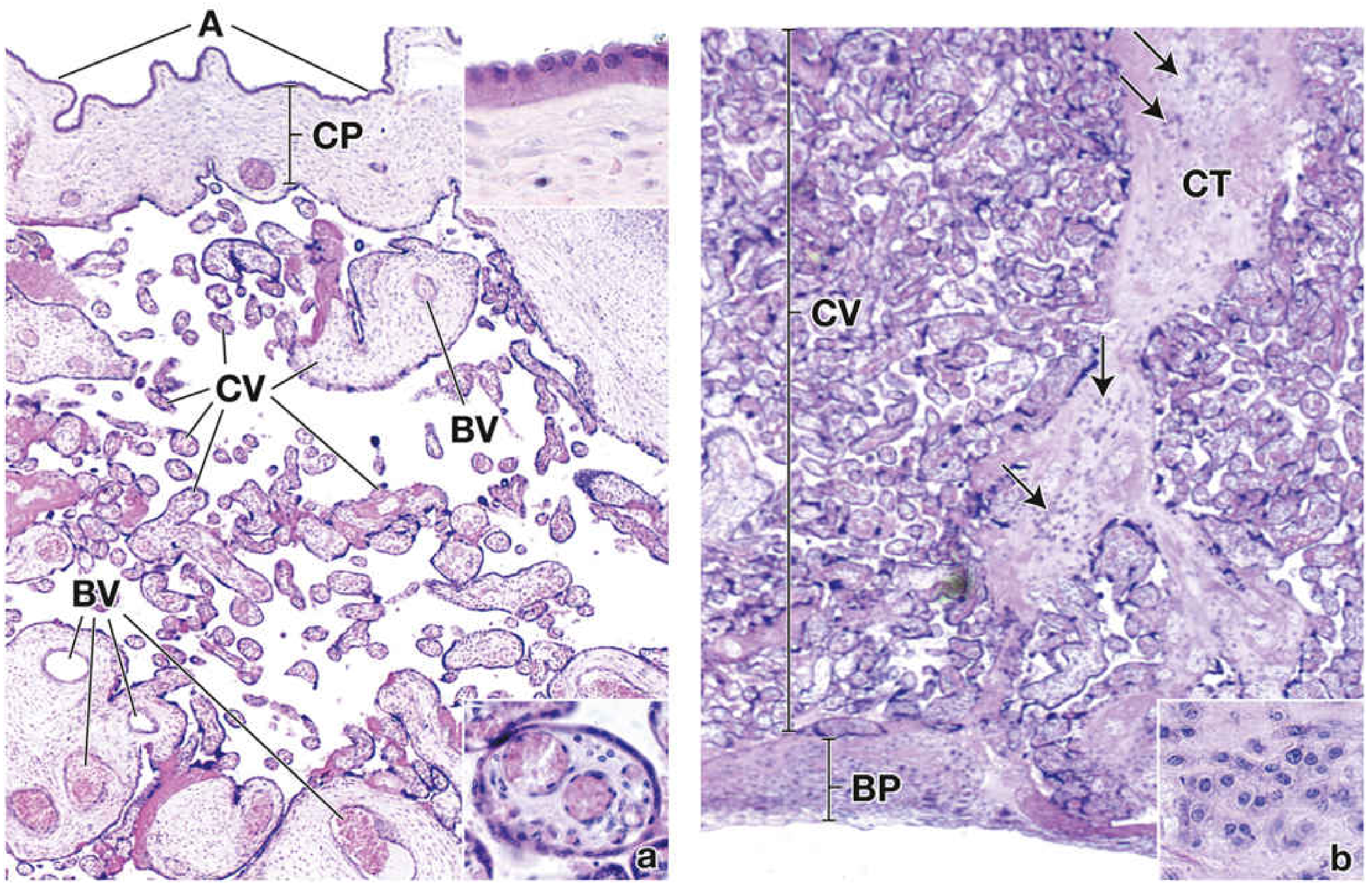
Structures visible histologically:
- Amnion (A): Simple cuboidal epithelium on the fetal surface
- Chorionic plate (CP): Dense connective tissue from which stem villi emerge
- Chorionic villi (CV): Tree-like branching units bathed in maternal blood; the smallest terminal villi carry fetal capillaries for exchange
- Blood vessels (BV): Present in larger villi; capillaries in terminal villi
- Basal plate (BP): Maternal side; anchors villi to the uterine wall
- Decidual cells: Large polygonal cells in the basal plate, arise from endometrial stroma under progesterone influence
In early pregnancy villi are large, edematous, covered by thick layers of both syncytiotrophoblast AND cytotrophoblast.
In late pregnancy the cytotrophoblast layer becomes discontinuous; syncytial nuclei cluster into syncytial knots; more fetal vessels appear.
Source: Histology - A Text and Atlas (Pawlina), p.2263-2264
Slide 9: Anatomy of the Mature Placenta (Late 2nd Trimester)
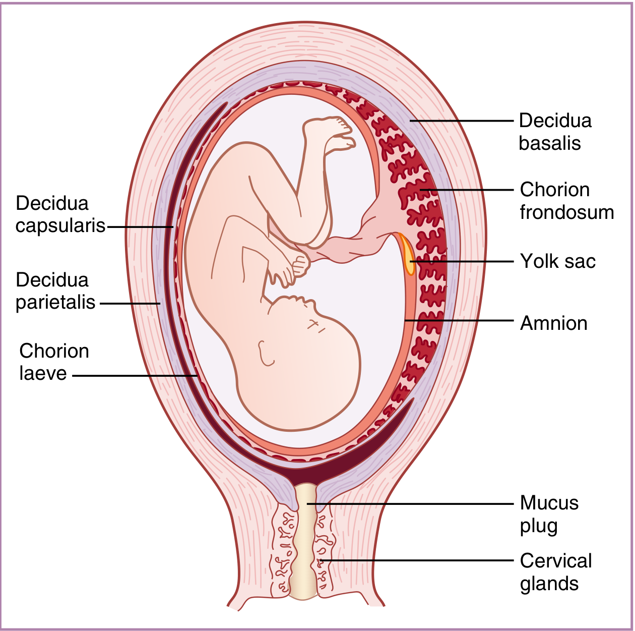
Decidual regions:
- Decidua basalis - beneath the implantation site; forms the maternal placenta
- Decidua capsularis - overlies the gestational sac (fuses with decidua parietalis later)
- Decidua parietalis - lines the rest of the uterine cavity
Chorionic regions:
- Chorion frondosum ("leafy chorion") - the thick, villous side that faces decidua basalis and forms the definitive fetal placenta
- Chorion laeve ("smooth chorion") - the bare, opposite side (villi regress here by week 8-10)
Source: Creasy & Resnik's Maternal-Fetal Medicine, p.74
Slide 10: Summary Timeline
| Time | Event |
|---|---|
| Day 1-3 | Fertilization → zygote → morula |
| Day 4-5 | Morula → blastocyst (ICM + trophoblast) |
| Day 6 | Zona pellucida dissolves; implantation begins |
| Day 7-8 | Trophoblast differentiates into cytotrophoblast + syncytiotrophoblast |
| Day 9 | Lacunae form in syncytiotrophoblast → future intervillous space |
| Day 11-13 | Primary chorionic villi (no mesenchyme, no vessels) |
| Day 14 | Primitive syncytium taps into endometrial glands - histiotrophic nutrition |
| Day 16 | Secondary chorionic villi (mesenchyme invades villi cores) |
| Week 3 | Tertiary chorionic villi (capillaries form in mesenchymal cores) |
| Weeks 4-10 | EVT plug spiral arteries; embryo grows under physiologic hypoxia |
| Weeks 10-12 | eEVT plugs dissolve; maternal blood flows freely into intervillous space |
| Week 16 onward | Chorion laeve forms; chorion frondosum expands |
| Term | Mature placenta ~500g, ~22cm disc, ~2.5cm thick |
Sources:
- Creasy & Resnik's Maternal-Fetal Medicine: Principles and Practice (Figs. 3.3-3.6)
- Histology: A Text and Atlas with Correlated Cell and Molecular Biology (Pawlina), Ch. 23
- The Developing Human: Clinically Oriented Embryology (Moore & Persaud), Ch. 4
This is a shared conversation. Sign in to Orris to start your own chat.