attachment
attachment

Answer for gross lesions of the images referred from ganti sastry and vegad

Reading File
Finding Sources
Finding Sources
Finding Sources
Searching Images

petechial haemorrhage heart cardiovascular gross lesion veterinary
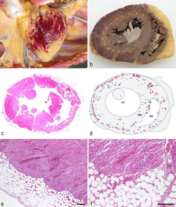
A multi-panel figure illustrating macroscopic and microscopic findings of the heart during autopsy. Figure 1a is a clinical photograph showing the posterior pericardium in situ with marked petechial hemorrhages. Figure 1b displays a cross-sectional gross specimen of the heart after fixation; yellow arrowheads indicate a circumferential black-red discoloration involving the pericardium and the superficial myocardium. Figure 1c provides a whole-mount scanning magnification of a transverse heart section stained with hematoxylin and eosin (H&E). Figure 1d is a schematic diagram mapping the pathology: red dots represent microscopic hemorrhage, blue rectangles indicate microthrombi, and green arcs signify areas of contraction band necrosis. Figures 1e and 1f provide low- and high-power H&E micrographs, respectively, demonstrating extensive hemorrhage within the epicardial fat and the adjacent cardiomyocytes of the myocardium. The pathology is characterized by multifocal microthrombi in small vessels and capillaries at the border of hemorrhagic regions, alongside contraction band necrosis, relevant to cardiovascular pathology and forensic medicine.
A multi-panel figure illustrating macroscopic and microscopic findings of the heart during autopsy. Figure 1a is a clinical photograph showing the posterior pericardium in situ with marked petechial hemorrhages. Figure 1b displays a cross-sectional gross specimen of the heart after fixation; yellow arrowheads indicate a circumferential black-red discoloration involving the pericardium and the superficial myocardium. Figure 1c provides a whole-mount scanning magnification of a transverse heart section stained with hematoxylin and eosin (H&E). Figure 1d is a schematic diagram mapping the pathology: red dots represent microscopic hemorrhage, blue rectangles indicate microthrombi, and green arcs signify areas of contraction band necrosis. Figures 1e and 1f provide low- and high-power H&E micrographs, respectively, demonstrating extensive hemorrhage within the epicardial fat and the adjacent cardiomyocytes of the myocardium. The pathology is characterized by multifocal microthrombi in small vessels and capillaries at the border of hemorrhagic regions, alongside contraction band necrosis, relevant to cardiovascular pathology and forensic medicine.

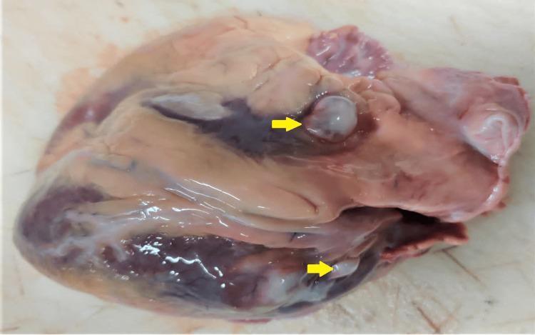
This clinical photograph shows a gross pathological specimen of a human heart during a postmortem examination. The image displays the epicardial surface, with two distinct cystic lesions highlighted by yellow arrows. These lesions appear as smooth, well-circumscribed, translucent to whitish-grey nodules protruding from the myocardium. The superior lesion is located near the base of the heart/left atrium, while the inferior lesion is situated along the ventricular wall. The surrounding cardiac tissue exhibits a mottled reddish-brown appearance with visible coronary grooves and areas of darker venous congestion. There is evidence of epicardial fat and focal pale, fibrous-appearing regions. The pathology is highly characteristic of cardiac cysticercosis, demonstrating the larval stage of Taenia solium (cysticercus cellulosae) embedded within the cardiac structure. This specimen serves as an educational example of parasitic infestation of the cardiovascular system and its macroscopic presentation during autopsy.
This clinical photograph shows a gross pathological specimen of a human heart during a postmortem examination. The image displays the epicardial surface, with two distinct cystic lesions highlighted by yellow arrows. These lesions appear as smooth, well-circumscribed, translucent to whitish-grey nodules protruding from the myocardium. The superior lesion is located near the base of the heart/left atrium, while the inferior lesion is situated along the ventricular wall. The surrounding cardiac tissue exhibits a mottled reddish-brown appearance with visible coronary grooves and areas of darker venous congestion. There is evidence of epicardial fat and focal pale, fibrous-appearing regions. The pathology is highly characteristic of cardiac cysticercosis, demonstrating the larval stage of Taenia solium (cysticercus cellulosae) embedded within the cardiac structure. This specimen serves as an educational example of parasitic infestation of the cardiovascular system and its macroscopic presentation during autopsy.

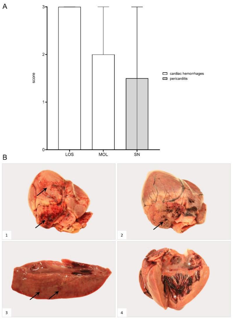
Educational visual regarding cardiac pathology, featuring a bar chart and gross anatomical specimens. Panel A presents a comparison chart of pathology scores (0-3) for cardiac hemorrhages and pericarditis across different geographical cohorts (LOS, MOL, SN), illustrating disease severity. Panel B contains four macroscopic clinical photographs of heart specimens. B1 and B2 demonstrate the epicardial surface with multifocal 'paintbrush-like' to coalescing hemorrhages (arrows), ranging from petechiae to larger ecchymoses. B3 shows a sectioned myocardium with scant, focal hemorrhages (arrows). B4 illustrates the endocardial surface of the opened heart, revealing multifocal endocardial hemorrhages (asterisks) that must be distinguished from post-mortem blood coagulum. The images collectively demonstrate the cardiovascular manifestations of systemic viral infection, such as African Swine Fever Virus (ASFV), focusing on hemorrhagic diathesis and inflammatory cardiac lesions. Key concepts include macroscopic lesion identification, distribution of hemorrhages (epicardial vs. endocardial), and pathological scoring in veterinary/human comparative pathology.

Educational visual regarding cardiac pathology, featuring a bar chart and gross anatomical specimens. Panel A presents a comparison chart of pathology scores (0-3) for cardiac hemorrhages and pericarditis across different geographical cohorts (LOS, MOL, SN), illustrating disease severity. Panel B contains four macroscopic clinical photographs of heart specimens. B1 and B2 demonstrate the epicardial surface with multifocal 'paintbrush-like' to coalescing hemorrhages (arrows), ranging from petechiae to larger ecchymoses. B3 shows a sectioned myocardium with scant, focal hemorrhages (arrows). B4 illustrates the endocardial surface of the opened heart, revealing multifocal endocardial hemorrhages (asterisks) that must be distinguished from post-mortem blood coagulum. The images collectively demonstrate the cardiovascular manifestations of systemic viral infection, such as African Swine Fever Virus (ASFV), focusing on hemorrhagic diathesis and inflammatory cardiac lesions. Key concepts include macroscopic lesion identification, distribution of hemorrhages (epicardial vs. endocardial), and pathological scoring in veterinary/human comparative pathology.

Searching Images

vegetative endocarditis gross lesion heart valve
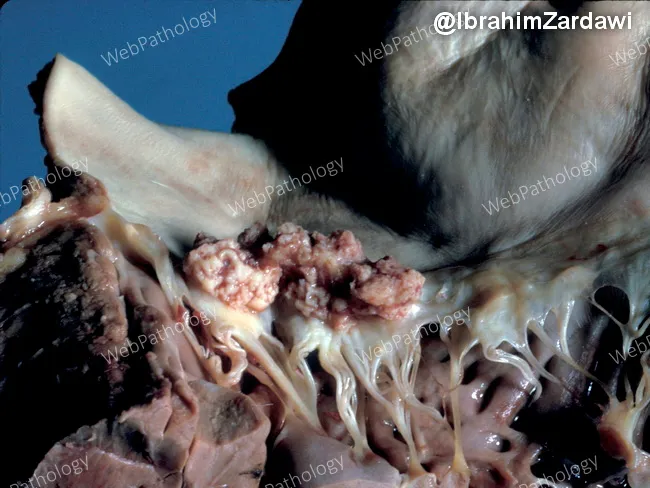
Gross pathology photograph of an explanted heart demonstrating left-sided infective endocarditis with mitral valve involvement. The image highlights a shaggy vegetative mass on the mitral valve, with thickened chordae tendineae and irregular nodularity of the cusp just to the right of the vegetation. The vegetations are friable and tan-pink, with adjacent valvular tissue showing edema and thickening. The chords appear markedly elongated and edematous, consistent with valvular destruction and tethering. The cusp shows focal thickening and rime-like nodules indicative of inflammatory deposition and possible microabscess formation. This gross morphology correlates with acute/subacute infective endocarditis, often due to bacterial pathogens such as Staphylococcus aureus or viridans streptococci, though fungal infections can also be implicated. Clinically, such findings signal bacteremia, embolic risk, and potential heart failure from valvular insufficiency. The image is valuable for education in cardiac pathology, surgical pathology, and infectious disease teaching; it demonstrates typical mitral valve vegetations, chordal involvement, and cusp irregularity useful for differential diagnosis with Libman-Sacks endocarditis, rheumatic disease, or marantic endocarditis. In practice, correlation with blood cultures, echocardiography, and systemic evaluation is essential for diagnosis and management. Documentation of vegetative morphology aids correlating radiographic and microbiological findings, informs prognosis, and guides rheology of therapeutic interventions management.
Gross pathology photograph of an explanted heart demonstrating left-sided infective endocarditis with mitral valve involvement. The image highlights a shaggy vegetative mass on the mitral valve, with thickened chordae tendineae and irregular nodularity of the cusp just to the right of the vegetation. The vegetations are friable and tan-pink, with adjacent valvular tissue showing edema and thickening. The chords appear markedly elongated and edematous, consistent with valvular destruction and tethering. The cusp shows focal thickening and rime-like nodules indicative of inflammatory deposition and possible microabscess formation. This gross morphology correlates with acute/subacute infective endocarditis, often due to bacterial pathogens such as Staphylococcus aureus or viridans streptococci, though fungal infections can also be implicated. Clinically, such findings signal bacteremia, embolic risk, and potential heart failure from valvular insufficiency. The image is valuable for education in cardiac pathology, surgical pathology, and infectious disease teaching; it demonstrates typical mitral valve vegetations, chordal involvement, and cusp irregularity useful for differential diagnosis with Libman-Sacks endocarditis, rheumatic disease, or marantic endocarditis. In practice, correlation with blood cultures, echocardiography, and systemic evaluation is essential for diagnosis and management. Documentation of vegetative morphology aids correlating radiographic and microbiological findings, informs prognosis, and guides rheology of therapeutic interventions management.

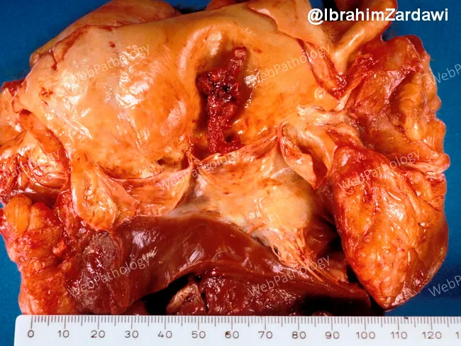
Gross photography of a surgically excised heart valve specimen showing a large, pedunculated vegetation attached to the non‑coronary cusp of the aortic valve. The lesion projects into the left ventricular outflow tract and measures several centimeters on the ruler included for scale. The vegetation has a polypoid, friable appearance with an irregular, lobulated surface and a tan-yellow to reddish hue consistent with fibrin, inflammatory debris, and possible microbial colonies not identifiable by gross inspection. The surrounding valve tissue is thickened and deformed, with partial leaflet destruction and altered commissural architecture. These macroscopic features are highly suggestive of an endocarditis-related vegetation, though definitive etiologic identification requires microbiologic culture and histopathology. The non-coronary cusp is the most involved site, with adjacent leaflet tissue showing edema and inflammatory changes. The image illustrates how infectious or inflammatory valvulopathy can disrupt valvular integrity, potentially leading to significant valvular insufficiency or outflow obstruction if not addressed surgically. Clinically, such a lesion prompts consideration of infective endocarditis (bacterial or fungal) or less likely nonbacterial thrombotic endocarditis, in the appropriate clinical setting. Correlation with echocardiography, blood cultures, and laboratory inflammatory markers is essential. This photograph serves as an educational example of valvular vegetation morphology and postoperative pathology. Teaching.

Gross photography of a surgically excised heart valve specimen showing a large, pedunculated vegetation attached to the non‑coronary cusp of the aortic valve. The lesion projects into the left ventricular outflow tract and measures several centimeters on the ruler included for scale. The vegetation has a polypoid, friable appearance with an irregular, lobulated surface and a tan-yellow to reddish hue consistent with fibrin, inflammatory debris, and possible microbial colonies not identifiable by gross inspection. The surrounding valve tissue is thickened and deformed, with partial leaflet destruction and altered commissural architecture. These macroscopic features are highly suggestive of an endocarditis-related vegetation, though definitive etiologic identification requires microbiologic culture and histopathology. The non-coronary cusp is the most involved site, with adjacent leaflet tissue showing edema and inflammatory changes. The image illustrates how infectious or inflammatory valvulopathy can disrupt valvular integrity, potentially leading to significant valvular insufficiency or outflow obstruction if not addressed surgically. Clinically, such a lesion prompts consideration of infective endocarditis (bacterial or fungal) or less likely nonbacterial thrombotic endocarditis, in the appropriate clinical setting. Correlation with echocardiography, blood cultures, and laboratory inflammatory markers is essential. This photograph serves as an educational example of valvular vegetation morphology and postoperative pathology. Teaching.

Searching Images

serous atrophy heart gelatinous fat arteriosclerosis aorta gross
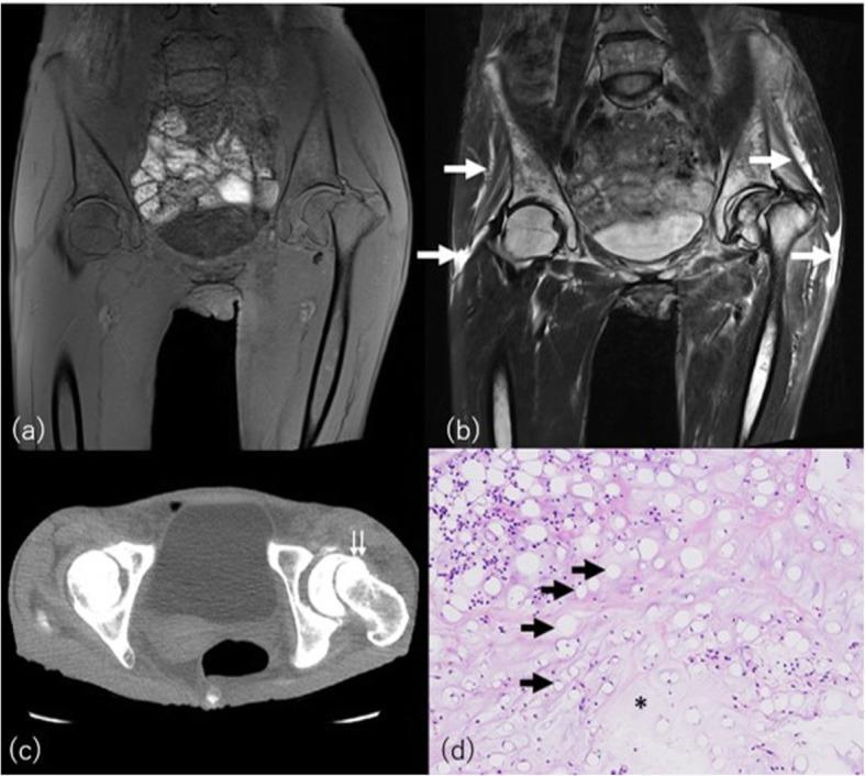
This multi-modal educational figure illustrates serous atrophy of bone marrow (gelatinous bone marrow transformation) and associated findings in a patient with bulimia nervosa. (a) Coronal T1-weighted MRI shows diffuse, abnormal hypointense marrow signal throughout the pelvis and proximal femora, along with a left femoral neck fracture. (b) Coronal STIR MRI reveals a corresponding hyperintense signal within the marrow space and highlights focal edema or serous fluid in the surrounding subcutaneous and intermuscular tissues (white arrows). (c) Axial CT image at the level of the femoral neck confirms the fracture (double arrow) and demonstrates increased attenuation (increased density) of the depleted subcutaneous fat. (d) Histopathological H&E stain (100x magnification) provides the cellular correlation, showing a marked reduction and shrinkage of adipocytes (black arrows), a decrease in hematopoietic cells, and the diagnostic deposition of abundant, amorphous, eosinophilic extracellular gelatinous substances (asterisk). The visual findings emphasize the characteristic imaging appearance of nutritional marrow depletion and the increased risk of insufficiency fractures in eating disorders.
This multi-modal educational figure illustrates serous atrophy of bone marrow (gelatinous bone marrow transformation) and associated findings in a patient with bulimia nervosa. (a) Coronal T1-weighted MRI shows diffuse, abnormal hypointense marrow signal throughout the pelvis and proximal femora, along with a left femoral neck fracture. (b) Coronal STIR MRI reveals a corresponding hyperintense signal within the marrow space and highlights focal edema or serous fluid in the surrounding subcutaneous and intermuscular tissues (white arrows). (c) Axial CT image at the level of the femoral neck confirms the fracture (double arrow) and demonstrates increased attenuation (increased density) of the depleted subcutaneous fat. (d) Histopathological H&E stain (100x magnification) provides the cellular correlation, showing a marked reduction and shrinkage of adipocytes (black arrows), a decrease in hematopoietic cells, and the diagnostic deposition of abundant, amorphous, eosinophilic extracellular gelatinous substances (asterisk). The visual findings emphasize the characteristic imaging appearance of nutritional marrow depletion and the increased risk of insufficiency fractures in eating disorders.

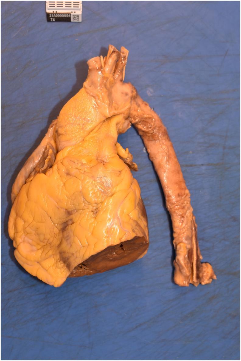
A clinical gross pathology specimen showing a human heart and a dissected segment of the thoracic and descending aorta during a necropsy study. The heart exhibits a prominent yellowish hue across the epicardial surface, consistent with significant epicardial fat accumulation. The surface texture appears lobulated with visible sulci corresponding to the anatomical pathways of the coronary arteries. A transverse cut at the inferior aspect of the ventricles reveals the dark brown, muscular myocardium of the ventricular wall. Attached to the superior aspect of the heart is the aortic arch, leading into a longitudinally dissected segment of the descending thoracic aorta. The aortic wall shows a transitioning color from pale tan at the root to a mottled reddish-brown along its length. The intimal surface of the aorta displays mild irregularities and roughness, indicative of early atherosclerotic changes, though no large aneurysms or obstructive thrombi are visible. The specimen is photographed against a blue surgical background with a scale marker for forensic and pathological documentation.

A clinical gross pathology specimen showing a human heart and a dissected segment of the thoracic and descending aorta during a necropsy study. The heart exhibits a prominent yellowish hue across the epicardial surface, consistent with significant epicardial fat accumulation. The surface texture appears lobulated with visible sulci corresponding to the anatomical pathways of the coronary arteries. A transverse cut at the inferior aspect of the ventricles reveals the dark brown, muscular myocardium of the ventricular wall. Attached to the superior aspect of the heart is the aortic arch, leading into a longitudinally dissected segment of the descending thoracic aorta. The aortic wall shows a transitioning color from pale tan at the root to a mottled reddish-brown along its length. The intimal surface of the aorta displays mild irregularities and roughness, indicative of early atherosclerotic changes, though no large aneurysms or obstructive thrombi are visible. The specimen is photographed against a blue surgical background with a scale marker for forensic and pathological documentation.

Searching Images

mulberry heart disease swine porcine cardiovascular
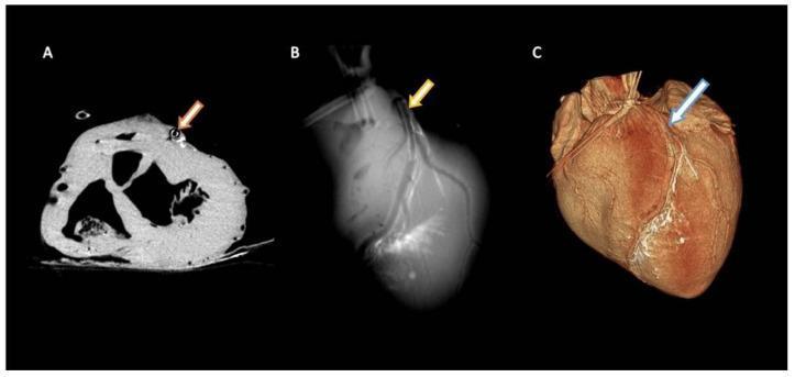
This composite diagnostic image displays three different radiological views of a porcine heart model used for cardiovascular research. Panel A (Axial view) shows a transverse section of the ventricles with an artificial atherosclerotic plaque inserted into the Left Anterior Descending (LAD) artery, appearing as a dense, hyper-attenuated ring-like structure (indicated by an orange arrow). Small, contrasted circular structures representing cardiac lymphatic vessels are visible adjacent to the LAD. Panel B (Maximum Intensity Projection - MIP) offers a two-dimensional projection where the LAD and its branching lymphatic network appear as thin, high-contrast linear structures (yellow arrow). Panel C (Volume-Rendering Technique - VRT) provides a three-dimensional anatomical reconstruction of the heart surface, highlighting the spatial distribution and network-like arrangement of the cardiac lymphatics (light blue arrow) as they track along the epicardial surface near the LAD. The image demonstrates the capabilities of advanced CT imaging in visualizing coronary pathology and the associated lymphatic system for preclinical procedural modeling.
This composite diagnostic image displays three different radiological views of a porcine heart model used for cardiovascular research. Panel A (Axial view) shows a transverse section of the ventricles with an artificial atherosclerotic plaque inserted into the Left Anterior Descending (LAD) artery, appearing as a dense, hyper-attenuated ring-like structure (indicated by an orange arrow). Small, contrasted circular structures representing cardiac lymphatic vessels are visible adjacent to the LAD. Panel B (Maximum Intensity Projection - MIP) offers a two-dimensional projection where the LAD and its branching lymphatic network appear as thin, high-contrast linear structures (yellow arrow). Panel C (Volume-Rendering Technique - VRT) provides a three-dimensional anatomical reconstruction of the heart surface, highlighting the spatial distribution and network-like arrangement of the cardiac lymphatics (light blue arrow) as they track along the epicardial surface near the LAD. The image demonstrates the capabilities of advanced CT imaging in visualizing coronary pathology and the associated lymphatic system for preclinical procedural modeling.

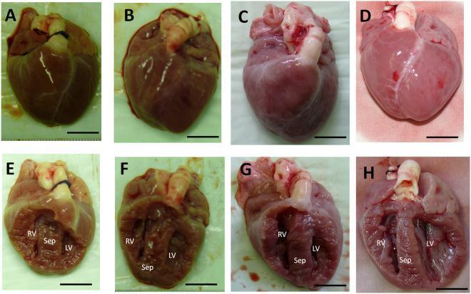
This image presents the gross morphology and coronal sections of neonatal porcine hearts in a comparative study of genome editing. Panels A-C show the external view of representative hearts from genome-edited animals (harboring the MYH7 R723G mutation), while Panel D shows a wildtype control heart. Panels E-G depict coronal sections of the genome-edited hearts, and Panel H shows the coronal section of the control heart. In the external views (A-D), the genome-edited specimens (A, B) appear more congested and darker in color compared to the paler control (D). In the coronal sections (E-H), the right ventricle (RV), left ventricle (LV), and interventricular septum (Sep) are labeled. Both the edited and control hearts exhibit symmetrical ventricular development consistent with neonatal anatomy, with no evidence of gross cardiac hypertrophy or wall thickening at this developmental stage (less than 24 hours of age). The image is part of a cardiovascular pathology study investigating Hypertrophic Cardiomyopathy (HCM) using a porcine model. Scale bars represent 1 cm.

This image presents the gross morphology and coronal sections of neonatal porcine hearts in a comparative study of genome editing. Panels A-C show the external view of representative hearts from genome-edited animals (harboring the MYH7 R723G mutation), while Panel D shows a wildtype control heart. Panels E-G depict coronal sections of the genome-edited hearts, and Panel H shows the coronal section of the control heart. In the external views (A-D), the genome-edited specimens (A, B) appear more congested and darker in color compared to the paler control (D). In the coronal sections (E-H), the right ventricle (RV), left ventricle (LV), and interventricular septum (Sep) are labeled. Both the edited and control hearts exhibit symmetrical ventricular development consistent with neonatal anatomy, with no evidence of gross cardiac hypertrophy or wall thickening at this developmental stage (less than 24 hours of age). The image is part of a cardiovascular pathology study investigating Hypertrophic Cardiomyopathy (HCM) using a porcine model. Scale bars represent 1 cm.

Searching Images

ball thrombus cardiac chamber mural thrombus
Transthoracic echocardiogram (TTE) in a grayscale B-mode view with overlaid color Doppler, demonstrating a mural thrombus within the left ventricle. The ventricular chamber is roughly conical with speckled, hyperechoic borders representing the myocardium. At the left ventricular apex, a focal, echogenic mass is visible, consistent with a mural thrombus. The thrombus appears as a region of similar or slightly increased echogenicity compared to the adjacent cardiac wall, with somewhat poorly defined margins. A central rectangular Color Doppler gate shows red signals, indicating blood flow velocity directed toward the ultrasound transducer. This diagnostic image highlights a common complication of myocardial infarction, where localized wall motion abnormalities (hypokinesis) facilitate stasis and subsequent thrombus formation, posing a significant risk for systemic thromboembolic events such as cerebral infarction.
Transthoracic echocardiogram (TTE) in a grayscale B-mode view with overlaid color Doppler, demonstrating a mural thrombus within the left ventricle. The ventricular chamber is roughly conical with speckled, hyperechoic borders representing the myocardium. At the left ventricular apex, a focal, echogenic mass is visible, consistent with a mural thrombus. The thrombus appears as a region of similar or slightly increased echogenicity compared to the adjacent cardiac wall, with somewhat poorly defined margins. A central rectangular Color Doppler gate shows red signals, indicating blood flow velocity directed toward the ultrasound transducer. This diagnostic image highlights a common complication of myocardial infarction, where localized wall motion abnormalities (hypokinesis) facilitate stasis and subsequent thrombus formation, posing a significant risk for systemic thromboembolic events such as cerebral infarction.

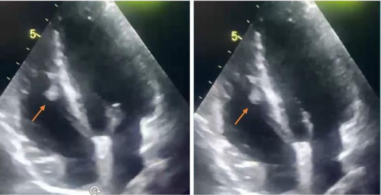
Two side-by-side frames of a transthoracic echocardiogram (TTE) in an apical four-chamber view, demonstrating a right ventricular mural thrombus. The right ventricle (RV) is prominently displayed on the left side of the ultrasound sector. An orange arrow in both panels points to a distinct, hyperechoic, lobulated mass attached to the endocardial surface of the right ventricular free wall. The mass exhibits a texture different from the surrounding myocardium and is situated within the ventricular cavity, appearing mobile and suggestive of a mural thrombus. The interventricular septum and left ventricle are also partially visualized. This clinical imaging illustrates an intracardiac complication often associated with Behçet's disease and other systemic inflammatory conditions or prothrombotic states. It serves as an educational example for identifying right-sided cardiac masses and assessing their echogenicity, mobility, and anatomical attachment via diagnostic ultrasound.

Two side-by-side frames of a transthoracic echocardiogram (TTE) in an apical four-chamber view, demonstrating a right ventricular mural thrombus. The right ventricle (RV) is prominently displayed on the left side of the ultrasound sector. An orange arrow in both panels points to a distinct, hyperechoic, lobulated mass attached to the endocardial surface of the right ventricular free wall. The mass exhibits a texture different from the surrounding myocardium and is situated within the ventricular cavity, appearing mobile and suggestive of a mural thrombus. The interventricular septum and left ventricle are also partially visualized. This clinical imaging illustrates an intracardiac complication often associated with Behçet's disease and other systemic inflammatory conditions or prothrombotic states. It serves as an educational example for identifying right-sided cardiac masses and assessing their echogenicity, mobility, and anatomical attachment via diagnostic ultrasound.

Searching Images

coronary atherosclerosis gross specimen aorta atherosclerotic plaques
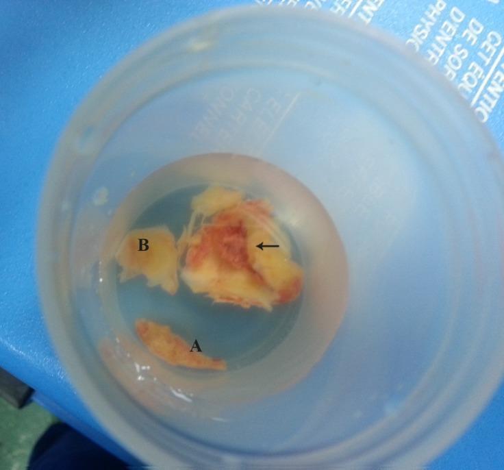
This clinical photograph displays a gross pathological specimen of resected atherosclerotic plaques from a human aorta, submerged in a clear preservative solution within a specimen container. The image shows three distinct fragments of varying size and morphology, labeled A, B, and one indicated by a horizontal arrow. Fragment A is the smallest, exhibiting a narrow, elongated, and relatively uniform appearance. Fragment B is intermediate in size with an amorphous, lumpy shape. The largest specimen, highlighted by the black horizontal arrow, demonstrates a complex, heterogeneous composition characteristic of an advanced unstable plaque. This major fragment displays multiple lobulations and distinct color variations, including yellowish-white calcified or lipid-rich areas and reddish-brown zones indicative of intraplaque hemorrhage or adherent thrombus. A central recessed area, marked by the arrow, identifies a significant ulceration on the plaque surface. These findings illustrate the macroscopic features of complex aortic atherosclerosis, which can serve as a source for systemic embolic events.
This clinical photograph displays a gross pathological specimen of resected atherosclerotic plaques from a human aorta, submerged in a clear preservative solution within a specimen container. The image shows three distinct fragments of varying size and morphology, labeled A, B, and one indicated by a horizontal arrow. Fragment A is the smallest, exhibiting a narrow, elongated, and relatively uniform appearance. Fragment B is intermediate in size with an amorphous, lumpy shape. The largest specimen, highlighted by the black horizontal arrow, demonstrates a complex, heterogeneous composition characteristic of an advanced unstable plaque. This major fragment displays multiple lobulations and distinct color variations, including yellowish-white calcified or lipid-rich areas and reddish-brown zones indicative of intraplaque hemorrhage or adherent thrombus. A central recessed area, marked by the arrow, identifies a significant ulceration on the plaque surface. These findings illustrate the macroscopic features of complex aortic atherosclerosis, which can serve as a source for systemic embolic events.

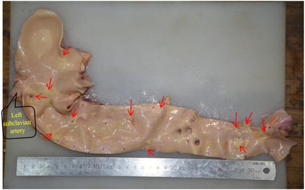
This clinical photograph displays a gross pathological specimen of a human aorta, dissected longitudinally to expose the intimal surface. The specimen includes the aortic arch, descending thoracic aorta, and abdominal aorta, positioned alongside a centimeter ruler for scale. The tissue exhibits a light tan to pinkish coloration with visible branching vessels, including a labeled 'Left subclavian artery' at the proximal end. Numerous red arrows highlight the focal distribution of atherosclerotic disease. Yellowish, irregular, and raised hardened plaques are clearly visible, demonstrating the progression from fatty streaks to more advanced calcified or fibrous lesions. These plaques are particularly concentrated around the ostia of vascular branches, such as the subclavian artery and distal bifurcation areas. The image serves as a teaching tool for cardiovascular pathology, illustrating the gross morphology of aortic atherosclerosis and its predilection for arterial branching points and the distal abdominal segment.

This clinical photograph displays a gross pathological specimen of a human aorta, dissected longitudinally to expose the intimal surface. The specimen includes the aortic arch, descending thoracic aorta, and abdominal aorta, positioned alongside a centimeter ruler for scale. The tissue exhibits a light tan to pinkish coloration with visible branching vessels, including a labeled 'Left subclavian artery' at the proximal end. Numerous red arrows highlight the focal distribution of atherosclerotic disease. Yellowish, irregular, and raised hardened plaques are clearly visible, demonstrating the progression from fatty streaks to more advanced calcified or fibrous lesions. These plaques are particularly concentrated around the ostia of vascular branches, such as the subclavian artery and distal bifurcation areas. The image serves as a teaching tool for cardiovascular pathology, illustrating the gross morphology of aortic atherosclerosis and its predilection for arterial branching points and the distal abdominal segment.

Only few simple lines to write on record

Here are brief, record-ready gross lesion descriptions for all 10 cardiovascular system topics (as per Ganti Sastry & Vegad - Veterinary Pathology):

Cardiovascular System - Gross Lesions

1. Petechial Haemorrhage Small, pin-point to pin-head sized, bright red hemorrhagic spots seen on epicardium, endocardium or pericardium. Common in septicaemia, toxaemia and viral infections.

2. Vegetative Endocarditis Cauliflower-like or warty, greyish-white to yellowish irregular vegetations on the valve cusps (commonly mitral and tricuspid valves). Valves are thickened and distorted. Seen in bacterial infections.

3. Dilatation of Heart Heart appears enlarged and flabby. Ventricular walls are thin. Chambers are dilated and filled with blood/clots. Overall weight is increased. Seen in chronic congestive heart failure.

4. Ball Thrombus Rounded, firm, smooth-surfaced, dark red to grey mass found free-floating or loosely attached inside the cardiac chamber (usually left atrium). May obstruct the atrioventricular orifice.

5. Serous Atrophy of Heart Epicardial fat is replaced by a transparent, gelatinous, jelly-like material (gelatinous atrophy). Heart appears small and shrunken. Seen in chronic wasting diseases and starvation.

6. Mulberry Heart Disease Heart appears dark red to purple, resembling a mulberry. Extensive hemorrhages seen on epicardium and myocardium. Pericardial sac contains excess blood-stained fluid. Seen in pigs due to Vitamin E / Selenium deficiency.

7. Persistent Aortic Arch Abnormal retention of the right 4th aortic arch (instead of left) forming a vascular ring around the oesophagus and trachea. Oesophagus appears dilated cranial to the constriction (megaoesophagus). Seen mainly in dogs.

8. Dextroposition of Aorta (Dextrotransposition) Aorta arises from the right ventricle instead of the left. Heart shows a ventricular septal defect. Aorta and pulmonary artery are transposed. Part of Tetralogy of Fallot complex in animals.

9. Arteriosclerosis Arterial wall appears thickened, hardened and inelastic. Intima shows white to yellowish irregular thickening. Lumen is narrowed. Calcification may be present. Common in aged animals and turkeys.

10. Coronary Atherosclerosis Coronary arteries show yellowish-white, raised, irregular plaques on the intimal surface. Lumen is narrowed. Plaques may show calcification or ulceration. Rarely seen in animals; common in humans and aged dogs/pigs.

All descriptions are based on Ganti Sastry & Vegad - Textbook of Veterinary Pathology gross morphology standards, suitable for practical record writing.Here are brief, record-ready gross lesion descriptions for both systems as per Ganti Sastry & Vegad:

Urinary System - Gross Lesions

1. Horse-shoe Kidney Both kidneys are fused at their poles (usually lower poles) forming a U-shaped or horse-shoe appearance. A band of renal tissue connects them across the midline. Congenital anomaly seen in cattle and other animals.

2. Urolithiasis Urinary bladder or urethra contains one or more calculi (stones). Stones are yellowish-white, hard, irregular or rounded. Mucosa of bladder shows congestion, haemorrhage and thickening. Obstruction may cause bladder distension.

3. Nephrolithiasis Kidney pelvis contains calculi of varying size, shape and colour (yellowish-white to brown). Renal pelvis is dilated. Surrounding parenchyma may show pressure atrophy and fibrosis.

4. Hydronephrosis Kidney is greatly enlarged and cyst-like. On cut section, pelvis and calyces are massively dilated and filled with clear watery fluid. Renal cortex and medulla are compressed and thinned. Caused by obstruction to urine outflow.

5. Pyemic Nephritis (Embolic Nephritis) Multiple small, pale-yellow to grey, circular abscesses scattered throughout the cortex and medulla on cut section. Kidney surface shows multiple white foci with red haemorrhagic margins. Caused by haematogenous spread of bacteria.

6. Pyelonephritis Kidney is enlarged. Pelvis is dilated and contains pus (yellow-grey turbid fluid). Mucosa of pelvis is thickened and ulcerated. Cortex shows grey-yellow streaks extending from medulla. Ascending bacterial infection.

7. Focal Interstitial Nephritis Kidney surface shows multiple small, pale-white, irregular foci (white spots) scattered throughout cortex. Cut section reveals same whitish foci. Kidney size is normal or slightly reduced. Seen in leptospirosis (white-spotted kidney).

8. Chronic Diffuse Interstitial Nephritis Kidney is small, firm and contracted. Surface is granular and irregular (granular contracted kidney). Cut section shows thinned cortex with indistinct corticomedullary junction. Fibrosis throughout parenchyma.

9. Polycystic Kidney Kidney is greatly enlarged. Entire parenchyma is replaced by numerous cysts of varying sizes filled with clear to straw-coloured fluid. Normal renal tissue is markedly reduced. Congenital condition seen in calves, foals and cats.

10. Transitional Cell Carcinoma Urinary bladder wall is thickened. Mucosa shows irregular, papillary or cauliflower-like projections, greyish-white in colour. Areas of haemorrhage, necrosis and ulceration present. Seen commonly in cattle (associated with bracken fern poisoning).


Reproductive System - Gross Lesions

1. Epididymitis Epididymis is enlarged, firm and swollen. Cut section shows thickened wall with caseous or purulent exudate in the lumen. Adhesions may be present with surrounding tissue. Seen in brucellosis and other bacterial infections.

2. Phimosis Prepuce opening is abnormally narrow, preventing protrusion of the penis. Prepuce appears constricted. Smegma and debris may accumulate inside. Congenital or acquired condition.

3. Paraphimosis Penis is protruded and cannot be retracted into the prepuce. Exposed penis appears swollen, congested, dark red to purple, oedematous and dry. Necrosis may develop in prolonged cases.

4. Cryptorchid Testis Testis is small, soft and underdeveloped, located in the inguinal canal or abdominal cavity instead of the scrotum. Cut section shows absence of normal spermatogenic tubules; replaced by fibrous or fatty tissue.

5. Balanoposthitis Glans penis and prepuce mucosa show redness, swelling, erosions, ulcers and necrosis. Exudate may be present (mucopurulent). Seen in sheep (pizzle rot due to Corynebacterium renale) and other species.

6. Freemartin Genital organs of the female co-twin (of male-female twins in cattle) are abnormal. Ovaries are small and rudimentary. Uterus is hypoplastic. Gonads may resemble testes. Vagina is shortened. External genitalia appear female but internal organs are intersex.

7. Uterus Didelphys Uterus is completely duplicated - two separate uterine horns, cervices and sometimes vaginas are present. Each horn is fully developed but smaller than normal. Congenital anomaly due to failure of fusion of Mullerian ducts.

8. Pyometra Uterus is greatly enlarged and distended with pus (thick, creamy yellow to grey fluid). Wall is thickened and flaccid. Mucosa is congested, haemorrhagic and ulcerated. Common in bitches, queens and cows.

9. Macerated Fetus Dead fetus retained in uterus undergoes maceration. Soft tissues are liquefied and bones are separated and disarticulated. Uterus contains reddish-brown, foul-smelling fluid with loose fetal bones. Occurs when cervix is open.

10. Retained Placenta Placenta (cotyledons/placentomes in ruminants) fails to be expelled after parturition. Cotyledons appear dark red, swollen, necrotic and foul-smelling. Caruncles and cotyledons are firmly adherent. Common in cattle after abnormal parturition or brucellosis.

All descriptions as per Ganti Sastry & Vegad - Veterinary Pathology gross morphology, suitable for practical record writing.
This is a shared conversation. Sign in to Orris to start your own chat.