
Formation of the intraembryonic cavity, Serous membranes and thoracic cavity explain to a first year mbbs student
Reading File
Finding Sources
Finding Sources
Finding Sources
Found good sources: The Developing Human (Moore) for intraembryonic coelom, and Langman's Medical Embryology for diaphragm/thoracic cavity. Let me read both relevant sections.
Reading File
Reading File
Reading File
Finding Sources
Finding Sources
Finding Sources
Let me read the key section in Moore around line 4850-5000 covering division of the coelom, pericardioperitoneal canals, and pleuropericardial membranes.
Reading File
Reading File
Reading File
Reading File
Searching Images
intraembryonic coelom division into pericardial pleural peritoneal cavities diagram embryology
Searching Images
pleuropericardial membrane pleuroperitoneal membrane formation diaphragm development diagram
Development of the Intraembryonic Cavity, Serous Membranes, and the Thoracic Cavity
(Based on Moore's "The Developing Human" and Langman's Medical Embryology)
This is one continuous story: one cavity (the intraembryonic coelom) → gets divided into three cavities (pericardial, pleural, peritoneal) → each cavity gets lined by a serous membrane → and specifically for the thorax, extra folds/membranes come in to separate the heart from the lungs.
1. Formation of the Intraembryonic Coelom
- In the trilaminar embryo, the lateral plate mesoderm splits into two layers early in the 4th week. Small, isolated clefts (coelomic spaces) appear within this mesoderm and in the cardiogenic mesoderm.
- These isolated spaces coalesce to form a single continuous, horseshoe-shaped cavity — the intraembryonic coelom.
- The bend of the horseshoe (at the cranial end) = future pericardial cavity.
- The two limbs running caudally = future pleural and peritoneal cavities (they are continuous with each other and, at their lateral edges, with the extraembryonic coelom).
By splitting the lateral mesoderm, the coelom creates two mesodermal layers:
| Layer | Position | Continuous with | Forms |
|---|---|---|---|
| Somatic (parietal) mesoderm | beneath ectoderm | extraembryonic mesoderm over the amnion | body wall (somatopleure) + parietal layer of serous membranes |
| Splanchnic (visceral) mesoderm | around endoderm | extraembryonic mesoderm over the yolk sac | gut wall (splanchnopleure) + visceral layer of serous membranes |
Why it matters functionally: this cavity gives the developing organs (heart, gut, lungs) room to grow, rotate, and move — e.g. it is what allows the midgut to physiologically herniate into the umbilical cord later.
2. Effect of Embryonic Folding
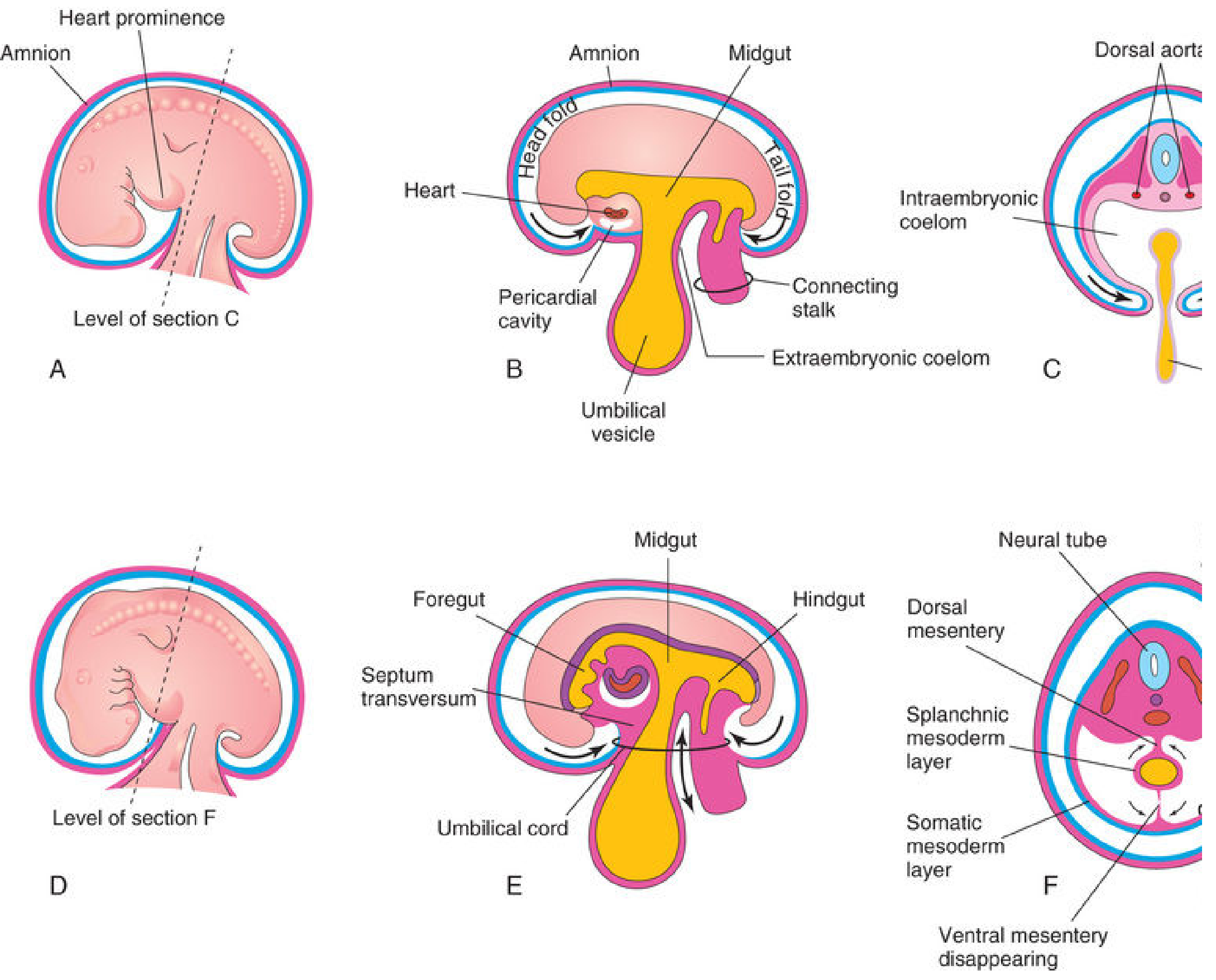
During folding (lateral body folds + head/tail folds, ~day 22-28):
- The two limbs of the coelom are brought together ventrally.
- The ventral mesentery in the region of the future peritoneal cavity degenerates, so the right and left peritoneal spaces become one continuous cavity.
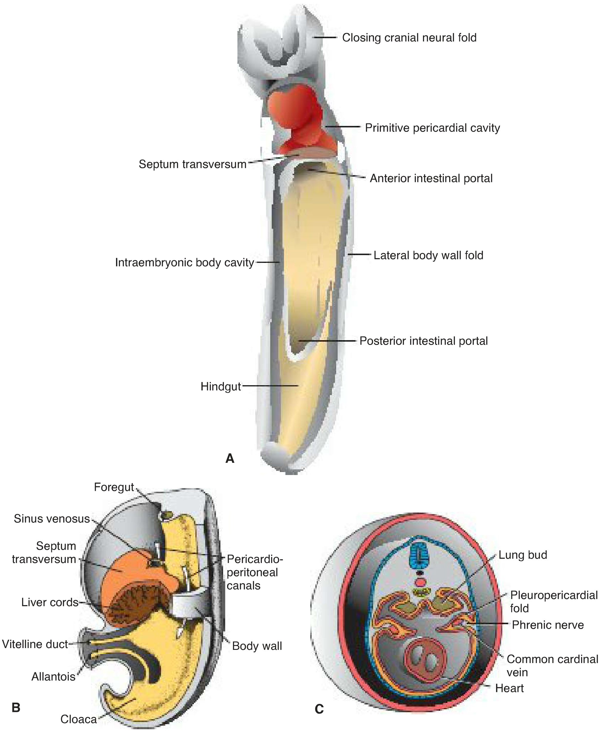
- The head fold carries the heart and pericardial cavity ventral to the foregut, so the pericardial cavity now opens dorsally into two channels called the pericardioperitoneal canals, which run on either side of the foregut and connect the pericardial cavity to the peritoneal cavity.
3. Formation of the Serous Membranes
Each of the three future cavities is lined by mesothelium derived from the two mesodermal layers described above:
- Parietal layer (from somatic mesoderm) → lines the body wall.
- Visceral layer (from splanchnic mesoderm) → covers the organ itself.
This same pattern produces all three serous membranes of the body:
| Cavity | Parietal layer | Visceral layer |
|---|---|---|
| Peritoneal | parietal peritoneum (lines abdominal wall) | visceral peritoneum (covers stomach, intestines, liver, etc.) |
| Pleural | parietal pleura (lines thoracic wall) | visceral pleura (covers the lung) |
| Pericardial | parietal (serous) pericardium | visceral pericardium / epicardium (covers the heart) |
So a "serous membrane" is really just the mesothelial covering of the coelom split around an organ, leaving a potential space between the two layers (peritoneal cavity, pleural cavity, pericardial cavity) that normally contains a thin film of lubricating serous fluid.
4. Partitioning into the Three Definitive Cavities — and Formation of the Thoracic Cavity
By the end of the 4th–7th weeks, the single coelom (via the pericardioperitoneal canals) is subdivided by two pairs of membranous folds. This is where the pericardial cavity, the two pleural cavities, and the peritoneal cavity become fully separated, and the thoracic cavity takes its final shape.
a) Septum transversum
A thick plate of splanchnic mesoderm that comes to lie between the pericardial cavity and the stalk of the yolk sac. It is the primordium of the central tendon of the diaphragm and partially (not completely) separates the thorax from the abdomen — leaving the pericardioperitoneal canals open on either side of the foregut.
b) Growth of the lung buds
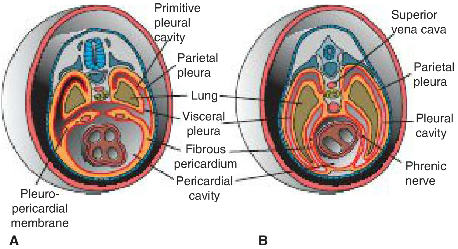
The lung buds grow laterally from the trachea into the pericardioperitoneal canals. As the lungs enlarge, the canals become too small, so the expanding pleural cavities burrow into the surrounding body wall mesenchyme — splitting it into an outer layer (future thoracic wall) and an inner layer.
c) Pleuropericardial membranes (separate pericardial cavity from pleural cavities)
- Ridges called pleuropericardial folds project into the primitive undivided thoracic cavity. Each fold carries the common cardinal vein and the phrenic nerve.
- As the pleural cavities expand ventrally around the heart, these folds enlarge into membranous partitions — the pleuropericardial membranes.
- By the 7th week they fuse with mesenchyme ventral to the esophagus, completely separating the pericardial cavity from the two pleural cavities.
- The inner layer left behind becomes the fibrous pericardium; the right membrane fuses slightly earlier than the left (which is why a rare congenital pericardial defect is more often left-sided).
- This is also why the phrenic nerve ends up running on the fibrous pericardium in the adult.
d) Pleuroperitoneal membranes (separate pleural cavities from the peritoneal cavity)
- Similarly, pleuroperitoneal folds enlarge at the caudal end of the pericardioperitoneal canals and become the pleuroperitoneal membranes.
- These fuse with the septum transversum and the dorsal mesentery of the esophagus, closing off each pleural cavity from the peritoneal cavity by about the 7th week.
- Together with the septum transversum and muscle ingrowth from the body wall, this completes the diaphragm, and finalizes the thoracic cavity as two separate pleural cavities (each housing a lung) plus the pericardial cavity, all now walled off from the abdomen.
5. Quick Summary (exam-style)
- Isolated coelomic clefts in lateral mesoderm → coalesce → horseshoe-shaped intraembryonic coelom.
- This coelom splits lateral mesoderm into somatic (→ parietal serosa) and splanchnic (→ visceral serosa) layers.
- Folding brings the two limbs together ventrally → one continuous cavity connected by pericardioperitoneal canals.
- Septum transversum partially separates thorax from abdomen.
- Pleuropericardial membranes (from pleuropericardial folds, carrying phrenic nerve + common cardinal vein) separate pericardial cavity from pleural cavities.
- Pleuroperitoneal membranes separate pleural cavities from the peritoneal cavity, completing the diaphragm.
- Net result: 1 pericardial cavity + 2 pleural cavities + 1 peritoneal cavity, each lined by parietal and visceral serous membranes.
Clinical correlation worth remembering: failure of the pleuroperitoneal membrane to close (usually left, posterolateral) causes congenital diaphragmatic hernia — abdominal viscera herniate into the thorax and can compress the developing lung. Failure of pleuropericardial membrane fusion causes a rare congenital pericardial defect.
- The Developing Human Clinically Oriented Embryology, Ch. 8 "Body Cavities, Mesenteries, and Diaphragm"
- Langman's Medical Embryology, Ch. 7 "Diaphragm and Thoracic Cavity"
This is a shared conversation. Sign in to Orris to start your own chat.