
Make detailed mbbs notes on maternal changes during pregnancy
Reading File
Reading File
Finding Sources
Finding Sources
Searching PubMed
"pregnancy"[MeSH Terms] AND "physiological adaptation"[MeSH Terms]
Reading File
Reading File
Reading File
Reading File
Searching Images
changes in cardiac output blood volume during pregnancy graph
Reading File
Reading File
Reading File
Searching Images
pregnancy cardiac output plasma volume red cell mass increase
Searching Images
uterus size fundal height at different weeks of gestation
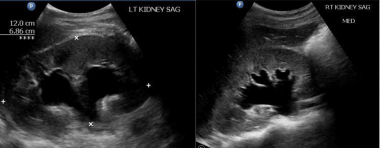
Maternal Changes During Pregnancy (MBBS Notes)
Pregnancy changes almost every organ system. The drivers are placental hormones (hCG, hPL, oestrogen, progesterone, relaxin), the mechanical effect of the growing uterus, and the extra metabolic demands of the fetus. Most changes are adaptive and reverse after delivery, but they can mimic disease or hide it. Normal values in pregnancy differ from non-pregnant values, which is why they matter clinically.
1. Hormonal drivers
| Hormone | Source | Key effects |
|---|---|---|
| hCG | Syncytiotrophoblast | Maintains the corpus luteum, peaks at 8-10 weeks, causes nausea and vomiting, has weak TSH-like action |
| Progesterone | Corpus luteum, then placenta (from about 8-10 weeks) | Smooth muscle relaxation, uterine quiescence, respiratory drive, raises body temperature |
| Oestrogen (mainly estriol) | Placenta (fetoplacental unit) | Uterine growth, breast duct growth, increased blood flow, raises binding globulins |
| hPL (human placental lactogen) | Placenta | Insulin resistance, lipolysis, diabetogenic effect |
| Relaxin | Corpus luteum | Ligament and cervical softening |
| Prolactin | Maternal pituitary | Prepares breast for lactation |
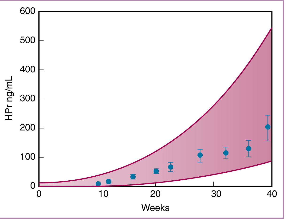
The pituitary enlarges in pregnancy through lactotroph hyperplasia. MRI volume rises by about 40%, 75% and 120% in the second trimester, third trimester and immediate postpartum period. Prolactin rises about 10-fold, while LH and FSH fall.
- Creasy & Resnik's Maternal-Fetal Medicine, p. 1526-1527
2. Reproductive system
Uterus
- Size: it grows from 7.5 x 5 x 2.5 cm to about 30 x 22 x 20 cm.
- Weight: it grows from 50-70 g to about 1000 g, roughly a 15-fold increase.
- Capacity: it grows from 10 mL to 5 L or more.
- The growth comes from hypertrophy of muscle cells (early hyperplasia, later stretching), mainly driven by oestrogen and progesterone.
- Position: the uterus stays in the pelvis until about 12 weeks. It reaches the umbilicus at about 20-24 weeks (textbooks differ) and the costal margin at 34-36 weeks.
- Fundal height (standard landmarks):
| Weeks | Level |
|---|---|
| 12 | Symphysis pubis |
| 16 | Midway between symphysis and umbilicus |
| 24 | Umbilicus |
| 28 | Between umbilicus and xiphisternum (lower third) |
| 32 | Upper third |
| 36 | Xiphisternum (highest) |
| 40 | Falls slightly with "lightening" as the head engages |
- Between 24 and 36 weeks, fundal height in cm is roughly equal to the weeks of gestation (McDonald's rule).
- Uterine blood flow: about 50 mL/min before pregnancy rises to 500-750 mL/min at term, roughly 10-12% of cardiac output. Guyton gives about 625 mL/min in the last month.
- Guyton and Hall Textbook of Medical Physiology, p. 1039
- Blood flow falls if the mother is hypovolaemic or hypotensive, because the uterine vessels are already maximally dilated and cannot autoregulate.
- Braxton Hicks contractions: painless, irregular contractions that begin from about 12 weeks.
- Shape and Hegar's sign: the uterus is pyriform early, then globular, then ovoid. The isthmus softens and elongates (Hegar's sign, 6-8 weeks) to form the lower uterine segment.
- Lower segment: it forms from the isthmus after about 28-30 weeks. The retraction ring separates it from the thick upper segment.
Cervix
- Softening: Goodell's sign.
- Colour: bluish due to vascularity (Chadwick's sign).
- Glands: the glands proliferate, with eversion of columnar epithelium (ectropion).
- Mucus plug: thick mucus (operculum) seals the canal.
- Ripening: collagen is remodelled before labour.
Vagina and vulva
- Appearance: the vaginal wall turns bluish-violet (Chadwick/Jacquemier sign) with increased vascularity.
- Discharge: it increases (leucorrhoea).
- pH: it becomes acidic (about 3.5-6) because of lactobacilli converting glycogen to lactic acid. This protects against infection.
- Osiander's sign: pulsation in the lateral fornices.
- Vulva: varicosities are common, with increased pigmentation.
Ovary and tubes
- Ovulation ceases.
- The corpus luteum of pregnancy supports the pregnancy until about 8-10 weeks. After that the placenta takes over (the luteoplacental shift).
- The ovary and tube are drawn up out of the pelvis with the uterus.
Breast
- Size increases and the veins become prominent.
- The nipple and areola darken, and a secondary areola appears.
- Montgomery's tubercles enlarge.
- Colostrum can be expressed from about 12 weeks.
- Oestrogen drives duct growth, progesterone drives alveolar growth, and prolactin and hPL prepare the gland for lactation. Milk secretion is held back until delivery by high oestrogen and progesterone levels.
3. Cardiovascular system
The changes begin as early as 6 weeks.
- Braunwald's Heart Disease, p. 1061-1062
| Parameter | Change |
|---|---|
| Plasma volume | Increases from 6 weeks |
| Blood volume | About +35-45% |
| Cardiac output | +30-50%, rising to about 6 L/min from 4.5 |
| Stroke volume | About +30% |
| Heart rate | +10-20 bpm (about 10-15 above baseline) |
| Systolic BP | Falls 2-4 mmHg |
| Diastolic BP | Falls 5-15 mmHg |
| Systemic vascular resistance | About -15% to -20% |
| Pulmonary vascular resistance | About -30% |
| Central venous pressure | Unchanged |
| Femoral venous pressure | Increased |
- Morgan and Mikhail's Clinical Anesthesiology, p. 1572
- Rosen's Emergency Medicine, p. 3424
Key points
- Cardiac output: it rises early (cardiac output reaches about 6 L/min by the second trimester) and is highest at about 28-32 weeks. Guyton notes a 30-40% rise by 27 weeks that then falls slightly in the last 8 weeks.
- Twins add another 10-15%.
- Early in pregnancy the rise comes mainly from stroke volume. Later it comes mainly from heart rate.
- Blood pressure: it falls in the first trimester, reaches its lowest point at about 20-24 weeks, and returns to non-pregnant values in the third trimester. The fall is due to progesterone and prostacyclin/NO vasodilation and the low-resistance placental circulation.
- Labour and delivery: cardiac output rises a further 60-80% around delivery. Contributing factors are catecholamines, autotransfusion from uterine contraction, and relief of IVC compression.
- Fluid mobilisation in the first week postpartum can cause heart failure in women with cardiomyopathy or severe outflow obstruction.
- Most changes resolve within 2 weeks but may take up to 6 months to settle fully.
- Braunwald's Heart Disease, p. 1062
- Supine hypotensive syndrome: after about 20 weeks the gravid uterus compresses the IVC and aorta when the woman lies supine. This reduces venous return and cardiac output, so BP falls and she feels faint. Nursing her in the left lateral position corrects it.
- Heart on examination:
- The diaphragm rises, so the heart is displaced upward and to the left. The apex beat moves outward.
- A grade I-II systolic ejection flow murmur is common and benign.
- A third heart sound may be present.
- A continuous venous hum or mammary souffle is possible.
- ECG: left axis deviation, small Q wave and inverted T in lead III, and ectopic beats.
- Clinical points:
- Normal pregnancy can mimic shock or mask early hypovolaemia. A pregnant woman can lose 1000-1500 mL before showing signs of shock.
- Dependent oedema, varicose veins and haemorrhoids are common. They are due to raised femoral venous pressure and pelvic venous obstruction.
4. Haematological changes
Blood volume and red cells
- Plasma volume: it rises by about 40-55%, from about 6 weeks, and peaks at 32-34 weeks.
- Red cell mass: it rises by about 20-30% (more with iron supplements).
- Plasma rises more than red cell mass, so there is haemodilution. This causes physiological anaemia of pregnancy:
- Haemoglobin falls (about -20% at maximum).
- Haematocrit falls.
- The lower limit of normal Hb is 11 g/dL in the first and third trimesters, and 10.5 g/dL in the second trimester.
- MCV rises slightly. The MCHC stays unchanged.
- The haemodilution is beneficial: it reduces blood viscosity and improves placental perfusion. It also reduces the impact of blood loss at delivery.
White cells, platelets and ESR
- WBC: it rises to 9,000-15,000/mm3. During labour it can reach 25,000-30,000/mm3 (neutrophilia).
- Platelets: a mild fall (about -10%), with gestational thrombocytopenia in 5-8%.
- ESR: it rises markedly (an unreliable marker in pregnancy).
Coagulation (pregnancy is a hypercoagulable state)
- Increased: fibrinogen (about 400-600 mg/dL), factors VII, VIII, IX, X, XII and von Willebrand factor. The overall rise ranges from 30% to 250% depending on the factor.
- Morgan and Mikhail's Clinical Anesthesiology, p. 1572
- Decreased: protein S, factors XI and XIII.
- Fibrinolysis: it is decreased, and PAI-1 and PAI-2 are raised.
- Venous thromboembolism risk is increased 4-5 fold, mainly in the puerperium. Contributing factors include stasis, hypercoagulability and vascular injury (Virchow's triad).
Iron needs
- The total extra requirement is about 1000 mg (red cell expansion about 500 mg, fetus and placenta about 300 mg, blood loss about 200 mg).
- Absorption increases, but supplementation is still usually needed.
- Folate requirement rises, and B12 levels fall slightly.
Plasma proteins
- Total protein and albumin fall (albumin from about 3.5 to 2.5 g/dL). This lowers oncotic pressure and contributes to oedema.
- Globulins rise (alpha and beta).
5. Respiratory system
| Parameter | Change |
|---|---|
| Tidal volume | +40% |
| Minute ventilation | +50% |
| Respiratory rate | Little change or slight rise (about +15%) |
| Oxygen consumption | +20-50% |
| FRC | -20% |
| Airway resistance | -35% |
| PaO2 | Increased (about 100-105 mmHg) |
| PaCO2 | Decreased (about 28-32 mmHg) |
| HCO3 | Decreased (about -15%, 18-21 mEq/L) |
| Vital capacity | Unchanged |
- Morgan and Mikhail's Clinical Anesthesiology, p. 1572
Key points
- Anatomical changes: the diaphragm rises by about 4 cm. The subcostal angle widens, the ribs flare and the transverse diameter of the chest increases by 2 cm. Chest circumference increases by 5-7 cm. Diaphragmatic excursion actually increases, so breathing remains largely diaphragmatic.
- Hyperventilation: progesterone increases the sensitivity of the respiratory centre to CO2. The result is a compensated respiratory alkalosis (pH 7.40-7.45, with renal excretion of bicarbonate). The low maternal PaCO2 helps CO2 move from the fetus to the mother.
- Lung volumes: the FRC is reduced because of residual volume and expiratory reserve volume falling. Total lung capacity falls slightly (about 5%).
- Physiological dyspnoea: about 70% of pregnant women notice breathlessness. It is due to hyperventilation and sensitivity to CO2, not to disease.
- Anaesthesia and airway:
- The reduced FRC combined with a higher oxygen consumption means rapid desaturation during apnoea.
- Capillary engorgement of the upper airway makes intubation more difficult, and a smaller tube is often needed.
- Rosen notes that in the third trimester a chest drain needs to be placed one or two interspaces higher than usual because of diaphragmatic elevation.
- Rosen's Emergency Medicine, p. 3424
- The MAC of inhalational agents falls by about 40%.
6. Renal and urinary system
Anatomy
- Kidney length increases by 1-1.5 cm and volume by up to 30%.
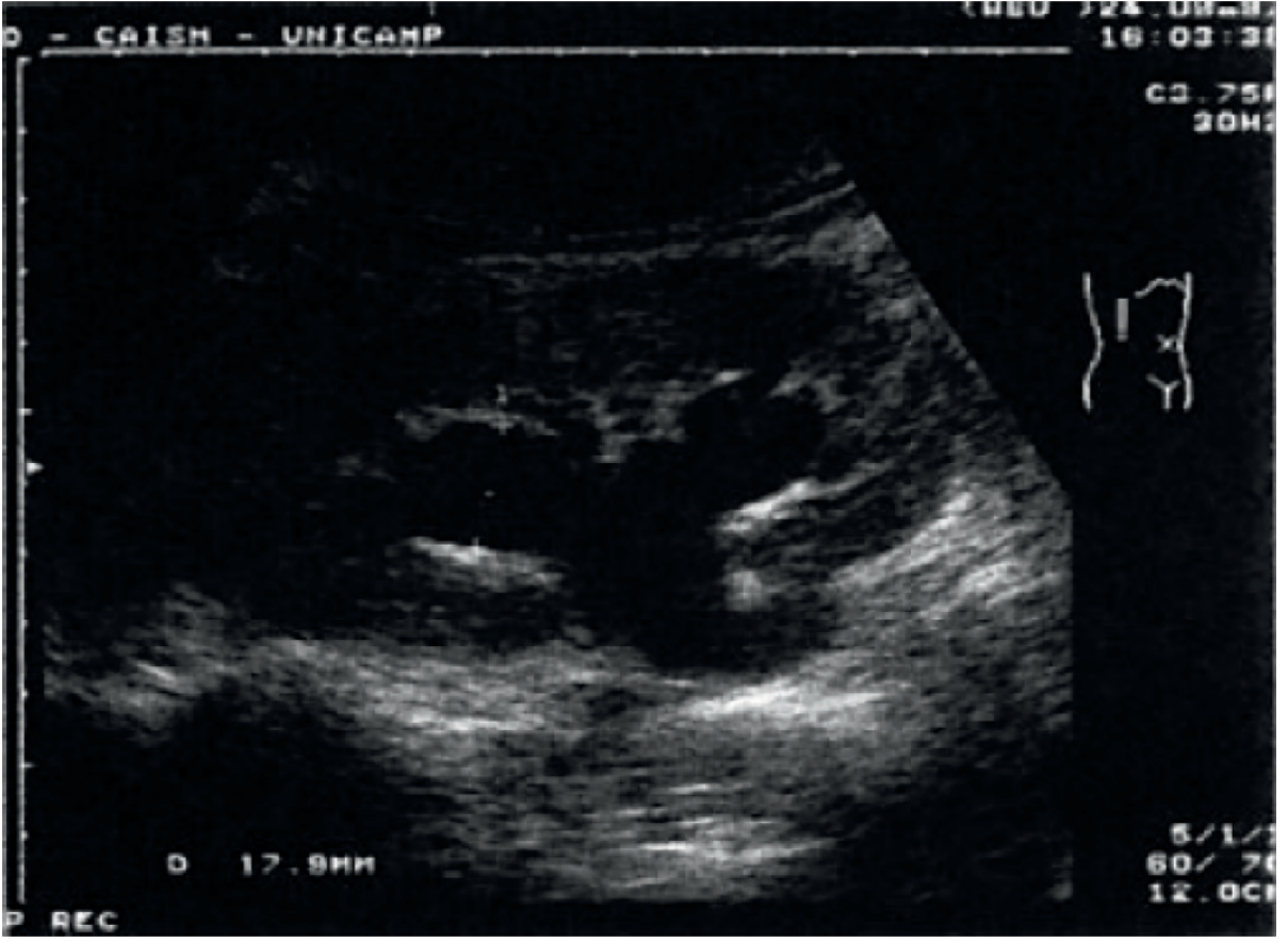
- Physiological hydronephrosis and hydroureter: the collecting system dilates in about 50% of pregnant women, more on the right than the left.
- Causes: progesterone-induced smooth muscle relaxation, mechanical compression of the ureters at the pelvic brim by the gravid uterus (the dextrorotated uterus and the right ovarian vein crossing the right ureter), and the cushioning effect of the sigmoid colon on the left.
- It is usually asymptomatic. This must be remembered when interpreting scans and when judging obstruction.
- Brenner and Rector's The Kidney, p. 2140-2141
Function
- GFR: it rises about 40-50% within weeks of conception and stays high until delivery. Renal plasma flow rises first (up to about 80%), then falls somewhat toward term. GFR stays high because of a lower plasma oncotic pressure and a higher Kf, so filtration fraction rises in late pregnancy.
- Brenner and Rector's The Kidney, p. 2140-2141
- Creatinine, urea and uric acid: they fall. Creatinine drops from about 0.8 to 0.5-0.6 mg/dL, so a value of 1.0 mg/dL (normal outside pregnancy) suggests renal impairment. BUN falls from about 13 to 8-10 mg/dL.
- Creatinine clearance rises to 150-200 mL/min.
- Glycosuria: it can be physiological, because the filtered glucose load exceeds tubular reabsorption. Glycosuria alone should not be used to diagnose diabetes.
- Proteinuria: protein excretion rises. The upper limit of normal is about 300 mg/24 h, and values above that raise concern for pre-eclampsia or renal disease.
- Sodium and water: the RAAS is activated, progesterone antagonises aldosterone, and the net result is about 900 mEq sodium and 6-8 L of water retained. The osmostat is reset (plasma osmolality falls about 10 mOsm/kg and sodium by about 4-5 mEq/L).
- Bladder: it is displaced upward and forward into the abdomen after about 12 weeks. The trigone is elevated and tone is reduced.
- Symptoms: frequency (early from pressure, late from head engagement), nocturia and stress incontinence.
- Urinary stasis and vesicoureteral reflux raise the risk of asymptomatic bacteriuria, UTI and pyelonephritis. Screening and treatment of bacteriuria is routine.
7. Gastrointestinal system and liver
- Mouth: gingivitis and bleeding gums, epulis of pregnancy (pyogenic granuloma), ptyalism, and altered taste (dysgeusia).
- Nausea and vomiting: "morning sickness" affects up to 70% and peaks at 8-10 weeks, correlating with hCG. It resolves by about 16-20 weeks in most.
- Oesophagus and stomach: progesterone relaxes the lower oesophageal sphincter and the stomach is displaced upward, which causes heartburn and reflux (especially third trimester). Gastric acid secretion is reduced early on.
- Gastric emptying: it is generally normal in pregnancy but is delayed in labour, so all labouring women are treated as having a full stomach for anaesthesia.
- Intestines: reduced motility causes constipation, increased water absorption and bloating. Haemorrhoids result from constipation and venous pressure.
- Appendix: it is displaced upward and laterally by the uterus, so appendicitis pain may be felt in the right flank or upper quadrant. Rosen notes that upward displacement of viscera also alters the localisation of pain and that stretched abdominal muscles blunt guarding and rebound.
- Rosen's Emergency Medicine, p. 3424
- Gallbladder: hypotonia and stasis, and progesterone-related cholesterol saturation of bile, increase gallstone risk.
- Liver: there is no morphological change and hepatic blood flow is unchanged.
- Alkaline phosphatase doubles (placental origin).
- Albumin falls.
- AST, ALT, bilirubin and GGT stay normal or slightly lower, so elevated transaminases are always abnormal.
- Cholesterol and triglycerides rise.
- Spider naevi and palmar erythema (oestrogen) are normal.
8. Endocrine system
- Pituitary: it enlarges. Prolactin rises about 10-fold. Placental GH replaces pituitary GH after the first trimester, and IGF-1 rises. LH and FSH fall.
- Creasy & Resnik's Maternal-Fetal Medicine, p. 1526-1527
- Thyroid:
- Oestrogen raises TBG about 2-fold, so total T4 and T3 rise, while free T4 stays normal.
- hCG has weak TSH activity, so TSH is lower in the first trimester (it can fall below the non-pregnant range).
- There is mild glandular enlargement, and iodine need increases.
- Trimester-specific reference ranges should be used.
- Adrenal:
- Placental CRH rises progressively in the second and third trimesters and peaks at delivery. ACTH rises 2-4 fold.
- Corticosteroid-binding globulin triples, so total cortisol and free cortisol (urinary, salivary) rise, hence a false-positive cortisol result for Cushing's if non-pregnancy ranges are used.
- Diurnal variation is blunted but retained.
- Placental 11-beta-HSD2 converts cortisol to cortisone, protecting the fetus.
- Aldosterone and renin rise (RAAS activation).
- Creasy & Resnik's Maternal-Fetal Medicine, p. 1537-1538
- Parathyroid and calcium: total calcium falls (low albumin), but ionised calcium is normal. 1,25-dihydroxyvitamin D rises, with increased intestinal calcium absorption. Calcium requirement is met by increased absorption.
- Pancreas and glucose metabolism:
- Early pregnancy: insulin sensitivity is normal or high, and fasting glucose is lower.
- Later pregnancy: hPL, cortisol, progesterone and placental GH cause insulin resistance, with postprandial hyperglycaemia. Pregnancy is called "diabetogenic" because gestational diabetes can unmask in women with limited beta-cell reserve. Screening is done at 24-28 weeks.
- Pregnancy is a state of "accelerated starvation": ketosis develops faster during fasting.
9. Metabolic changes and weight gain
- Weight gain: about 11-12 kg overall (recommended 10-12 kg in India, depending on pre-pregnancy BMI), with most gained in the second and third trimesters (about 0.4-0.5 kg per week).
| Component | Approx. weight |
|---|---|
| Fetus | 3.3 kg |
| Placenta | 0.6 kg |
| Liquor | 0.8 kg |
| Uterus | 0.9 kg |
| Breasts | 0.4 kg |
| Maternal blood volume | 1.5 kg |
| Extracellular fluid | 2 kg |
| Fat stores | 3-4 kg |
- BMR: it rises by about 15-20% by term, and oxygen consumption by 20%.
- Protein: a positive nitrogen balance, with an extra requirement of about 15 g/day.
- Fat: lipolysis in late pregnancy. Maternal cholesterol and triglycerides rise (fat stored in early pregnancy is a reserve for lactation).
- Water: total body water increases by about 6-8 L, causing physiological oedema in the legs.
- Minerals: an increased need for iron, calcium (extra 1 g/day during lactation), and folate (400 microgram/day, 4-5 mg for high-risk women).
- Extra calories: about 300-350 kcal/day in the second and third trimesters.
10. Skin, musculoskeletal and other changes
Skin and hair
- Hyperpigmentation: it is due to MSH and oestrogen/progesterone. It produces linea nigra, chloasma/melasma ("mask of pregnancy"), darkened areola and nipples, and darkening of scars.
- Striae gravidarum: stretch marks on the abdomen, breasts and thighs, due to collagen disruption and cortisol.
- Vascular: spider naevi, palmar erythema, and varicose veins.
- Hair: in pregnancy hair is in a prolonged anagen phase. After delivery, there is telogen effluvium (postpartum hair loss at about 3 months).
- Nails and glands: nails become brittle, and sweat and sebaceous gland activity increases.
Musculoskeletal
- Ligament laxity (relaxin and progesterone): the symphysis pubis widens by about 3-4 mm, and the sacroiliac joints become mobile. Rosen warns that a pubic symphysis diastasis can be mistaken for a pelvic fracture on imaging.
- Rosen's Emergency Medicine, p. 3424
- Posture: increased lumbar lordosis shifts the centre of gravity, causing the characteristic "waddling" gait ("pride of pregnancy"), backache and pelvic girdle pain.
- Rectus abdominis: diastasis recti may occur.
- Calcium and bone: transient bone mineral loss is possible, and it recovers after weaning.
Nervous system
- Carpal tunnel syndrome (fluid retention), meralgia paraesthetica, Bell's palsy (higher risk in third trimester and puerperium), and sleep disturbance.
- Reduced MAC (-40%) from progesterone and endorphins.
- Epidural venous engorgement and reduced epidural space volume mean lower local anaesthetic doses are needed.
Eyes and immunity
- Eyes: corneal thickness and sensitivity change, and contact lenses may become uncomfortable. IOP falls.
- Immunity: there is a shift toward Th2 and reduced cell-mediated immunity so the fetus is tolerated. This makes some infections more severe (influenza, varicella, malaria, hepatitis E, listeriosis).
11. Summary table: normal values in pregnancy
| Parameter | Non-pregnant | Pregnancy |
|---|---|---|
| Hb | 12-15 g/dL | 10.5-11 g/dL or above |
| Serum creatinine | 0.8 mg/dL | 0.5-0.6 mg/dL |
| BUN | 13 mg/dL | 8-10 mg/dL |
| GFR | Baseline | +40-50% |
| PaCO2 | 40 mmHg | About 30 mmHg |
| HCO3 | 24 mEq/L | 18-21 mEq/L |
| Cardiac output | 4.5 L/min | About 6 L/min |
| Fibrinogen | 200-400 mg/dL | 400-600 mg/dL |
| Alkaline phosphatase | Baseline | About 2-fold |
| Albumin | 3.5-5 g/dL | About 2.5-3.5 g/dL |
| Total T4 | Baseline | About 1.5 fold |
| Total cortisol | Baseline | About 3-fold |
| Prolactin | Baseline | About 10-fold |
12. Clinical correlations and exam points
- Supine hypotension: manage with left lateral tilt.
- Physiological anaemia: Hb below 11 g/dL is anaemia. Do not attribute a low Hb to dilution without checking iron status.
- Creatinine of 1.0 mg/dL in pregnancy suggests renal impairment.
- Hydronephrosis on ultrasound is usually physiological and right-sided.
- ALP is raised in normal pregnancy. Raised ALT or AST is not normal.
- Hypercoagulable state: DVT/PE risk is increased, highest postpartum.
- Compensated respiratory alkalosis: a "normal" PaCO2 of 40 mmHg in a breathless pregnant woman suggests impending respiratory failure.
- Cardiac disease: peak risk is at 28-32 weeks, during labour, and in the first week postpartum.
- Glycosuria can be physiological, so test blood glucose.
- Anaesthesia: aspiration risk, difficult airway, rapid desaturation and a reduced MAC.
13. Quick mnemonic and viva questions
Remember: "Everything goes UP except..." Blood volume, cardiac output, GFR, minute ventilation, clotting factors, ESR and ALP all go up. Haemoglobin, creatinine, BP (briefly), FRC, PaCO2, albumin and gastrointestinal motility go down.
Likely viva questions
- Why is there physiological anaemia of pregnancy? (Plasma volume rises more than red cell mass.)
- When is cardiac output highest? (28-32 weeks; further surge in labour and immediately postpartum.)
- Why is hydronephrosis commoner on the right?
- What is the respiratory acid-base status of pregnancy? (Compensated respiratory alkalosis.)
- What is the luteoplacental shift? (Placenta takes over progesterone production from the corpus luteum at about 8-10 weeks.)
Notes on sources
- Values come from the library textbooks cited above, with supporting details (fundal height landmarks, ranges for reference values, mnemonics) from standard obstetric teaching. These reference values vary slightly between textbooks (for example, fundal height at the umbilicus is 20 weeks in Rosen and 24 weeks in Indian teaching), so use the figures your own university text prescribes.
- A quick PubMed check for recent systematic reviews on pregnancy physiology found only reviews on gait biomechanics and taste/smell changes (PMIDs 40311235, 38569400, 37890603). None of them contradict the material above.
I can follow this with a focused note on one system (for example cardiovascular or haematological changes), a set of MCQs, or short-answer questions. Tell me if you want any of those.
This is a shared conversation. Sign in to Orris to start your own chat.