Whiplash injury

10 mark pg answer with flow chart and images

Reading File
Finding Sources
Finding Sources
Searching the Web

whiplash injury mechanism pathophysiology classification WAD grades management

Reading File
Reading File
Reading File
Reading File
Reading File
Reading File
Searching PubMed

whiplash associated disorder management treatment

Loading Image
Loading Image
Reading File
Reading File
Generating Image

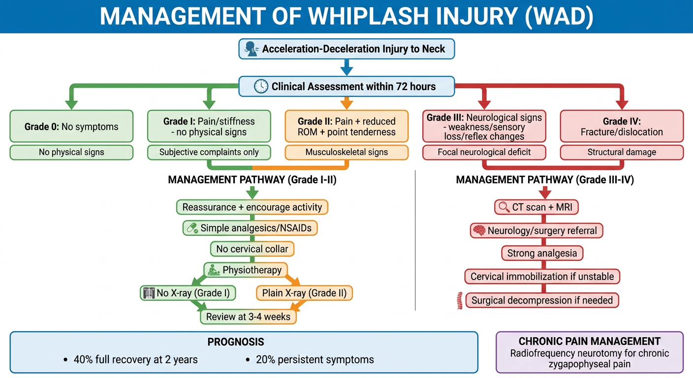
A detailed clinical flowchart titled "Management of Whiplash Injury (WAD)" showing the following pathway: At the top - "Acceleration-Deceleration Injury to Neck" leading down to "Clinical Assessment within 72 hours" then branching into 5 WAD grades (Grade 0: No symptoms, Grade I: Pain/stiffness - no physical signs, Grade II: Pain + reduced ROM + point tenderness, Grade III: Neurological signs - weakness/sensory loss/reflex changes, Grade IV: Fracture/dislocation). Below the grading - two management pathways: Left branch (Grade I-II) showing "Reassurance + encourage activity, Simple analgesics/NSAIDs, No cervical collar, Physiotherapy, No X-ray (Grade I) / Plain X-ray (Grade II), Review at 3-4 weeks". Right branch (Grade III-IV) showing "CT scan + MRI, Neurology/surgery referral, Strong analgesia, Cervical immobilization if unstable, Surgical decompression if needed". Bottom section showing "Prognosis: 40% full recovery at 2 years, 20% persistent symptoms" and a chronic pain box showing "Radiofrequency neurotomy for chronic zygapophyseal pain". Clean medical infographic style with color coding: green for mild grades, orange for moderate, red for severe.

Here is a comprehensive 10-mark answer on Whiplash Injury:

WHIPLASH INJURY (Whiplash-Associated Disorder - WAD)


DEFINITION

Whiplash injury is an indirect injury to the neck caused by an acceleration-deceleration incident resulting in a flexion-extension injury of the cervical spine, without direct trauma. The term "whiplash" implies amplification of relatively small forces into larger ones. The resulting functional impairment is termed Whiplash-Associated Disorder (WAD).
Scott-Brown's Otorhinolaryngology Head & Neck Surgery, Vol 2

MECHANISM OF INJURY

The traditional view of simple hyperextension is overly simplistic. Modern biomechanical studies have revealed a more complex sequence:
In a rear-end collision:
  1. The lower cervical spine is thrust upward and forward by inertial loading
  2. The lower segments undergo extension while upper segments remain in relative flexion
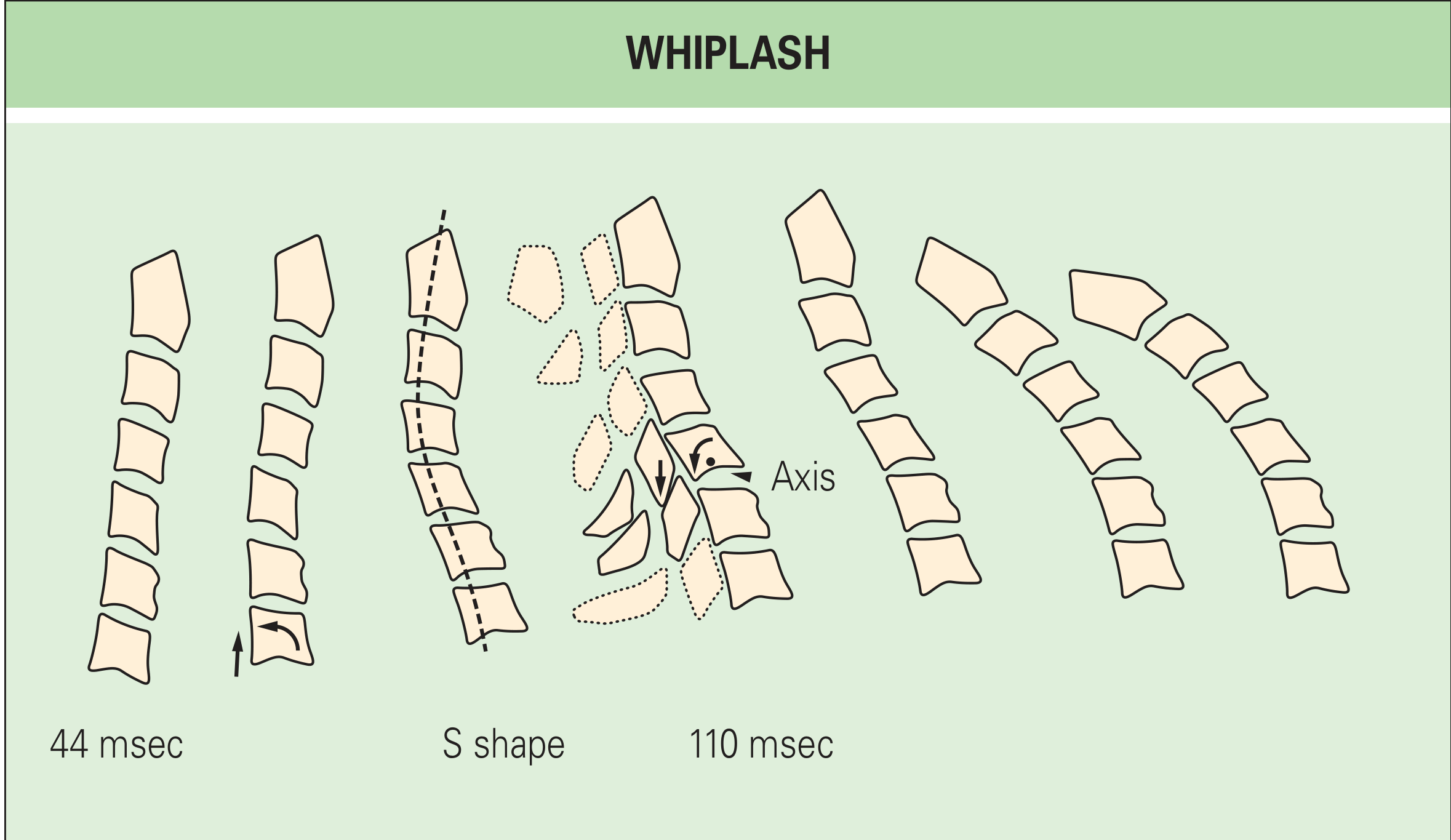
  3. This creates a characteristic "S-shape" deformity at ~100 msec
  4. Movement occurs around abnormal axes of rotation (pathologic axes higher than normal)
  5. The anterior end of the vertebra separates from the body below; posteriorly, the inferior articular process chisels into the superior articular process
  6. Thereafter, all segments undergo full extension and the head is thrown forward
The zygapophyseal (facet) joints are particularly susceptible due to rotation through these abnormal axes.
Sequential cervical spine radiographs showing S-shape deformity during the extension phase of whiplash, with abnormal axis at 110 msec
Sequential radiographs of the cervical spine during whiplash. At 110 msec, C5 rotates about an abnormally high axis, separating anteriorly from C6 while the inferior articular process chisels into the superior articular process of C6. - Rheumatology, 2-Volume Set (Elsevier 2022)

PATHOLOGY

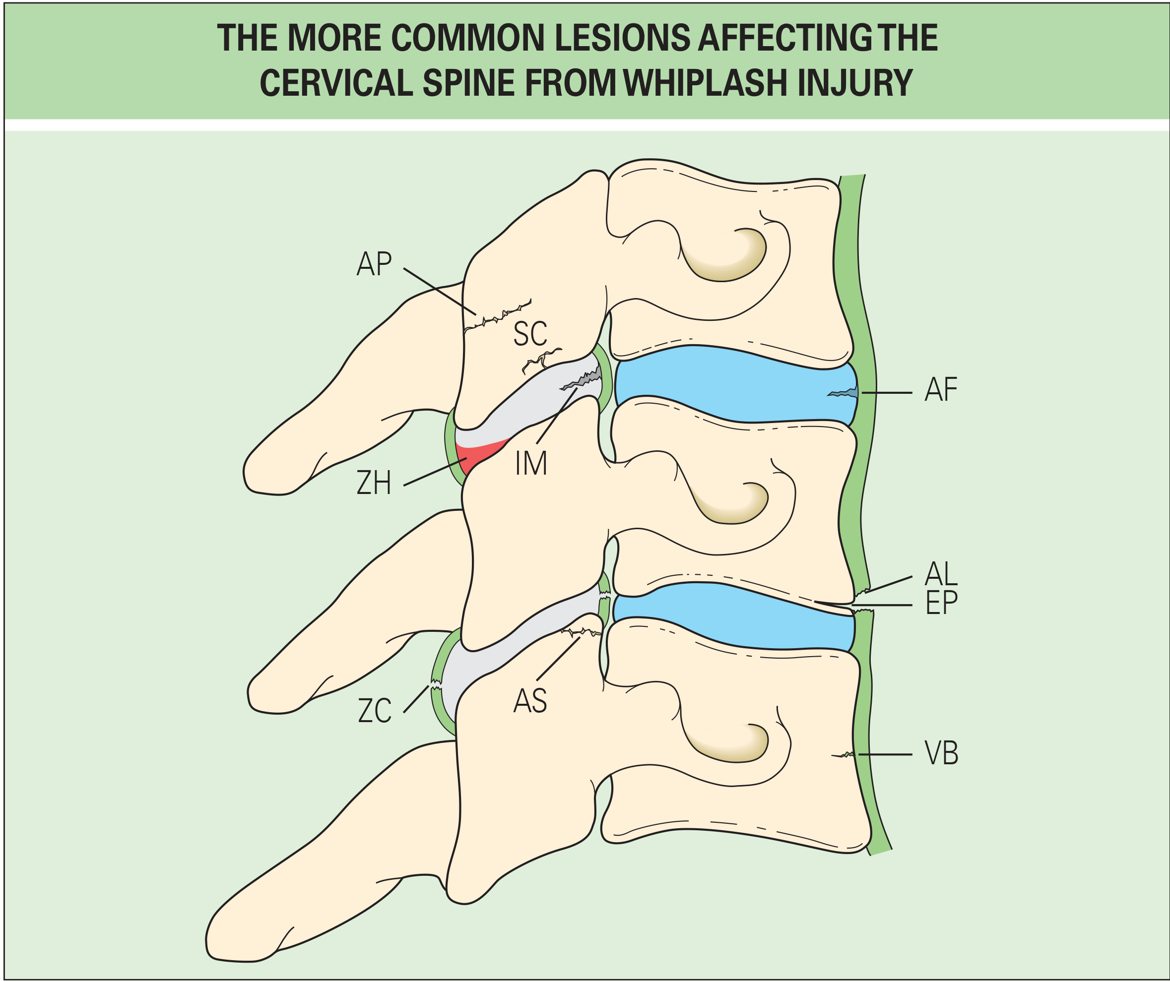
The common structural lesions from whiplash injury are:
Anatomical diagram showing cervical spine lesions from whiplash: AF = annulus fibrosus tear, AL = anterior longitudinal ligament tear, AP = articular pillar fracture, AS = articular surface fracture, EP = endplate avulsion, IM = intraarticular meniscus contusion, SC = subchondral plate fracture, VB = vertebral body fracture, ZC = zygapophyseal capsule rupture, ZH = zygapophyseal hemarthrosis
Common lesions of the cervical spine in whiplash. Key structures: AF = annulus fibrosus tear; ZC = zygapophyseal joint capsule rupture; ZH = hemarthrosis; IM = intraarticular meniscus contusion; EP = endplate avulsion; VB = vertebral body fracture - Rheumatology, 2-Volume Set (Elsevier 2022)
Plain radiographs detect only ~4 out of 245 injuries even under optimal conditions - they are profoundly insensitive to all but the most severe bony injuries.

EPIDEMIOLOGY

  • Incidence: 70-300 per 100,000 population
  • Over 1 million new cases per year in the US
  • 85% of cases are from rear-end motor vehicle collisions
  • UK: 250,000 patients per year at an estimated cost of £3 billion
  • Risk increases with seat belt use (paradoxically - 268% increase after mandatory seat belt law)

CLASSIFICATION - Quebec Task Force (QTF, 1995)

Symptoms/signs must occur within 72 hours to be attributable to the trauma.
GradeSymptoms and Signs
0No symptoms, no physical signs
IPain/stiffness/tenderness on motion - NO physical signs on examination
IIPain + musculoskeletal signs: reduced range of motion, point tenderness
IIIGrade II + neurological signs: weakness, sensory loss, absent/reduced reflexes, long-tract signs
IVFracture and/or dislocation of the cervical spine
  • 90% of all whiplash claims fall under Grade I and II
  • Grade III involves nerve root compression (e.g., disc herniation)

CLINICAL FEATURES

Acute symptoms (within 72 hours):
  • Neck pain and stiffness (most common)
  • Headache (occipital, often referred)
  • Shoulder and arm pain
  • Restricted cervical range of motion
  • Dizziness/vertigo (particularly in Grade III)
  • Visual disturbances
  • Dysphagia
Signs by grade:
  • Grade I: normal examination
  • Grade II: reduced ROM, point tenderness, muscle spasm
  • Grade III: reduced/absent deep tendon reflexes, weakness, dermatomal sensory loss
Less common features:
  • Cognitive symptoms (~20% of patients)
  • Tinnitus (not attributable to minor Grade I-II injury; tinnitus is not a persisting feature in Grade I-II)
  • Subjective hearing loss (13% of cases)
  • Benign Paroxysmal Positional Vertigo (most common vestibular complaint, usually Grade III)

INVESTIGATIONS / IMAGING

GradeImaging Recommendation
INo X-ray (exceptions: age >65, concurrent skeletal disease, prior neck surgery)
IIPlain X-ray or CT (CT mandatory if any nerve root or cord symptoms)
IIICT mandatory + MRI often indicated
IVCT + MRI (urgent surgical assessment)
Adapted from NICE guidelines and Jansen et al. - Scott-Brown's Otorhinolaryngology, Vol 2

MANAGEMENT FLOWCHART

Management flowchart for Whiplash-Associated Disorder showing grade-based pathway from initial assessment through treatment to prognosis, with Grade I-II managed conservatively and Grade III-IV requiring imaging and specialist referral

TREATMENT

Acute WAD (Grade I-II):

  • Encourage activity - activation is superior to rest; cervical collars and passive modalities are inferior and should be avoided
  • Simple analgesics (paracetamol); NSAIDs for 3 weeks maximum
  • Opioids not recommended for Grade I; only for VAS >8 in Grade II-III and for <2 weeks
  • Muscle relaxants: no proven efficacy
  • Systemic steroids: no benefit over placebo
  • Reassurance and psychological support
  • Physiotherapy with active exercise

Acute WAD (Grade III):

  • NSAIDs + nonopioid analgesics
  • Short-term opioids if severe pain
  • Neurology/surgical referral
  • CT + MRI imaging

WAD Grade IV:

  • Urgent orthopedic/neurosurgical referral
  • Cervical immobilization
  • Surgical decompression if indicated

Chronic Whiplash:

  • Percutaneous radiofrequency neurotomy - the single most promising treatment for constant, debilitating zygapophyseal joint pain (palliative but valuable)
  • Exercise therapy
  • Cognitive behavioral therapy for psychological components
  • Acupuncture: a 2024 systematic review (Lee et al., BMJ Open, PMID 38233056) found benefit for whiplash-related pain

PROGNOSIS

OutcomeGrade IGrade IIGrade III
Neck pain >6 months44%81%Up to 90%
Headache >6 months37%37%70%
  • ~40% of patients report complete recovery at 2 years
  • ~45% continue to have major complaints at 2 years
  • ~20% have persistent symptoms overall
  • Grade I-II: 97-100% recover within 1 year
  • Whiplash injury is extremely unlikely at speeds under 3 mph (4.8 km/h)
  • Compensation/litigation significantly worsens prognosis

MEDICOLEGAL IMPORTANCE

  • Whiplash is one of the most common causes of personal injury claims
  • The QTF emphasizes it is essentially a benign condition but a refractory minority accounts for disproportionate costs
  • Studies from Lithuania (where litigation is uncommon) showed far less symptomatology than comparable US accidents - highlighting the role of psychological and compensation factors
  • Cognitive symptoms claimed by ~20% are likely functional or malingering in many cases
Bradley and Daroff's Neurology in Clinical Practice; Rheumatology, 2-Volume Set (Elsevier 2022); Scott-Brown's Otorhinolaryngology Head & Neck Surgery, Vol 2

Key takeaway: Whiplash/WAD is classified Grade 0-IV by the Quebec Task Force (1995). The S-shape biomechanics explain zygapophyseal joint vulnerability. Treatment for Grade I-II centers on active mobilization, not rest or collar, and prognosis is generally good, though a significant minority develop chronic symptoms requiring radiofrequency neurotomy.

Create a comparison table of whiplash grades with imaging findings and prognosis

I have all the data needed from the previous research. Here is the comprehensive comparison table:

Whiplash-Associated Disorder (WAD) - Grade Comparison Table

Quebec Task Force Classification (1995)

FeatureGrade 0Grade IGrade IIGrade IIIGrade IV
SeverityNoneMildModerateSevereCritical
SymptomsNoneNeck pain, stiffness, or tenderness on motionNeck pain + stiffness + tendernessNeck pain + neurological symptoms (numbness, tingling, weakness)Neck pain + signs of fracture/dislocation
Physical Signs on ExamNoneNone (subjective complaints only)Reduced ROM + point tenderness + muscle spasmReduced/absent deep tendon reflexes, muscle weakness, dermatomal sensory loss, long-tract signsStructural instability; may have spinal cord compression signs
Nerve InvolvementNoneNoneNoneNerve root compression (e.g., disc herniation)Spinal cord or nerve root compromise
Structural DamageNoneSoft tissue micro-injury (undetectable)Soft tissue injury - muscle, ligament, zygapophyseal joint capsuleDisc herniation, nerve root compression, facet fractureCervical fracture and/or dislocation
Proportion of Cases-~50% of claims~40% of claims<10%Rare

Imaging Findings by Grade

FeatureGrade 0Grade IGrade IIGrade IIIGrade IV
X-Ray IndicationNot indicatedNot indicatedPlain X-ray (AP, lateral, odontoid views)Not sufficient aloneMandatory
X-Ray FindingsNormalNormalUsually normal; may show loss of cervical lordosis, prevertebral soft tissue swellingMay show subtle instability, disc space narrowingFracture, dislocation, malalignment visible
CT ScanNot indicatedNot indicatedMandatory if any nerve root or cord symptoms developMandatoryMandatory (urgent)
CT FindingsNormalNormalNormal or subtle soft tissue swellingDisc herniation, foraminal narrowing, facet fractureFracture fragments, dislocation, cord compression
MRI IndicationNot indicatedNot indicatedOnly if CT inconclusive or cord symptomsOften indicated (after CT)Mandatory
MRI FindingsNormalNormalSoft tissue edema, ligament tears, disc bulgeDisc herniation with cord/root contact, nerve root edema, cord signal changeCord contusion/transection, severe disc herniation, epidural hematoma
Key Imaging Rule-No imaging (exception: age >65, rheumatoid arthritis, prior neck surgery)Plain X-ray or CT; CT mandatory if neurological symptoms appearCT + MRICT + MRI urgently
Source: NICE guidelines and Jansen et al., adapted from Scott-Brown's Otorhinolaryngology Head & Neck Surgery, Vol 2

Prognosis by Grade

Prognostic FactorGrade 0Grade IGrade IIGrade IIIGrade IV
Symptom OnsetNoneWithin 72 hoursWithin 72 hoursWithin 72 hoursImmediate
Neck Pain >6 Months0%44%81%Up to 90%Persistent (variable with surgery)
Headache >6 Months0%37%37%70%Common
Recovery within 1 Year100%~97-100%~80%<50%Depends on surgery
Full Recovery at 2 Years100%~60%~40%~10-20%Poor without surgery
Persistent Symptoms OverallNoneMinority~20%MajorityMajority
Major Complaints at 2 YearsNoneLow~45%HighVery high
Chronicity RiskNoneLowModerate-highHighVery high
Return to WorkImmediateUsually within weeksWeeks to monthsMonths to yearsUncertain
Recommended TreatmentNoneReassurance, active mobilization, simple analgesia, no collarNSAIDs (≤3 weeks), physiotherapy, active exercise, no collarNSAIDs + opioids (short-term if VAS >8), neurology referral, CT + MRIUrgent surgical referral, immobilization, decompression

Factors That Worsen Prognosis (Any Grade)

FactorEffect
Ongoing litigation / compensation claimSignificantly prolongs symptoms and delays recovery
High initial pain intensityPredicts chronicity
Pre-existing neck diseaseWorsens outcome
Psychological distress / anxiety / depressionStrong predictor of chronic WAD
Older ageSlower recovery
High-speed impactGreater structural damage
Female sexSlightly worse prognosis in some studies
Passive coping (collar, bed rest)Promotes chronicity

Treatment Summary by Grade

TreatmentGrade IGrade IIGrade IIIGrade IV
Simple analgesics (paracetamol)YesYesYesYes
NSAIDsNot neededYes (≤3 weeks)YesYes
OpioidsNoOnly if VAS >8, ≤2 weeksShort-termYes
Cervical collarNoNoPossible short-termYes (if unstable)
Active mobilization/physioYes - first lineYes - first lineGuided physioPost-surgical rehab
Muscle relaxantsNoNo proven benefitNo proven benefit-
Systemic steroidsNoNo benefit vs placeboNo-
CT/MRI imagingNoOnly if neuro signs developYesYes (urgent)
Surgical interventionNoNoOnly if cord compressionYes
Radiofrequency neurotomyNoChronic cases onlyChronic cases-
Cognitive behavioral therapyConsider if persistentYesYesYes

Clinical pearl: Plain radiographs are profoundly insensitive - in one cadaveric study, only 4 of 245 structural injuries were detected on X-ray even under optimal conditions. CT and MRI are the investigations of choice for Grade III and IV. For Grade I-II, the most effective intervention is encouraging activity over rest - activation programs consistently outperform collars, passive modalities, and bed rest.
Sources: Rheumatology 2-Volume Set (Elsevier 2022); Scott-Brown's Otorhinolaryngology Head & Neck Surgery Vol 2; Bradley and Daroff's Neurology in Clinical Practice; NICE Guidelines
This is a shared conversation. Sign in to Orris to start your own chat.