
Genitourinary tract microbiology full chapter refer all text books
Reading File
Now let me search across all available textbooks for genitourinary tract microbiology content in parallel.
Finding Sources
Finding Sources
Finding Sources
Excellent - the key microbiology textbook is Sherris & Ryan's Medical Microbiology. Let me now read the primary microbiology chapter and search for additional relevant content in parallel.
Finding Sources
Finding Sources
Reading File
Good start. Now let me pull the full GU microbiology chapter from Sherris & Ryan, plus Jawetz microbiology GU content, in parallel.
Finding Sources
Finding Sources
Reading File
Let me now search for the dedicated GU infection chapters from Jawetz and other microbiology texts, and also look for specific topics like UTI pathogens, STIs, etc.
Finding Sources
Finding Sources
Finding Sources
I've found the key sources. Now let me read the most important textbook sections in parallel - the Sherris GU chapter, Jawetz GU infections, and Medical Microbiology 9e chapters.
Reading File
Reading File
Reading File
Excellent content. Let me now read more sections - Sherris Gonorrhea continued, Jawetz Chlamydia, and Jawetz Syphilis chapters.
Reading File
Reading File
Reading File
Excellent - I have rich content now. Let me read more from Sherris (gonorrhea clinical, UTI section) and Jawetz syphilis continued and UTI pathogens.
Reading File
Reading File
Reading File
I now have rich content from multiple sources. Let me read more sections on UTI pathogens, Candida, HSV, and the vaginal flora sections.
Reading File
Reading File
Reading File
I now have comprehensive content from multiple textbooks. Let me fetch the key textbook images and read one more critical section on UTI pathogens.
Reading File
Loading Image
Loading Image
Excellent - I have beautiful textbook images. Now let me fetch two more important images and read the UTI pathogens section from Jawetz.
Loading Image
Reading File
I now have comprehensive, rich content from multiple textbooks. Let me update the todos and compile the full comprehensive chapter.Now I have all the content needed to produce a comprehensive, well-organized chapter. Here it is:
Genitourinary Tract Microbiology - Comprehensive Chapter
Sourced from: Sherris & Ryan's Medical Microbiology (8th Ed), Jawetz Melnick & Adelberg's Medical Microbiology (28th Ed), Medical Microbiology 9e (Murray), Goldman-Cecil Medicine, Bailey & Love's Surgery
SECTION 1: NORMAL FLORA OF THE GENITOURINARY TRACT
1.1 Urinary Tract
The urinary tract is sterile in health above the distal 1 cm of the urethra. Urine in the bladder, ureters, and renal pelvis is normally free of organisms. The distal urethra harbors a scanty flora derived from the perineum, which may include coagulase-negative staphylococci, diphtheroids, and some Gram-negative organisms.
(Sherris & Ryan's Medical Microbiology, 8th Ed, p. 37)
1.2 Vaginal Flora
Vaginal flora varies profoundly with hormonal status:
| Life Stage | Dominant Flora | pH |
|---|---|---|
| Pre-puberty | Mixed, scanty; skin and colonic organisms | Neutral |
| Childbearing years | Lactobacillus spp. (anaerobic/microaerophilic) dominant | 4–5 (acidic) |
| Post-menopause | Mixed, non-specific; reduced Lactobacillus | Neutral |
Mechanism of acidification: Estrogen stimulates glycogen deposition in vaginal epithelial cells. Lactobacilli metabolize this glycogen to lactic acid, producing a pH of 4–5. This acidic environment is optimal for Lactobacillus survival but inhibits many pathogens.
Other organisms present during reproductive years (in smaller numbers):
- Anaerobic Gram-negative rods
- Gram-positive cocci
- Candida spp. (yeasts)
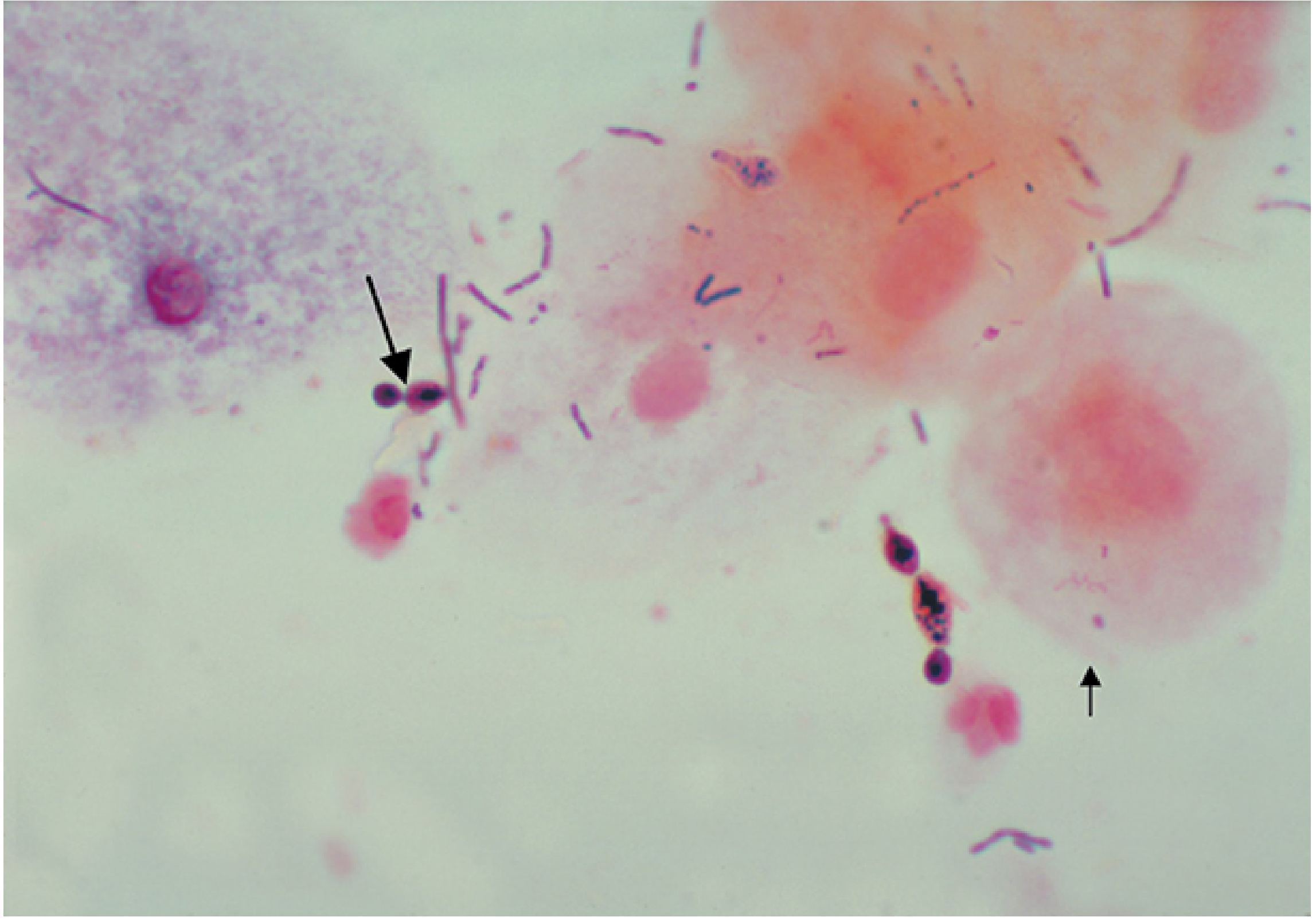
(Sherris & Ryan's Medical Microbiology, 8th Ed, p. 37)
1.3 Bacterial Vaginosis (BV)
Definition: Bacterial vaginosis is a common syndrome (not a true infection) characterized by a shift in the vaginal microbiota away from the normal acidic Lactobacillus-dominant flora.
Key Features:
- Vaginal discharge with a characteristic "fishy" (amine) odor
- Clue cells - epithelial cells coated with bacteria visible on microscopy
- Higher pH (>4.5) and greater microbial diversity
- Increased anaerobes
Organisms implicated:
- Gardnerella vaginalis (most associated)
- Mobiluncus spp.
- Anaerobes (Prevotella, Peptostreptococcus)
- Mycoplasma hominis
Whiff test (KOH): Positive - amine odor released when 10% KOH added to discharge.
Diagnosis (Amsel's criteria - 3 of 4 required):
- Homogeneous, thin, white discharge
- Clue cells on wet mount
- pH >4.5
- Positive whiff test
(Sherris & Ryan's Medical Microbiology, 8th Ed, p. 38)
SECTION 2: URINARY TRACT INFECTIONS (UTIs)
2.1 Epidemiology and Pathogenesis
UTIs are among the most common bacterial infections. They occur more frequently in women due to the shorter urethra and proximity of the urethral meatus to the perineum/anus.
Route of infection: Ascending route is the most common - organisms from the perineal/perianal flora ascend the urethra into the bladder.
Host factors predisposing to UTI:
- Female sex
- Sexual activity (introduction of organisms)
- Pregnancy (ureteral dilation, bladder atony)
- Urologic abnormalities (obstruction, reflux, calculi)
- Catheterization (bypasses urethral defenses)
- Diabetes mellitus
- Immunosuppression
- Incomplete bladder emptying
2.2 Classification
| Type | Definition |
|---|---|
| Uncomplicated UTI | Infection in structurally/neurologically normal urinary tract |
| Complicated UTI | Associated with functional/structural abnormality, or special host factors (pregnancy, male sex, immunosuppression) |
| Cystitis | Infection limited to bladder (lower UTI) |
| Pyelonephritis | Infection of renal parenchyma and pelvis (upper UTI) |
| Asymptomatic bacteriuria | ≥10⁵ CFU/mL on 2 consecutive specimens, no symptoms |
2.3 Causative Organisms
Most common causative organisms in community-acquired UTI:
| Organism | Frequency | Notes |
|---|---|---|
| Escherichia coli | ~80% community UTI | Most important; UPEC strains with specific virulence factors |
| Staphylococcus saprophyticus | ~5-15% in young women | Second most common in young sexually active women |
| Klebsiella pneumoniae | ~5% | More common in complicated/hospital UTI |
| Proteus mirabilis | ~2-5% | Associated with struvite stones; urease-positive |
| Enterococcus faecalis | Common in catheter-associated/hospital-acquired | |
| Pseudomonas aeruginosa | Hospital-acquired, catheter-associated | |
| Staphylococcus aureus | Hematogenous spread; unusual for ascending |
Hospital/Catheter-Associated UTI (CAUTI):
- E. coli, Klebsiella, Pseudomonas, Enterococcus, Candida spp.
- Polymicrobial infections more common
2.4 Virulence Factors of Uropathogenic E. coli (UPEC)
| Virulence Factor | Function |
|---|---|
| Type 1 fimbriae (FimH) | Attach to uroplakins on bladder epithelium |
| P fimbriae (Pap) | Bind to P blood group antigen glycolipids on uroepithelium; associated with pyelonephritis |
| S fimbriae | Bind sialic acid-containing structures |
| Hemolysin (HlyA) | Lyses erythrocytes and leukocytes; releases iron |
| Aerobactin/Siderophores | Iron acquisition |
| Capsular antigen (K antigen) | Resistance to complement and phagocytosis |
| Lipopolysaccharide (LPS/O antigen) | Endotoxin, contributes to inflammation |
2.5 Diagnosis
Urine Collection:
- Midstream clean-catch specimen
- Catheterized specimen
- Suprapubic aspirate (gold standard - any growth significant)
Significant Bacteriuria (Kass criteria):
- ≥10⁵ CFU/mL in midstream specimen: significant
- ≥10³ CFU/mL in symptomatic women: diagnostic
- Any growth in suprapubic aspirate: significant
Dipstick analysis:
- Leukocyte esterase: Indicates pyuria (WBCs in urine)
- Nitrite test: Positive if nitrate-reducing bacteria (E. coli, Klebsiella, Proteus) present; NOT positive for Enterococcus, Pseudomonas, S. saprophyticus
Microscopy: >5 WBCs/HPF (pyuria) or bacteria on unspun specimen
Culture and Sensitivity: Gold standard; required for complicated UTI and treatment failures.
2.6 Treatment
| Condition | First-Line Agents |
|---|---|
| Uncomplicated cystitis (women) | Trimethoprim-sulfamethoxazole, nitrofurantoin, fosfomycin |
| Pyelonephritis (outpatient) | Fluoroquinolone (ciprofloxacin) or oral cephalosporin |
| Pyelonephritis (inpatient) | IV ceftriaxone, fluoroquinolone, aminoglycoside |
| Complicated UTI | Based on culture/sensitivity |
| CAUTI | Remove/replace catheter; treat based on culture |
SECTION 3: SEXUALLY TRANSMITTED INFECTIONS (STIs) - BACTERIAL
3.1 Gonorrhea (Neisseria gonorrhoeae)
Bacteriology
N. gonorrhoeae (gonococcus) are Gram-negative diplococci (kidney/coffee bean-shaped, pairs with concave surfaces facing each other). They are:
- Oxidase positive, catalase positive
- Acid from glucose only (not maltose - differentiates from N. meningitidis)
- Grow best on chocolate agar (or Thayer-Martin/NYC/ML selective media) supplemented with CO₂
- Colonies: small, smooth, non-pigmented; appear at 18-24 hours; 2-4 mm at 48 hours
- More fragile than meningococci; lack a polysaccharide capsule
(Jawetz, 28th Ed, p. 301 - Biochemical Reactions Table 20-1)
Differential (glucose oxidation table):
| Organism | Glucose | Maltose | Lactose | Sucrose |
|---|---|---|---|---|
| N. gonorrhoeae | + | - | - | - |
| N. meningitidis | + | + | - | - |
| N. lactamica | + | + | + | - |
Outer Membrane Virulence Factors
| Component | Function |
|---|---|
| Type IV Pili | Initial attachment to nonciliated epithelium; "twitching motility"; transformation (DNA uptake) |
| Opa proteins | Tight binding to host cell receptors (CD46, CD66, integrins); mediate microcolony cohesion |
| Por proteins (Por1BA, Por1BB) | Trigger host cell signaling; channel formation in outer membrane |
| LOS (Lipooligosaccharide) | Endotoxin; sheds vesicles; LOS sialylation binds Factor H - resists complement |
| Rmp (MOMP III) | Blocks bactericidal antibody |
| IgA1 protease | Cleaves secretory IgA at mucosal surfaces |
| Transferrin/lactoferrin-binding proteins | Iron acquisition |
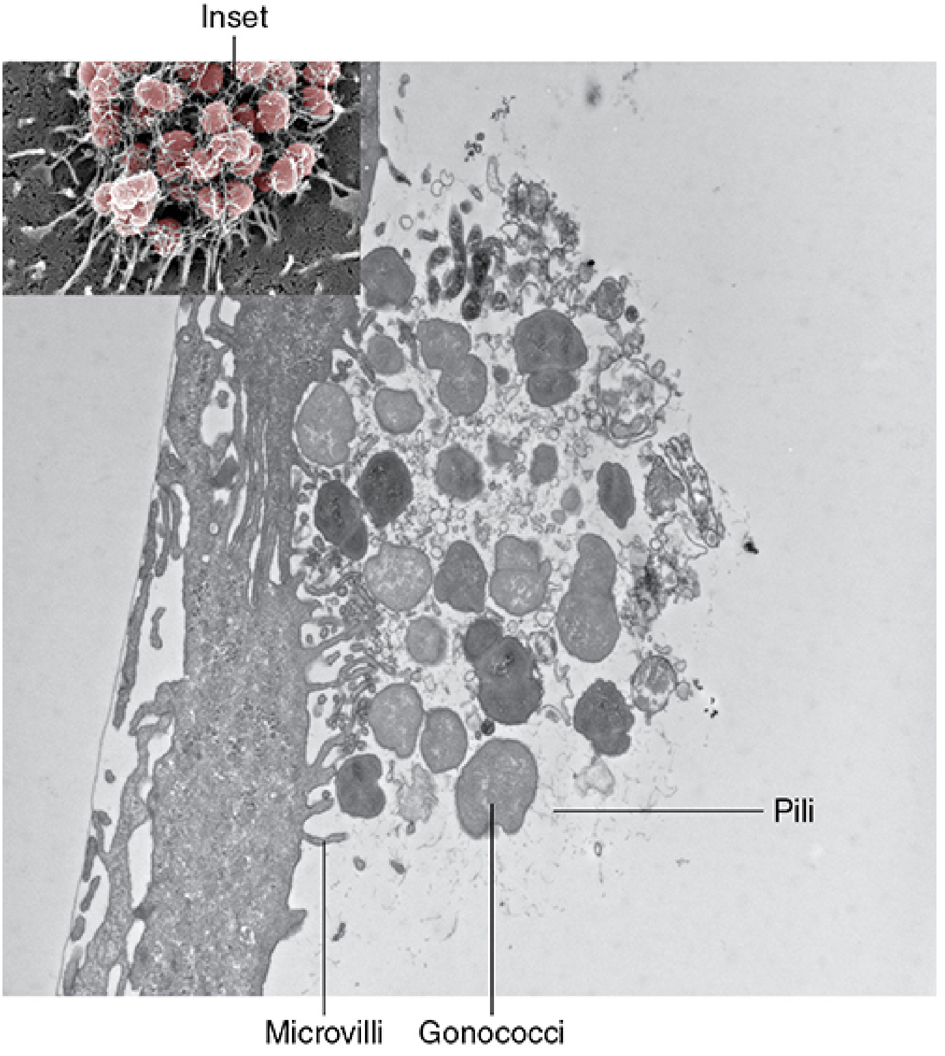
Pathogenesis
- Attachment: Pili and Opa proteins attach to receptors (CD46, CD66, integrins) on nonciliated epithelial cells
- Active twitching motility of pili moves microcolonies across the cell surface
- Phagocytosis induction: Por1BA triggers signaling cascades → microfilaments/microtubules engulf bacteria
- Transcytosis: Bacteria pass through the host cell and exit through the basal membrane into the submucosa
- Survival: LOS sialylation binds Factor H → blocks C3b deposition; pili + Opa interfere with phagocytic killing; catalase upregulation resists oxidative killing
(Sherris & Ryan's Medical Microbiology, 8th Ed, pp. 1200-1201)
Clinical Diseases
In Men:
- Urethritis: Incubation 2-5 days; dysuria and purulent urethral discharge (yellow-green); 10% asymptomatic
- Epididymitis: Extension from urethra; unilateral scrotal pain and swelling
- Prostatitis: Fever, perineal pain, urinary symptoms
- Proctitis (MSM): rectal pain, discharge
In Women:
- Cervicitis: Most common presentation; mucopurulent endocervical discharge; often asymptomatic
- Urethritis: Dysuria, frequency; may coexist with cervicitis
- Bartholin gland abscess: Swollen, painful unilateral labial mass
- Pelvic Inflammatory Disease (PID): Extension up fallopian tubes causing salpingitis; fever, lower abdominal pain, cervical motion tenderness; complications: infertility and ectopic pregnancy from tubal scarring
- Tubo-ovarian abscess: Serious complication of PID
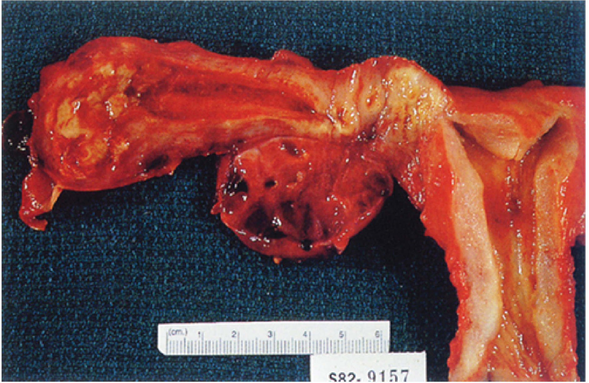
Important: PID may also be caused by Chlamydia trachomatis, anaerobes, or mixed organisms ascending the same route.
Extragenital and Systemic:
- Pharyngitis: Exudative or asymptomatic; from orogenital contact
- Conjunctivitis (Ophthalmia Neonatorum): In neonates exposed during delivery; severe purulent conjunctivitis that can lead to blindness; prevented by 1% silver nitrate or erythromycin eye drops at birth
- Disseminated Gonococcal Infection (DGI):
- Occurs in ~1-3% of untreated infections
- Bacteremic phase: fever, migratory polyarthralgia, petechial/maculopapular/pustular skin rash
- Risk factors: complement deficiency (C5-C9 deficiency)
- Septic arthritis: Purulent arthritis affecting large joints (elbow, knee); gonococci culturable from joint pus
- Rare: endocarditis, meningitis
(Sherris & Ryan's Medical Microbiology, 8th Ed, pp. 1206)
Diagnosis
| Test | Specimen | Sensitivity/Specificity |
|---|---|---|
| Gram smear | Urethral discharge (men) | >95% sensitive AND specific in symptomatic men |
| Gram smear | Cervical specimens | Only 50-70% sensitive; less specific due to normal flora |
| Culture | All sites | Sensitive and specific; needed for antibiotic susceptibility |
| NAAT (PCR/TMA) | Urine, urethral/cervical swabs | Highest sensitivity; replaces culture in most labs |
Gram smear shows Gram-negative diplococci (bean-shaped pairs) inside neutrophils - pathognomonic in urethral specimens from symptomatic men.
(Medical Microbiology 9e; Sherris & Ryan's Medical Microbiology, 8th Ed)
Treatment & Resistance
- Current treatment of choice: Ceftriaxone (IM/IV) + azithromycin (dual therapy due to co-infection with Chlamydia and rising resistance)
- Antimicrobial resistance: Progressive resistance - to penicillin (via β-lactamase - PPNG), tetracycline (TRNG), fluoroquinolones, and now emerging high-level resistance to cephalosporins and azithromycin
- No effective vaccine available due to antigenic variation of pili and Opa proteins
- Contact tracing is essential
(Medical Microbiology 9e, p. 1040-1047)
3.2 Syphilis (Treponema pallidum subsp. pallidum)
The Organism
T. pallidum is a thin spirochete:
- Dimensions: 0.2 μm wide × 5-15 μm long
- Regular spiral coils spaced 1 μm apart
- Actively motile - rotates around endoflagella (periplasmic flagella)
- Cannot be cultured on artificial media, eggs, or tissue culture (unique among human pathogens)
- Survives 3-6 days at 25°C in suspending fluid; 24 hours in blood at 4°C
- Killed rapidly by drying and temperatures above 42°C
- Penicillin-sensitive (no resistance documented); slow killing rate due to slow division time (~30 hours)
Staining:
- Does NOT stain well with aniline dyes (Gram stain useless)
- Visible with dark-field microscopy or immunofluorescence
- In tissues: visualized with silver impregnation methods (Warthin-Starry, Levaditi stains)
Genome: ~1.138 million base pairs (circular); small genome, no transposable elements - explains conserved penicillin sensitivity; limited metabolic genes → obligate dependence on host for energy and nutrients.
(Jawetz, 28th Ed, pp. 344-345)
Pathogenesis and Virulence
- Hyaluronidase: Breaks down hyaluronic acid in tissue ground substance, facilitating invasion
- Outer membrane proteins with lipid anchors: poorly accessible to antibodies - explains immune evasion
- Endoflagella: In periplasmic space; mediate motility
Clinical Stages of Syphilis
Primary Syphilis:
- Incubation: 10-90 days (average ~21 days)
- Chancre: Painless, indurated ulcer with clean base and raised edges at inoculation site
- Accompanied by regional non-tender lymphadenopathy
- Spontaneous resolution in 3-6 weeks
- Highly infectious
Secondary Syphilis (6-8 weeks after primary):
- Spirochetemia; dissemination throughout body
- Skin rash: Classic palmoplantar involvement (maculopapular); affects palms and soles
- Condylomata lata: Moist, flat, highly infectious wart-like lesions in moist intertriginous areas
- Mucous patches: Highly infectious whitish plaques on oral/genital mucosa
- Systemic: fever, malaise, lymphadenopathy, alopecia, hepatitis, nephritis, meningitis
- Spontaneous resolution in weeks to months
Latent Syphilis:
- Early latent (<1 year): Still infectious; 25% relapse with secondary lesions
- Late latent (>1 year): Non-infectious; may persist for life
Tertiary Syphilis (after years to decades):
- Gummas: Granulomatous lesions in skin, bone, liver; few organisms
- Cardiovascular syphilis: Aortitis → aortic aneurysm (ascending aorta), aortic regurgitation
- Neurosyphilis:
- Meningovascular: stroke-like syndromes
- General paresis: dementia, psychiatric changes
- Tabes dorsalis: posterior column degeneration; Argyll Robertson pupil, lightning pains, Romberg sign
Congenital Syphilis:
- Spirochetes cross placenta after 16th week of gestation
- Early: Rhinitis ("snuffles"), maculopapular rash, hepatosplenomegaly, osteitis
- Late: Hutchinson's triad (interstitial keratitis, deafness, notched incisors), saddle nose, saber shins
Serological Diagnosis
Non-Treponemal Tests (screening, quantitative):
- VDRL (Venereal Disease Research Laboratory) and RPR (Rapid Plasma Reagin)
- Detect antibodies to cardiolipin-cholesterol-lecithin complex (reagin antibodies)
- Quantitative (titer reflects disease activity; falls with successful treatment)
- False positives: SLE, antiphospholipid syndrome, pregnancy, viral infections, drug use
- Used for screening and monitoring treatment response
Treponemal Tests (confirmation, qualitative):
- FTA-ABS (Fluorescent Treponemal Antibody Absorbed)
- TP-PA (T. pallidum Particle Agglutination)
- TPHA / MHA-TP
- EIA/CIA (automated, increasingly used for high-throughput screening)
- Remain positive for life regardless of treatment (cannot be used to monitor response)
- Positive FTA-ABS in newborn CSF indicates congenital infection
(Jawetz, 28th Ed, pp. 346-347)
Reverse algorithm (now increasingly used): Screen with EIA/CIA treponemal → confirm positive with RPR → if RPR negative, confirm with TP-PA.
Treatment
- Penicillin G benzathine (IM) is the drug of choice for all stages
- Primary/secondary/early latent: Single dose
- Late latent/tertiary: 3 doses over 3 weeks
- Neurosyphilis: IV aqueous penicillin G for 10-14 days
- Penicillin allergy: Doxycycline (non-pregnant); desensitize and treat with penicillin in pregnancy
3.3 Chlamydial Infections (Chlamydia trachomatis)
The Organism
Chlamydiae are obligate intracellular pathogens that require host cells because of their limited metabolic genome. They have a unique biphasic developmental cycle:
| Form | Description | Function |
|---|---|---|
| Elementary Body (EB) | Small (0.3 μm), electron-dense, cell wall present, metabolically inactive | Infectious extracellular form |
| Reticulate Body (RB) | Large (0.8-1 μm), metabolically active, replicates intracellularly | Non-infectious replicative form |
Developmental cycle:
- EB attaches to and enters host cell by endocytosis
- EB differentiates into RB within membrane-bound inclusion
- RB replicates by binary fission (36-48 hour cycle)
- RB condenses back to EB
- Inclusion ruptures, releasing EBs to infect adjacent cells
Staining:
- EBs: Stain purple with Giemsa stain
- RBs: Stain blue with Giemsa stain
- C. trachomatis inclusions: Stain brown with iodine (glycogen matrix) - distinguishes from C. pneumoniae and C. psittaci
- Immunofluorescence: Inclusions glow bright yellow-green with labeled antibodies
- Gram-negative or variable - Gram stain NOT useful
(Jawetz, 28th Ed, pp. 373-374)
Serovars and Clinical Syndromes:
| Serovars | Disease |
|---|---|
| A, B, Ba, C | Trachoma (chronic keratoconjunctivitis → blindness) |
| D-K | Urogenital infections, inclusion conjunctivitis, neonatal pneumonia |
| L1, L2, L3 | Lymphogranuloma venereum (LGV) |
Genital Tract Infections (Serovars D-K)
In Men:
- Non-gonococcal urethritis (NGU): Most common STI in men; dysuria, mucopurulent urethral discharge (often watery/clear vs. the yellow discharge of gonorrhea); many asymptomatic
- Epididymitis in young men
- Proctitis (MSM)
- Reiter's syndrome (Reactive arthritis): Urethritis + arthritis + conjunctivitis/uveitis; triggered by chlamydial infection
In Women:
- Cervicitis: Mucopurulent discharge; hypertrophic cervix ("strawberry cervix"); often asymptomatic
- Urethritis: Dysuria, "sterile pyuria" (WBCs but negative standard culture)
- Pelvic Inflammatory Disease (PID): Ascending infection; salpingitis; infertility and ectopic pregnancy
- Perihepatitis (Fitz-Hugh-Curtis syndrome): Extension of PID to liver capsule; right upper quadrant pain; violin-string adhesions between liver and abdominal wall
Neonatal Infections:
- Inclusion conjunctivitis: Purulent conjunctivitis appearing 5-14 days after birth; treated with erythromycin eye drops
- Neonatal pneumonia: Afebrile, staccato cough; bilateral interstitial infiltrates; at 2-12 weeks of age
Lymphogranuloma Venereum (LGV) - Serovars L1, L2, L3
(Jawetz, 28th Ed, pp. 377-378)
Pathogenesis: LGV serovars are more invasive than D-K; invade lymphatics and macrophages.
Clinical Stages:
- Primary lesion: Small, transient, painless papule or vesicle on genitalia; often unnoticed; heals in days
- Secondary (Inguinal) stage: Painful inguinal lymphadenopathy (buboes); nodes coalesce, become fluctuant, suppurate; overlying skin turns purplish; "groove sign" (nodes above and below Poupart's ligament); systemic symptoms: fever, headache, arthralgias
- Tertiary (Genitoanorectal) stage: Fibrosis, lymphatic obstruction, rectal strictures; elephantiasis of penis, scrotum, or vulva
In Women and MSM: Perirectal nodes prominently involved; proctitis, bloody mucopurulent anal discharge; progressive rectal strictures and fistulas.
Diagnosis:
- NAAT (PCR) on swabs - detects but cannot differentiate LGV from D-K serovars
- Culture in McCoy cells
- Serology: CF titer >1:64 or rising titer
- Biopsy showing granulomatous inflammation
Treatment: Doxycycline 100 mg BD × 21 days (first-line); or erythromycin/azithromycin
Diagnosis of Urogenital Chlamydia (D-K)
| Test | Notes |
|---|---|
| NAAT (PCR/SDA/TMA) | Gold standard; highest sensitivity (~95%); preferred for urogenital specimens |
| Cell culture (McCoy cells, cyclohexamide-treated) | Centrifuge inoculation; visualize inclusions by IF or iodine; high specificity |
| DFA (Direct Fluorescent Antibody) | Requires experienced microscopist; acceptable sensitivity |
| Antigen detection (EIA) | Less sensitive than NAAT |
| Serology (MIF) | Useful for LGV; limited utility for urogenital infections |
Treatment: Azithromycin 1g single dose OR doxycycline 100 mg BD × 7 days; treat partners.
3.4 Herpes Simplex Virus - Genital Herpes (HSV-2 > HSV-1)
- Primary infection: Painful vesicles on genital/perianal areas that ulcerate; bilateral tender inguinal lymphadenopathy; systemic symptoms (fever, malaise); lesions last 2-3 weeks
- Recurrences: HSV-2 recurs more frequently than HSV-1; often milder; preceded by prodromal tingling/burning
- Asymptomatic shedding: Major source of transmission
- Neonatal herpes: Acquired during delivery from active maternal lesions; can cause severe disseminated infection, encephalitis, or localized skin/eye disease
- Diagnosis: PCR (most sensitive), viral culture (cell lines), DFA, Tzanck smear (multinucleated giant cells - not specific)
- Treatment: Acyclovir, valacyclovir, famciclovir; daily suppressive therapy reduces recurrences and transmission
3.5 Trichomonas vaginalis
(Jawetz, 28th Ed, pp. 889-893)
Classification: A flagellated protozoan (NOT a bacterium or fungus)
- Exists only as trophozoite (no cyst stage)
- Pear-shaped (pyriform), ~20 μm long × 10 μm wide
- Four free anterior flagella + one flagellum forming an undulating membrane
- Sexually transmitted
Clinical Features:
- Women: Profuse, frothy, yellow-green vaginal discharge; vulval pruritus; burning; local tenderness; cervical petechiae ("strawberry cervix")
- Men: Usually asymptomatic; ~10% have thin white urethral discharge; may infect prostate, seminal vesicles
- Incubation period: 5-28 days
- Estimated 3.7 million infections in US; only 30% symptomatic
Diagnosis:
- Wet mount: Motile pear-shaped flagellated protozoa (sensitivity ~60-70%)
- NAAT: Highest sensitivity (>95%)
- Culture (CPLM or Diamond's medium)
- Vaginal pH >4.5
Treatment: Metronidazole (or tinidazole) - both partners must be treated simultaneously
3.6 Other Important STIs
Chancroid (Haemophilus ducreyi)
- Painful soft genital ulcer (vs. painless chancre of syphilis) with undermined edges
- Tender suppurative inguinal lymphadenopathy (bubo)
- Common in developing countries; co-factor for HIV transmission
- Diagnosis: Culture on special media (difficult); clinical
- Treatment: Azithromycin 1g single dose or ceftriaxone
Granuloma Inguinale (Donovanosis) (Klebsiella granulomatis)
- Chronic painless genital ulcer with beefy-red granulation tissue; progressive tissue destruction
- Donovan bodies: Intracellular organisms in mononuclear cells on Giemsa or Wright stain (pathognomonic)
- Treatment: Doxycycline × 3 weeks or azithromycin
Human Papillomavirus (HPV)
- Condylomata acuminata (genital warts): Types 6, 11 (low risk)
- Cervical cancer: Types 16, 18 (high risk oncogenic)
- Diagnosis: Clinical/colposcopy; Pap smear cytology; HPV DNA testing
- Treatment: Podophyllin, TCA, cryotherapy, imiquimod; vaccines (Gardasil 9 - 9-valent)
Genital Mycoplasmas
- Mycoplasma genitalium: Increasingly recognized as cause of NGU in men and cervicitis/PID in women; resistance to azithromycin emerging
- Ureaplasma urealyticum: Urease-producing; associated with NGU, preterm labor, neonatal disease
- M. hominis: Associated with BV, PID; highly prevalent in women with multiple sexual partners
SECTION 4: INFECTIONS OF SPECIFIC STRUCTURES
4.1 Prostatitis
| Type | Causative Organisms | Clinical Features |
|---|---|---|
| Acute bacterial | E. coli, Klebsiella, Pseudomonas, Enterococcus | Fever, rigors, perineal/low back pain, dysuria, urinary retention; tender boggy prostate |
| Chronic bacterial | Same as above | Recurrent UTI with same organism; pelvic/perineal discomfort; normal/mildly tender prostate |
| Chronic pelvic pain syndrome (CPPS) | No bacteria isolated | Most common; pain without infection |
| STI-related | N. gonorrhoeae, C. trachomatis | Young sexually active men |
4.2 Epididymo-orchitis
| Age Group | Likely Organism |
|---|---|
| <35 years (sexually active) | C. trachomatis, N. gonorrhoeae |
| >35 years / older men | E. coli, Klebsiella, Pseudomonas (gram-negative enteric organisms) |
| Any age | Mycobacterium tuberculosis (granulomatous epididymo-orchitis) |
4.3 Pelvic Inflammatory Disease (PID)
PID is polymicrobial in most cases. Primary pathogens triggering initial infection:
- Neisseria gonorrhoeae
- Chlamydia trachomatis
Secondary organisms (from vaginal flora that ascend):
- Anaerobes (Bacteroides, Peptostreptococcus, Prevotella)
- Gardnerella vaginalis
- Enteric Gram-negative rods
CDC Treatment for PID:
- Inpatient: Cefoxitin/cefotetan + doxycycline, OR clindamycin + gentamicin
- Outpatient: Ceftriaxone IM + doxycycline ± metronidazole × 14 days
SECTION 5: UROGENITAL TUBERCULOSIS
- Mycobacterium tuberculosis can seed the kidneys and genital tract hematogenously from primary lung focus
- Renal TB: "Sterile pyuria" (WBCs in urine but negative standard cultures); granulomatous inflammation with caseous necrosis; calcification
- Epididymitis - painless or mildly tender; classic "beaded" appearance on palpation
- Fallopian tube TB: Major cause of infertility in endemic regions
- Diagnosis: Early morning urine × 3 for AFB smear and culture (Lowenstein-Jensen medium); PCR; histology
- Treatment: Standard anti-TB regimen (2HRZE/4HR)
SECTION 6: FUNGAL INFECTIONS OF THE GENITOURINARY TRACT
6.1 Vulvovaginal Candidiasis (VVC)
- Causative organism: Candida albicans (75-80%); C. glabrata, C. tropicalis (non-albicans increasing)
- Predisposing factors: Antibiotics (disrupt Lactobacillus), diabetes, pregnancy, immunosuppression, OCPs
- Clinical features: Vulval pruritus, burning; thick "cottage cheese" (curd-like) white discharge; satellite lesions; vulvovaginal erythema
- Diagnosis:
- Wet mount with 10% KOH: Pseudohyphae and budding yeast cells
- Culture on Sabouraud's dextrose agar
- pH: <4.5 (distinguishes from BV and Trichomoniasis)
- Treatment: Azole antifungals (fluconazole 150mg PO single dose or topical azoles); recurrent VVC requires longer courses
6.2 Candidal UTI
- Common in catheterized patients, ICU patients, diabetics, post-antibiotics
- Often asymptomatic; treat if symptomatic or immunosuppressed
- Treatment: Fluconazole (preferred for C. albicans); amphotericin B bladder irrigation for resistant cases
SECTION 7: SUMMARY TABLE - GU PATHOGENS AT A GLANCE
| Disease | Key Pathogen(s) | Key Diagnostic Feature | Treatment |
|---|---|---|---|
| Gonorrhea | N. gonorrhoeae | GN diplococci in PMNs (Gram smear); NAAT | Ceftriaxone + azithromycin |
| Syphilis | T. pallidum | Dark-field microscopy; RPR/VDRL + TP-PA | Penicillin G benzathine |
| Chlamydia (NGU/cervicitis) | C. trachomatis D-K | NAAT; sterile pyuria | Azithromycin or doxycycline |
| LGV | C. trachomatis L1-L3 | NAAT; CF titer >1:64; bubo | Doxycycline × 21 days |
| Trichomoniasis | Trichomonas vaginalis | Motile flagellates on wet mount; NAAT | Metronidazole (both partners) |
| Genital Herpes | HSV-2 (HSV-1) | PCR; viral culture; Tzanck smear | Acyclovir/valacyclovir |
| Chancroid | H. ducreyi | Painful ulcer; culture on special media | Azithromycin/ceftriaxone |
| BV | G. vaginalis + anaerobes | Clue cells; Amsel's criteria; whiff test | Metronidazole gel/oral |
| VVC | Candida albicans | KOH prep: pseudohyphae + budding yeast | Fluconazole |
| UTI (community) | E. coli (~80%) | Culture ≥10⁵ CFU/mL; dipstick (LE/nitrite) | TMP-SMX, nitrofurantoin |
| Genital TB | M. tuberculosis | Sterile pyuria; AFB culture; EMU | 2HRZE/4HR |
| Donovanosis | K. granulomatis | Donovan bodies on smear | Doxycycline × 21 days |
SECTION 8: IMPORTANT CLINICAL POINTS
-
Urethral discharge differential:
- Yellow-green purulent → N. gonorrhoeae
- Watery/mucopurulent, clear → C. trachomatis
- Always co-treat for both (30-50% co-infection rate)
-
Painless ulcer → Syphilis (chancre); Granuloma inguinale Painful ulcer → Chancroid (H. ducreyi); Herpes simplex
-
Sterile pyuria (WBCs in urine, negative standard culture):
- C. trachomatis urethritis
- Urogenital tuberculosis
- Trichomonas vaginalis
-
Newborn protection:
- Against gonococcal ophthalmia: Silver nitrate 1% or erythromycin eye drops at birth
- Against chlamydial conjunctivitis: Erythromycin eye drops
-
Complement deficiency (C5-C9): Predisposes to recurrent Neisseria infections (both gonococcal DGI and meningococcemia)
-
Antibiotic resistance: N. gonorrhoeae has developed resistance to penicillin (PPNG - β-lactamase producing), tetracycline (TRNG), fluoroquinolones, and now cephalosporins - a major public health threat; M. genitalium now shows high azithromycin resistance
-
PID complications: Infertility, ectopic pregnancy, chronic pelvic pain, tubo-ovarian abscess - all related to fallopian tube scarring
Sources consulted:
- Sherris & Ryan's Medical Microbiology, 8th Edition (Genitourinary Tract, Ch. 1 Normal Flora + Ch. 30 Neisseria)
- Jawetz, Melnick & Adelberg's Medical Microbiology, 28th Edition (Chlamydia, Treponema, Trichomonas chapters)
- Medical Microbiology, 9th Edition (Murray) (Neisseria, UTI pathogens)
- Goldman-Cecil Medicine (Genitourinary Tract Infections)
- Bailey and Love's Surgery (Genitourinary Tract section)
---Here is the complete, comprehensive chapter on Genitourinary Tract Microbiology, synthesized from all available textbooks in the library. Here's a quick orientation of what's covered:
Chapter Summary
The chapter above covers 8 major sections drawn from Sherris & Ryan's Medical Microbiology (8th Ed), Jawetz Melnick & Adelberg's Medical Microbiology (28th Ed), Medical Microbiology 9e (Murray), Goldman-Cecil Medicine, and Bailey & Love's Surgery.
What's covered:
| Section | Content |
|---|---|
| 1. Normal Flora | Urinary tract sterility; vaginal flora by hormonal stage; mechanism of Lactobacillus-mediated acidification; Bacterial Vaginosis (Amsel's criteria) |
| 2. Urinary Tract Infections | Epidemiology, classification (cystitis/pyelonephritis), causative organisms (E. coli ~80%), UPEC virulence factors, diagnostic criteria, treatment |
| 3. STIs - Bacterial | Gonorrhea, Syphilis (all 4 stages + serology), Chlamydia (serovars, LGV, genital infections, neonatal disease) |
| 4. STIs - Parasitic/Viral | Trichomonas vaginalis, Herpes simplex, Chancroid, Donovanosis, HPV, Mycoplasma |
| 5. Specific Structure Infections | Prostatitis, Epididymo-orchitis, PID (polymicrobial nature and treatment) |
| 6. Urogenital TB | Sterile pyuria, EMU cultures, reproductive complications |
| 7. Fungal GU Infections | VVC (Candida - pseudohyphae on KOH, pH <4.5), candidal UTI |
| 8. Summary Tables + Clinical Pearls | Ulcer differential (painful vs painless), sterile pyuria, neonatal prophylaxis, antibiotic resistance |
Key Textbook Images Included:
- Vaginal Gram smear (Sherris & Ryan, p. 37) - showing Lactobacilli, yeast, and mixed flora
- N. gonorrhoeae electron micrograph - pili + attachment to urethral epithelial cells
- Tubo-ovarian abscess gross specimen - PID complication
This is a shared conversation. Sign in to Orris to start your own chat.