Bills bar
Asking for Preferences
Internal auditory meatus
Reading File
Finding Sources
Finding Sources
Reading File
Reading File
Reading File
Loading Image
Internal Auditory Meatus (IAM) and Bill's Bar
The Internal Auditory Meatus
The IAM is a short canal (~1 cm long) lined with dura that runs laterally through the petrous part of the temporal bone from the cerebellopontine angle. It transmits:
- CN VII (facial nerve)
- CN VIII - cochlear and vestibular divisions
- Internal auditory artery and vein (labyrinthine artery, a branch of the AICA)
The canal is closed at its outer (lateral) end by a perforated bony plate called the fundus.
The Fundus and Its Landmarks
The fundus has two key bony landmarks that divide it into quadrants:
| Landmark | Description |
|---|---|
| Transverse crest (crista falciformis) | A horizontal ridge dividing the fundus into an upper and lower half |
| Bill's bar (vertical crest) | A small vertical ridge in the upper compartment, dividing it anterior from posterior |
Bill's Bar - the Key Landmark
Bill's bar is the vertical crest at the lateral end (fundus) of the IAM that separates:
- Anteriorly - the facial nerve canal (CN VII, entering the labyrinthine segment)
- Posteriorly - the superior vestibular nerve area (with foramina to the superior/lateral semicircular canals, utricle, and part of the saccule)
It is the surgical landmark for the labyrinthine segment of the facial nerve. In Cummings Otolaryngology, it is listed explicitly: "Labyrinthine segment - Vertical crest (Bill's bar)".
Contents by Quadrant (at the Fundus)
| Quadrant | Content |
|---|---|
| Upper anterior (above transverse crest, anterior to Bill's bar) | Facial nerve (CN VII) |
| Upper posterior (above transverse crest, posterior to Bill's bar) | Superior vestibular nerve |
| Lower anterior (below transverse crest) | Cochlear nerve (via spiral foramina + tractus spiralis foraminosus) |
| Lower posterior (below transverse crest) | Inferior vestibular nerve + foramen singulare (posterior semicircular canal ampulla) |
Surgical Importance
Bill's bar is the critical landmark used in the middle fossa approach to the IAC. During vestibular neurectomy or acoustic neuroma surgery, the IAC roof is drilled away and the dissection is carried to the lateral end to identify Bill's bar - this confirms the identity of the facial nerve (anterior) vs. the superior vestibular nerve (posterior), preventing inadvertent facial nerve injury.
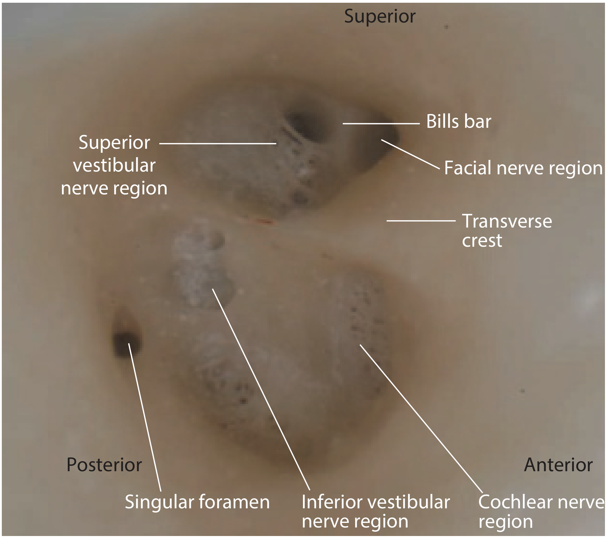
Endoscopic view of the left IAM fundus showing Bill's bar separating the facial nerve region (anterior) from the superior vestibular nerve region (posterior), with the transverse crest visible below - Scott-Brown's Otorhinolaryngology
- Scott-Brown's Otorhinolaryngology Head & Neck Surgery Vol. 2, "The internal auditory meatus"
- Cummings Otolaryngology Head and Neck Surgery, Table 126.1 - Surgical Landmarks of Facial Nerve by Segment
- Shambaugh Surgery of the Ear, "Vestibular Neurectomy" chapter
Course of arteries and nerves in internal auditory meatus
Finding Sources
Finding Sources
Your credits are exhausted. Your daily credits reset tomorrow, or you can purchase a top-up for more.
This is a shared conversation. Sign in to Orris to start your own chat.