How to reduce body acne and remove it's old acne marks?

Reading File
Finding Sources
Finding Sources
Searching the Web

how to treat body acne and fade acne marks scars dermatologist recommendations

Reading File
Reading File
Reading File
Reading File
Searching PubMed

"acne" AND "postinflammatory hyperpigmentation" AND treatment

I have enough to give a well-grounded answer, supported by dermatology textbooks and confirmed by recent systematic reviews. Here it is:

Reducing Body Acne

Body acne (chest, back, shoulders - "bacne") responds to the same principles as facial acne, adjusted for thicker skin and larger surface area.
First-line, at-home:
  • Benzoyl peroxide (BPO) wash - leave on skin for about 2 minutes before rinsing; effective for mild truncal acne and doesn't cause bacterial resistance. Use daily or every other day if it irritates skin - Andrews' Diseases of the Skin, p. 3549.
  • Topical retinoid (adapalene, tretinoin) applied at night to affected areas, not just individual spots - it takes 8-12 weeks to judge effect - Dermatology 2-Volume Set, p. 728.
  • Salicylic acid (up to 2% OTC) as a comedolytic, useful in body washes since it rinses over large areas easily - Dermatology 2-Volume Set, p. 728.
  • Wear loose, breathable clothing, shower promptly after sweating, and avoid heavy oil-based lotions/sunscreens on the trunk (noncomedogenic products only).
If OTC measures fail (moderate/severe truncal acne):
  • Oral antibiotics (e.g., doxycycline) combined with BPO and a topical retinoid - never as monotherapy, and typically limited to about 3 months to limit resistance.
  • Hormonal therapy (combined oral contraceptives, spironolactone) in women with hormonally-driven acne.
  • Isotretinoin for severe, nodular, scarring, or treatment-resistant acne - this is the most effective option and the one most likely to prevent new scarring - Andrews' Diseases of the Skin, Box 13.1.
Avoid scrubbing or harsh astringents/antibacterial soaps, as friction and irritation worsen acne rather than help it.

Fading Old Acne Marks

It matters which type of mark you have, because they're treated differently:
Flat dark/brown-red marks (post-inflammatory hyperpigmentation, PIH) - these are not true scars and usually fade on their own over months once active acne is controlled, but you can speed it up:
  • Daily sunscreen is the single most important step - UV exposure darkens PIH and prolongs it significantly.
  • Topical retinoids (tretinoin) - also help lighten PIH, in addition to treating acne itself - Andrews' Diseases of the Skin, p. 3547.
  • Hydroquinone, often combined with tretinoin, for more stubborn hyperpigmentation - most useful for marks that darken under a Wood's lamp - Andrews' Diseases of the Skin, p. 614.
  • Azelaic acid and vitamin C serums are gentler adjuncts that also help even tone.
  • Chemical peels (glycolic/salicylic acid) can help, but in darker skin tones these carry a real risk of worsening pigmentation if not done carefully by a professional - use caution and ideally dermatologist supervision.
Textured/indented scars (true atrophic scars: ice-pick, boxcar, rolling) - these do not resolve with skincare alone and need procedural treatment:
  • Microneedling (with or without PRP)
  • Fractional laser resurfacing
  • Chemical peels (deeper, professional-grade)
  • Subcision, punch excision, or punch elevation for specific scar types
  • These are generally combined for best results, and a dermatologist should assess scar type first since technique choice depends on it.
A recent 2024 systematic review on PIH treatment confirms this approach: combination therapy (topical depigmenting agents plus procedures like lasers or peels) outperforms any single treatment alone, though results vary by skin tone and evidence quality is still limited (Kashetsky et al., JEADV 2024, PMID 37843491). In skin of color specifically, retinoids and cautious laser use are supported, but aggressive procedures need extra care to avoid triggering more pigmentation (PMID 39075672, PMID 39953770).
Bottom line: control active acne first (this stops new marks from forming), use sunscreen and a retinoid/hydroquinone routine to fade discoloration, and see a dermatologist for textured scars or if OTC measures plateau after 2-3 months - especially before trying peels or lasers if you have darker skin, since improper treatment can worsen pigmentation.

Create a visual guide comparing acne scar types and their treatments

Finding Sources
Finding Sources
Searching Images

ice pick boxcar rolling acne scars comparison
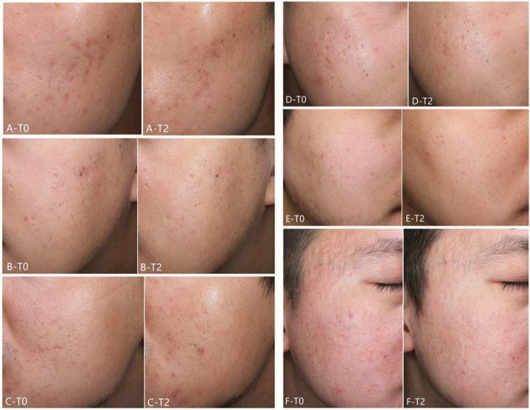
This clinical comparison chart presents side-by-side photographs of six patients (A–F) demonstrating the therapeutic effects of fractional laser treatments on atrophic acne scars. The images are categorized into two groups: Patients A, B, and C received 1927 nm fractional thulium laser (FTL) treatment, while Patients D, E, and F received 2940 nm fractional Er:YAG laser (FEL) treatment. Each pair compares the baseline (T0) to the clinical appearance 12 weeks after the final treatment session (T2). At baseline, the photographs show various morphologies of atrophic scarring, including ice-pick, boxcar, and rolling scars, often accompanied by post-inflammatory erythema and irregular skin texture. The T2 follow-up images demonstrate a visible reduction in scar depth, smoothing of the skin surface, and an overall improvement in skin texture and pigmentation. This visual comparison illustrates the efficacy of fractional resurfacing lasers in remodeling dermal collagen to treat facial acne scarring within a dermatology or aesthetic medicine context.
This clinical comparison chart presents side-by-side photographs of six patients (A–F) demonstrating the therapeutic effects of fractional laser treatments on atrophic acne scars. The images are categorized into two groups: Patients A, B, and C received 1927 nm fractional thulium laser (FTL) treatment, while Patients D, E, and F received 2940 nm fractional Er:YAG laser (FEL) treatment. Each pair compares the baseline (T0) to the clinical appearance 12 weeks after the final treatment session (T2). At baseline, the photographs show various morphologies of atrophic scarring, including ice-pick, boxcar, and rolling scars, often accompanied by post-inflammatory erythema and irregular skin texture. The T2 follow-up images demonstrate a visible reduction in scar depth, smoothing of the skin surface, and an overall improvement in skin texture and pigmentation. This visual comparison illustrates the efficacy of fractional resurfacing lasers in remodeling dermal collagen to treat facial acne scarring within a dermatology or aesthetic medicine context.

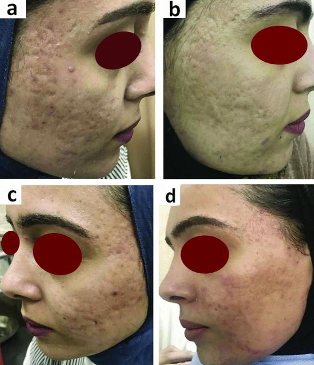
Clinical photographs showing a comparison of facial atrophic acne scars before and after treatment with Platelet-Rich Fibrin (PRF). The image consists of four panels: (a) and (b) depict the right cheek, while (c) and (d) depict the left cheek. Panels (a) and (c) establish baseline clinical features, including high density atrophic scarring—specifically ice-pick, boxcar, and rolling scars—accompanied by visible post-inflammatory hyperpigmentation (PIH) and uneven skin texture. Panels (b) and (d) demonstrate post-treatment outcomes. The right side (b) was treated with combined fluid PRF and skin needling, showing a significant reduction in scar depth and improved skin contour. The left side (d) was treated with intradermal PRF injection alone, illustrating a notable decrease in erythema and PIH with smoother epidermal texture. The set serves as a comparative study in dermatology for evaluating non-surgical interventions for acne-induced skin remodeling and the efficacy of different PRF delivery methods.
Clinical photographs showing a comparison of facial atrophic acne scars before and after treatment with Platelet-Rich Fibrin (PRF). The image consists of four panels: (a) and (b) depict the right cheek, while (c) and (d) depict the left cheek. Panels (a) and (c) establish baseline clinical features, including high density atrophic scarring—specifically ice-pick, boxcar, and rolling scars—accompanied by visible post-inflammatory hyperpigmentation (PIH) and uneven skin texture. Panels (b) and (d) demonstrate post-treatment outcomes. The right side (b) was treated with combined fluid PRF and skin needling, showing a significant reduction in scar depth and improved skin contour. The left side (d) was treated with intradermal PRF injection alone, illustrating a notable decrease in erythema and PIH with smoother epidermal texture. The set serves as a comparative study in dermatology for evaluating non-surgical interventions for acne-induced skin remodeling and the efficacy of different PRF delivery methods.

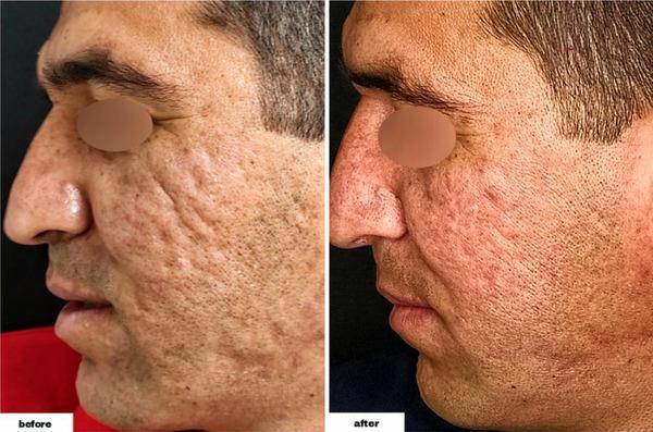
Comparison clinical photographs of a 34-year-old male patient's profile, showing the 'before' and 'after' results of an acne scar revision procedure. The 'before' image displays severe atrophic acne scarring on the left cheek, characterized by a combination of deep ice-pick, rolling, and boxcar lesions that create an undulating, pitted skin texture. Multiple hyperpigmented macules and enlarged pores are visible throughout the malar region. The 'after' image demonstrates a significant improvement in dermal contour and skin surface regularity. There is a visible reduction in the depth and shadow of the depressed scars, alongside a decrease in the number of active-appearing inflammatory lesions and pigmented blemishes. The treated area shows mild post-procedural erythema and a more uniform skin tone. This comparison serves as a clinical illustration of therapeutic efficacy in treating persistent facial scarring and post-inflammatory changes in adult male skin.

Comparison clinical photographs of a 34-year-old male patient's profile, showing the 'before' and 'after' results of an acne scar revision procedure. The 'before' image displays severe atrophic acne scarring on the left cheek, characterized by a combination of deep ice-pick, rolling, and boxcar lesions that create an undulating, pitted skin texture. Multiple hyperpigmented macules and enlarged pores are visible throughout the malar region. The 'after' image demonstrates a significant improvement in dermal contour and skin surface regularity. There is a visible reduction in the depth and shadow of the depressed scars, alongside a decrease in the number of active-appearing inflammatory lesions and pigmented blemishes. The treated area shows mild post-procedural erythema and a more uniform skin tone. This comparison serves as a clinical illustration of therapeutic efficacy in treating persistent facial scarring and post-inflammatory changes in adult male skin.

Reading File
Reading File
Generating Image

A medical infographic poster comparing the four main types of acne scars: Ice Pick Scars (narrow, deep, V-shaped, less than 2mm wide, tapering into the skin), Boxcar Scars (wide, sharply demarcated, U-shaped, box-like depressions), Rolling Scars (wide, shallow, undulating, wave-like depression with normal-appearing skin), and Hypertrophic/Keloid Scars (raised, thickened, firm bumps above the skin surface). Include a cross-section diagram of skin showing the depth profile of each scar type, plus small icons representing corresponding treatments: TCA CROSS and punch excision for ice pick scars, laser resurfacing and subcision for boxcar scars, subcision and dermal fillers for rolling scars, and intralesional steroid injection or laser for hypertrophic/keloid scars. Clean clinical dermatology style layout with labeled sections for each scar type in a grid.

Visual Guide: Acne Scar Types & Treatments
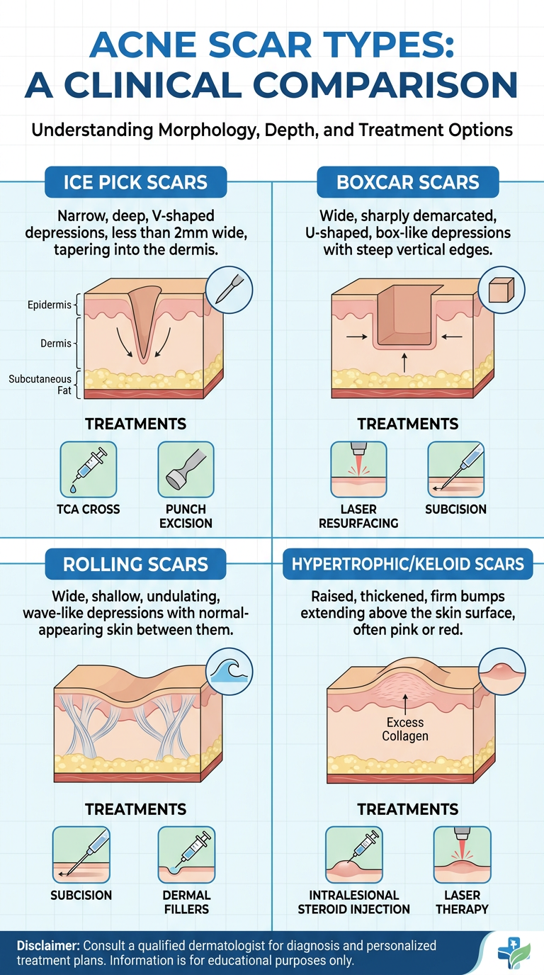
Acne scar types comparison infographic
Acne scars fall into two broad categories: atrophic (depressed, tissue loss) and hypertrophic/keloid (raised, excess tissue). Atrophic scars are further split by shape into ice pick, boxcar, and rolling - Fitzpatrick's Dermatology, p. 1423.

Comparison Table

Scar TypeAppearanceDepth/SizeBest Treatments
Ice pickNarrow, deep, V-shaped; widest at surface, tapers to a point in the dermisUsually <2 mm wide, but deepTCA CROSS (focal high-concentration trichloroacetic acid), punch excision, punch grafting - often needs these done before resurfacing since they're too deep for lasers/peels alone
BoxcarWide, sharply demarcated, U-shaped; does not taper at the base1.5-4 mm wideLaser resurfacing (fractional CO2/Er:YAG), dermabrasion, punch elevation (for the base), chemical peels
RollingWide, shallow, undulating "wave-like" depressions caused by fibrous tethering bands under the skinOften >4-5 mmSubcision (breaks the fibrous tethering bands) is first-line, often combined with dermal fillers, fat grafting, or dermabrasion
Hypertrophic/keloidRaised, firm, thickened scar tissue above skin surfaceVariable, can extend beyond original lesion (especially keloid)Intralesional corticosteroid injection, silicone sheeting, laser (pulsed dye), or surgical excision (keloid excision alone often recurs, so usually combined with steroid/radiation)

Treating Post-Inflammatory Hyperpigmentation (the flat marks, not true scars)

These are not structural scars, just discoloration, and are treated differently: daily sunscreen, topical retinoids, hydroquinone (for cases that darken under Wood's lamp examination), and azelaic acid - Andrews' Diseases of the Skin, p. 614, 3547.

Practical Notes from the Literature

  • Deeper ice-pick and fibrotic scars often need a "lifting" procedure (subcision, punch techniques) 4-6 weeks before resurfacing procedures like dermabrasion or laser, because resurfacing alone can't reach that depth - Dermatology 2-Volume Set, p. 3121.
  • Most patients have a mix of scar types, so combination therapy (e.g., subcision + laser + TCA CROSS) usually outperforms any single technique.
  • In darker skin tones, aggressive peels/lasers carry real risk of triggering new post-inflammatory hyperpigmentation - a systematic review in JEADV (2024, PMID 37843491) found combination approaches worked best but recommended caution and dermatologist supervision in skin of color.
Here's a real clinical example showing atrophic scarring before and after treatment with fractional laser:
Clinical comparison of ice-pick, boxcar, and rolling scars before/after fractional laser treatment
Bottom line: identifying which scar type(s) you have determines the right procedure - a dermatologist typically examines under raking light to classify scars before recommending subcision, TCA CROSS, laser, or filler.

Does coffee drinking has anything related to acne?

Finding Sources
Searching PubMed

coffee caffeine acne

Searching the Web

does coffee drinking cause or worsen acne

Coffee and Acne: What the Evidence Shows

There's no direct evidence in the dermatology literature that coffee or caffeine itself causes acne. My search of PubMed for "coffee/caffeine and acne" turned up studies on caffeine's relationship to skin cancer risk and rosacea, but nothing establishing a direct causal link to acne vulgaris specifically. The dermatology textbooks in the medical library also don't single out coffee as an acne trigger - they focus on diet more broadly.
That said, several indirect mechanisms are well-documented and plausibly connect coffee habits to acne:
1. High-glycemic diet link (established) Textbook sources note that high-glycemic-load diets can worsen acne, and this is one of the better-supported dietary associations - Andrews' Diseases of the Skin, p. 3416; Fitzpatrick's Dermatology, p. 6574. Sugar or syrup added to coffee contributes to this glycemic load, which can spike insulin and IGF-1, increasing sebum production and inflammation.
2. Dairy link (weak but noted) Milk (especially skim milk) has a weak documented association with acne, per Fitzpatrick's Dermatology (p. 593-606, citing Adebamowo et al.'s study on dietary dairy and teenage acne) and Dermatology 2-Volume Set, p. 2552. If you take your coffee with milk, that's the more likely contributor than the coffee itself.
3. Cortisol and stress (plausible, less direct evidence) Caffeine can raise cortisol levels somewhat, and chronically elevated cortisol is linked to increased sebum production and inflammatory skin changes. This mechanism is biologically reasonable but not specifically proven for coffee-triggered acne in controlled trials.
4. Sleep disruption Caffeine late in the day can impair sleep quality, and poor sleep is associated with higher stress and worse acne control in some individuals - though again, this is an indirect pathway rather than a direct effect.

Bottom line

Black coffee in moderation is unlikely to cause acne on its own. If you notice breakouts tracking with your coffee habit, the more likely culprits are the milk, sugar, or flavored syrups added to it, or excessive intake affecting sleep/stress - rather than the coffee itself. If you want to test it, try switching to black coffee or a non-dairy alternative for a few weeks and see if your skin changes; this is a reasonable self-experiment given the weak-to-moderate evidence for dairy and high-glycemic intake as acne factors.
I did not find a systematic review or meta-analysis specifically on coffee-and-acne to confirm or contradict this, so treat this as best-available inference from related diet-acne research rather than a settled finding.
This is a shared conversation. Sign in to Orris to start your own chat.