
Why mteabolic acidosis dont occur in ringer lactate
Reading File
Finding Sources
Searching PubMed
"Ringer's Lactate"[MeSH Terms] AND "acidosis"
Finding Sources
Reading File
Reading File
Ringer lactate (RL) doesn't cause metabolic acidosis because its lactate is converted to bicarbonate after infusion. The result is a fluid that is acid-base neutral.
1. Lactate is a bicarbonate precursor, not an acid load
- RL contains lactate as the sodium salt (sodium lactate), not lactic acid.
- Once infused, the liver metabolizes lactate. Gluconeogenesis and oxidation consume H⁺ and generate HCO₃⁻.
- Mulholland and Greenfield's Surgery says lactate "is mobilized and metabolized to bicarbonate in a single pass through the liver." (Mulholland and Greenfield's Surgery, 7e, Crystalloids, p. 557)
- Rosen's Emergency Medicine says the same: lactate is metabolized to bicarbonate, and RL is "a neutral fluid that does not induce either acidosis or alkalosis."
2. Why normal saline does cause acidosis
- Normal saline has a chloride concentration of 154 mEq/L, much higher than plasma (about 100 mEq/L).
- Large volumes raise plasma Cl⁻ and dilute bicarbonate. In the Stewart model, they also lower the strong ion difference (SID). The result is hyperchloremic acidosis.
- RL has less chloride (about 109 mEq/L) and contains a metabolizable anion.
- Miller's Anesthesia, 10e, says this change is "not seen with solutions containing non-Cl⁻ anions which are metabolized after infusion, such as lactated Ringer solution."
3. Stewart (SID) view
- In the bag, RL's SID is about zero.
- After the liver metabolizes lactate, the effective in vivo SID is about 29 mEq/L (Miller's Anesthesia, 10e). This is slightly below plasma SID. It is enough to offset the dilutional effect on weak acids (A_tot), so pH is maintained.
4. Why it doesn't worsen existing lactic acidosis
- The lactate in RL is the L-isomer (in some formulations a racemic mixture, with D-lactate handled more slowly).
- It is cleared quickly by the liver, and to a lesser extent by the kidney and muscle.
- As perfusion is restored, lactate clearance improves.
- Studies in shock show RL does not aggravate the lactic acidosis. Large-volume resuscitation can even cause mild metabolic alkalosis 1 to 2 days later (Mulholland and Greenfield's Surgery, 7e).
Caveats: when lactate may not be cleared
- Severe hepatic failure or cirrhosis, and severe hypoperfusion with poor hepatic blood flow, can impair lactate metabolism. Some clinicians prefer other fluids here, although the textbook notes that normal hepatic function is what makes RL acid-base neutral.
- Metformin toxicity and other causes of impaired lactate clearance are further reasons for caution.
- Measured serum lactate can rise slightly after RL, but this is not true acidosis.
Recent evidence check: My PubMed search (last 2 years, systematic reviews and meta-analyses) found nothing that contradicts the above. The results were about RL in acute pancreatitis (PMID 38101616), in labor (PMID 37994865), and in ophthalmic surgery (PMID 41092155), not acid-base physiology.
Why normal saline causes acidosis
Reading File
Finding Sources
Searching PubMed
"Saline Solution"[MeSH Terms] AND "Acidosis, Lactic" OR "hyperchloremic acidosis"
Normal saline (0.9% NaCl) causes a normal anion gap, hyperchloremic metabolic acidosis. The cause is its high chloride content, and it is not an acid load. Two models explain it.
Normal saline vs plasma (mEq/L)
| Na⁺ | Cl⁻ | Bicarbonate or buffer anion | |
|---|---|---|---|
| Plasma | about 140 | about 100 | HCO₃⁻ about 24 |
| 0.9% saline | 154 | 154 | none |
Saline has a Na⁺ to Cl⁻ ratio of 1:1, while plasma has about 1.4:1. Saline therefore gives a large chloride load and no buffer.
1. Traditional (Henderson-Hasselbalch) view: dilution of bicarbonate
- Saline rapidly expands the extracellular volume with a bicarbonate-free, chloride-rich fluid.
- Plasma HCO₃⁻ falls in proportion to the volume infused because it is diluted. The fall is balanced by a rise in Cl⁻.
- This is a dilutional acidosis. Morgan and Mikhail's Clinical Anesthesiology, 7e, says: "Plasma [HCO₃⁻] decreases in proportion to the amount of fluid infused as extracellular HCO₃⁻ is diluted, and this fall in [HCO₃⁻] is compensated by a rise in [Cl⁻]."
- Mulholland and Greenfield's Surgery, 7e, calls large-volume saline resuscitation "the most common example of a dilutional acidosis."
2. Stewart (strong ion difference) view
- The strong ion difference (SID) is (Na⁺ + K⁺ + Ca²⁺ + Mg²⁺) minus (Cl⁻ + lactate). Normal plasma SID is about 40 mEq/L.
- Saline is fully ionized, so its SID is zero. Infusing it progressively dilutes the plasma SID.
- A lower SID means more H⁺ is generated from water dissociation, so plasma pH falls.
- Rosen's Emergency Medicine says saline infusion raises chloride relative to the strong cations, which lowers the SID and causes acidosis. The result is a normal anion gap hyperchloremic metabolic acidosis.
- Miller's Anesthesia, 10e, notes this appears with large volumes, for example 30 mL/kg/h of 0.9% saline. The Cl⁻ concentration of the fluid is higher than that of plasma.
3. Contributing mechanisms
- Renal handling: The kidney excretes the chloride load slowly. This prolongs the hyperchloremia, so the acidosis can persist.
- Dilution of weak acids: Dilution of albumin and phosphate (A_tot) tends to cause alkalosis. Saline has no counterbalancing anion that gets metabolized, so the strong-ion effect wins.
- Bicarbonate loss: The acidosis comes from net loss or dilution of bicarbonate, not from added acid. That is why the anion gap stays normal.
Why it matters clinically
- Miller's Anesthesia, 10e, lists physiologic effects of saline-induced hyperchloremic acidosis: renal vasoconstriction, reduced GFR, reduced renin activity, and reduced renal cortical perfusion in healthy volunteers. Coagulopathy and GI dysfunction have also been suggested.
- Mortality in hyperchloremic acidosis is lower than in lactic acidosis or strong ion gap acidosis. In the cohort Miller's cites, it was 29% for hyperchloremic acidosis vs 56% for lactic acidosis. Hyperchloremia can still cause clinically significant organ dysfunction.
- It occurs mainly with large volumes, such as massive resuscitation, rather than small boluses.
- Balanced crystalloids such as Ringer lactate, Plasma-Lyte, or Normosol have lower chloride. They contain metabolizable anions (lactate, acetate, gluconate) that become bicarbonate, so they largely avoid this acidosis. This ties back to your previous question about Ringer lactate.
Recent evidence check: My PubMed search (last 2 years, systematic reviews and meta-analyses) found nothing that contradicts this mechanism. The one tangentially relevant result was a 2025 meta-analysis on dysregulated serum chloride in critically ill adults (PMID 41325394). It is related to hyperchloremia outcomes but does not change the explanation above.
Why serum na is low in siadh
Reading File
Finding Sources
Searching PubMed
"Inappropriate ADH Syndrome"[MeSH Terms] AND "hyponatremia"
Serum sodium is low in SIADH because of water retention, not sodium loss. Excess ADH makes the kidney hold water, and that water dilutes the sodium in the extracellular fluid.
Mechanism, step by step
- Inappropriate ADH release. ADH is secreted despite low plasma osmolality (hypotonicity), when it should be suppressed. Causes include CNS disease, lung disease such as small cell cancer, drugs, and pain or nausea. (Symptom to Diagnosis, 4e)
- ADH acts on the collecting duct. It inserts aquaporin-2 channels via V2 receptors, so the kidney reabsorbs free water even though the body is already water-overloaded.
- Total body water rises and plasma is diluted. Serum sodium falls and plasma osmolality drops. Henry's Clinical Diagnosis describes SIADH as "a euvolemic hypoosmolal hyponatremia associated with hyperosmolar urine."
- Urine stays concentrated. Urine osmolality is inappropriately high, usually above 100 mOsm/kg and often above plasma osmolality, because ADH keeps concentrating it. Fischer's Mastery of Surgery describes "hyponatremia, diminished plasma osmolality, and increased urine osmolality."
Why the patient looks euvolemic, not edematous
- The retained water expands the extracellular volume slightly.
- This triggers natriuresis. ANP and other signals reduce renal sodium reabsorption, and aldosterone is suppressed.
- Sodium is therefore also lost in urine, with urine Na usually above 30 to 40 mEq/L. This adds to the hyponatremia and keeps the patient close to euvolemic.
- Symptom to Diagnosis, 4e says: "Despite water retention, patients appear clinically euvolemic. Subtle increase in volume leads to urinary sodium loss."
- The sodium is low because of dilution, and the total body sodium is roughly normal. This is why SIADH is called dilutional hyponatremia. Adams and Victor's Principles of Neurology also describes "dilutional hyponatremia and water retention without edema."
Why normal saline can make it worse
- In SIADH the kidney excretes the sodium in the saline but keeps the water, because ADH is still acting. Urine is concentrated, so the water stays and sodium falls further.
- Symptom to Diagnosis, 4e warns that isotonic saline without furosemide "may worsen hyponatremia."
- Fluid restriction, salt tablets or urea, loop diuretics, and vasopressin antagonists are the usual treatments.
Diagnostic points
- Hypotonic hyponatremia (serum osmolality below about 275 mOsm/kg).
- Euvolemia, with no edema and no hypovolemia.
- Urine osmolality above 100 mOsm/kg and urine Na above 30 to 40 mEq/L.
- Normal thyroid, adrenal, and renal function. The Tietz textbook says other causes of dilutional hyponatremia must be excluded first: heart failure, renal insufficiency, nephrotic syndrome, cirrhosis, and hypothyroidism.
- Cerebral salt wasting looks similar in the labs, but those patients are volume-depleted, because the problem there is primary sodium loss. (Mulholland and Greenfield's Surgery, 7e)
Recent evidence check: Recent meta-analyses are consistent with this mechanism and mainly cover treatment: urea for SIADH (PMID 39362395), low-dose tolvaptan (PMID 40288608), and hyponatremia after subarachnoid hemorrhage (PMID 39732459). None contradict the explanation above.
Water derivation test in polydypsia
Reading File
Finding Sources
Searching PubMed
"polyuria-polydipsia syndrome" OR "diabetes insipidus" AND copeptin
Searching Images
water deprivation test diabetes insipidus primary polydipsia urine osmolality
Reading File
In primary polydipsia, the water deprivation test shows the urine concentrating normally once water is withheld. That separates it from diabetes insipidus (DI), where the urine stays dilute.
Why it works
- In primary (dipsogenic or psychogenic) polydipsia, the problem is excess drinking. ADH secretion and renal response are intact.
- Once water is withheld, plasma osmolality rises, ADH is released, and the kidney concentrates the urine.
- In central DI there is too little ADH. In nephrogenic DI the kidney does not respond to ADH. In both, the urine stays dilute despite dehydration.
Before the test
- Document polyuria (urine volume above 2.5 L/day in adults) and exclude glycosuria and uncontrolled diabetes mellitus. Correct hypokalemia and hypercalcemia, and make sure thyroid and adrenal function are normal.
- Avoid nicotine, alcohol, and caffeine, which affect ADH.
- Do not do the test if the patient is dehydrated at baseline or has renal insufficiency.
- Screen first. Primary polydipsia is unlikely if plasma osmolality is above 295 mOsm/kg or serum Na is above 145 mEq/L. Serum Na below 137 mEq/L with polyuria usually suggests primary polydipsia. (Tietz Textbook of Laboratory Medicine, 7e; Textbook of Family Medicine, 9e)
Procedure (Tietz)
- The patient is usually supervised in hospital, because of the risk of profound hypotension. The test starts in the morning after an overnight fast.
- The patient stays fasting and nil by mouth for water.
- At baseline, measure serum electrolytes, urea, creatinine, glucose, calcium, and serum osmolality. Also measure urine osmolality and specific gravity, body weight, and vital signs.
- Repeat these measurements at intervals. Stop when urine osmolality plateaus, when weight falls by about 3 to 5%, or when serum osmolality or Na reaches the upper range (my own addition, not from the retrieved text).
- At the end, if urine osmolality is above 400 mOsm/kg, give 5 U aqueous vasopressin subcutaneously (minimum 1 U/m²).
- Remeasure urine osmolality after the vasopressin.
Interpretation
| Condition | Urine osmolality after deprivation | Response to exogenous ADH |
|---|---|---|
| Normal or primary polydipsia | Rises above about 750 to 800 mOsm/kg | No further concentration |
| Central DI | Stays low (below 300 mOsm/kg) | Rises by more than 10% (up to 50% or more in complete DI) |
| Nephrogenic DI | Stays low | No rise |
- The textbooks give these cut-offs:
- Normal subjects reach 800 mOsm/kg or more, and exogenous vasopressin produces no further concentration in primary polydipsia (Textbook of Family Medicine, 9e).
- A urine osmolality above 750 mmol/L after water deprivation rules out DI (Bradley and Daroff's Neurology in Clinical Practice).
- A urine to plasma osmolality ratio below 1.5 at the end makes primary polydipsia unlikely (Tietz).
- A rise of more than 10% after ADH means central DI is probable. No rise means nephrogenic DI is highly probable (Tietz).
- Campbell-Walsh Urology states: "A patient with primary (dipsogenic or psychogenic) polydipsia will have normal urine osmolality on water deprivation tests."
Pitfall: partial results
- Chronic overdrinking washes out the medullary concentration gradient and suppresses ADH.
- Some patients with psychogenic polydipsia therefore fail to concentrate fully unless deprivation is prolonged. This can mimic partial DI (Tietz).
- When results are equivocal, measure baseline and post-deprivation serum AVP (Textbook of Family Medicine, 9e). Hypertonic saline stimulated copeptin is now widely used instead, from my own knowledge rather than the retrieved text.
Recent evidence check: A 2024 systematic review and meta-analysis found copeptin useful in children with polyuria-polydipsia syndrome (PMID 39409072). This supports copeptin as an alternative to the deprivation test and does not contradict the interpretation above.
I did not find a suitable illustrative image in the image library for this topic.
Give me a diagram pf liver with ivc , hepatic vein and protal vein
Reading File
Searching Images
liver anatomy inferior vena cava hepatic veins portal vein diagram
query_images_db
Loading Image
Here is a schematic of the liver showing the inferior vena cava (IVC), hepatic veins, and portal vein.
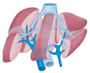
Source: Bailey and Love's Short Practice of Surgery, 28th Edition, p. 1213. This is a stylized anterior view with semi-transparent liver lobes.
How to read the diagram
- IVC: The large light-blue vessel at the top is the intrahepatic IVC. A second light-blue segment at the bottom is the infrahepatic IVC.
- Hepatic veins: The light-blue branches fan out from the IVC into the liver lobes. They are the right, middle, and left hepatic veins, and they drain blood out of the liver into the IVC.
- Portal vein: The dark-blue vessel at the lower center enters the liver at the porta hepatis. It divides into right and left branches that keep branching into smaller vessels within the liver.
Blood flow through the liver
- Portal vein: brings nutrient-rich blood from the gut and spleen, about 75% of liver blood flow. The hepatic artery supplies oxygenated blood, about 25%.
- Blood passes through the sinusoids, where hepatocytes process it.
- Central veins then join into the hepatic veins.
- The hepatic veins empty into the IVC just below the diaphragm, and blood returns to the right atrium.
Why this matters clinically
- Prehepatic portal hypertension is caused by portal vein thrombosis.
- Intrahepatic portal hypertension includes cirrhosis, which is sinusoidal.
- Posthepatic portal hypertension involves the hepatic veins or IVC, as in Budd-Chiari syndrome and congestive hepatopathy.
I can make a custom labeled diagram with text labels on each vessel if you want one.
Why specific gravity is low in diabetes insipidus
Reading File
Finding Sources
Searching PubMed
"Diabetes Insipidus"[MeSH Terms] AND "diagnosis"
Specific gravity is low in diabetes insipidus (DI) because the kidney cannot concentrate urine, so large volumes of dilute urine are passed.
What specific gravity measures
- Specific gravity (SG) compares the density of urine with water (SG 1.000). It rises with the amount of dissolved solute, mainly urea, sodium, and chloride.
- Concentrated urine has a high SG and dilute urine a low one.
- A low SG (below 1.010) reflects hydration, real or perceived by the body. A high SG (above 1.020) reflects dehydration. (Smith and Tanagho's General Urology, 19e)
Why DI causes dilute urine
- Normal physiology. ADH (vasopressin) acts on the V2 receptors of the collecting duct. It inserts aquaporin-2 water channels, so water moves out of the tubule into the hypertonic medulla. The urine becomes concentrated.
- Central DI. ADH is deficient because of damage to the hypothalamus or posterior pituitary. No ADH means no aquaporin-2, so the collecting duct stays water-impermeable.
- Nephrogenic DI. ADH is present, but the tubules do not respond to it. The result is the same.
- Result. Water is not reabsorbed in the collecting duct. The kidney passes a large volume of water-like urine with very little solute per mL.
- Consequence. Urine osmolality is low and SG is low. Serum osmolality and sodium rise as free water is lost.
Robbins says both forms "include the excretion of large volumes of dilute urine with a low specific gravity." Miller's Anesthesia, 10e, lists the main DI features: polydipsia, polyuria with dilute urine (SG below 1.003), increased serum osmolality, and hypernatremia.
Typical values
- Normal urine SG is about 1.003 to 1.030.
- In DI the SG can be as low as 1.001, and urines below 1.007 are called hyposthenuric (Henry's Clinical Diagnosis).
- DI is suggested by SG below 1.005 to 1.010 together with urine osmolality below 300 mOsm/kg and high or high-normal serum osmolality. (The 300 mOsm/kg cut-off is from Tietz, as discussed earlier.)
Why SG stays low even when the patient is dehydrated
- In DI, SG fails to rise even as plasma osmolality increases. Normally dehydration would trigger ADH and raise SG.
- That is why the water deprivation test works. SG and urine osmolality stay low in DI and rise in primary polydipsia.
- A morning SG of 1.010 or more suggests adequate ADH secretion (Tietz). Henry's notes that in children a first-morning SG below 1.015 identifies those who may benefit from ADH supplementation.
Other causes of low SG (not DI)
- Primary polydipsia: the urine is dilute because of excess water intake, but the kidney can still concentrate when water is restricted.
- Acute tubular injury or chronic renal insufficiency: SG is fixed at about 1.010, the same as plasma (Campbell-Walsh Urology).
- Significant intracranial trauma can lower SG by causing ADH deficiency (Smith and Tanagho's).
Recent evidence check: My PubMed search (last 2 years, systematic reviews and meta-analyses) found nothing that changes this mechanism. The results covered central DI after pituitary tumour surgery (PMID 38996052) and gestational DI (PMID 40440645).
This is a shared conversation. Sign in to Orris to start your own chat.