
Pectinate line is known as the watershed line of anal canal
The pectinate (dentate) line marks the junction between the upper and lower anal canal and serves as a key anatomical watershed because structures above and below it differ in arterial supply, venous drainage, lymphatic drainage, nerve supply, and epithelial type.
Searching sources...
Reading File
Finding Sources
Finding Sources
Reading File
Reading File
Reading File
Finding Sources
Reading File
Reading File
Now let me fetch the key anatomical diagram for visual reference:
Loading Image
This is an excellent and clinically important anatomical concept. Here is a full breakdown:
Pectinate (Dentate) Line - The Watershed Line of the Anal Canal
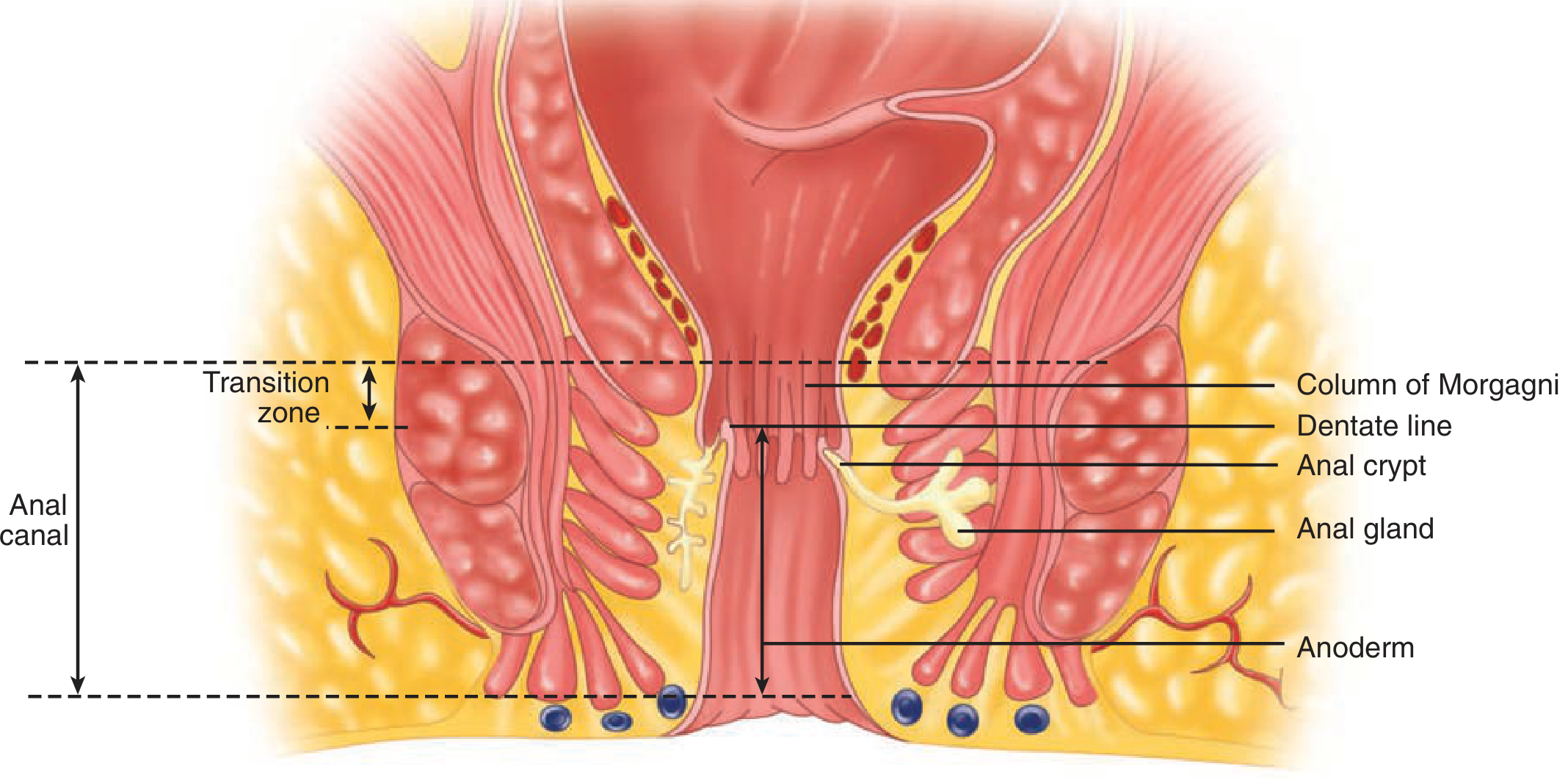
The anal canal showing the dentate (pectinate) line as the central landmark - Schwartz's Principles of Surgery, 11e
What is the Pectinate Line?
The pectinate (dentate) line is a saw-toothed, irregular mucosal junction located approximately 2-3 cm above the anal verge. It is formed by the bases of the anal valves (connecting the lower ends of the columns of Morgagni). It represents the embryological fusion point between the endodermal hindgut (above) and the ectodermal proctodeum (below), which is why virtually everything changes across it.
The "Watershed" - 5 Key Differences
The term "watershed" is used because the pectinate line separates two zones with completely different anatomy in five dimensions:
1. Epithelium
| Above Pectinate Line | Below Pectinate Line |
|---|---|
| Columnar (simple columnar/transitional zone) | Stratified squamous (non-keratinized - the anoderm) |
| Insensate to pain (visceral innervation) | Highly sensitive (somatic innervation) |
"The dentate (pectinate) line is the 'saw-toothed' junction between these 2 segments, each of which differs in venous and lymphatic drainage, nerve supply, and epithelial lining." - Sleisenger & Fordtran's GI and Liver Disease
2. Arterial Supply
| Above | Below |
|---|---|
| Superior rectal artery (branch of inferior mesenteric artery) | Inferior rectal artery (branch of internal pudendal artery) |
| Also middle rectal artery (from internal iliac) | - |
3. Venous Drainage
| Above | Below |
|---|---|
| Internal hemorrhoidal plexus → Superior rectal vein → Inferior mesenteric vein → Portal system | Inferior rectal vein → Internal pudendal vein → Internal iliac vein → Systemic (caval) system |
This is clinically important: internal hemorrhoids (above) are painless because of visceral innervation, while external hemorrhoids (below) are painful due to somatic innervation. Portal hypertension can cause internal hemorrhoids due to porto-systemic anastomosis at this watershed.
4. Lymphatic Drainage
| Above | Below |
|---|---|
| Internal iliac nodes and inferior mesenteric nodes | Superficial inguinal lymph nodes |
Clinical implication: Anal canal cancer above the pectinate line spreads to pelvic lymph nodes, while cancer below it spreads to the inguinal nodes. This fundamentally changes staging and management.
5. Nerve Supply
| Above | Below |
|---|---|
| Autonomic (sympathetic from L1-L3 via hypogastric plexus; parasympathetic from S2-S4 via nervi erigentes) | Somatic (inferior rectal branch of pudendal nerve, S2-S4) |
| Pain-insensitive (only senses pressure/distension) | Pain-sensitive (sharp, well-localized pain) |
"While the rectum is relatively insensate, the anal canal below the dentate line receives somatic innervation." - Schwartz's Principles of Surgery, 11e
Embryological Basis
The pectinate line marks the site of the cloacal membrane in fetal life. The upper two-thirds of the anal canal derive from endoderm (hindgut), while the lower third derives from ectoderm (proctodeum). This is why the transitions in epithelium, blood supply, nerve supply, and drainage all occur at this line simultaneously.
Clinical Relevance Summary
| Feature | Above Pectinate Line | Below Pectinate Line |
|---|---|---|
| Hemorrhoids | Internal (painless, bleed bright red) | External (painful, thrombose) |
| Pain | None (visceral innervation) | Severe (somatic innervation) |
| Venous drainage | Portal (can develop varices in portal hypertension) | Systemic |
| Lymph nodes | Internal iliac / pelvic | Inguinal |
| Anal fissures | Rare above this line | Common below (especially posterior midline) |
| Biopsy/banding | Can be done without anesthesia | Requires local anesthesia |
| Cancer spread | To pelvic nodes | To inguinal nodes |
Sources: Fischer's Mastery of Surgery, 8e | Schwartz's Principles of Surgery, 11e | Sleisenger & Fordtran's GI and Liver Disease | Gray's Anatomy for Students
Take reference from bdc
Searching sources...
Finding Sources
Finding Sources
BD Chaurasia's Human Anatomy is not available in the library database. The library has Gray's Anatomy for Students and Netter's Atlas, which are the closest anatomy references available. Let me pull the anal canal content from Gray's (which is the standard anatomy reference in the library) and present it clearly.
Finding Sources
Finding Sources
Reading File
Reading File
Finding Sources
Loading Image
I need to be transparent: BD Chaurasia's Human Anatomy is not available in the textbook library. The library contains Gray's Anatomy for Students, which covers the same content and is the reference used below. The BDC-style format (point-wise, concise) is preserved in the presentation.
Pectinate Line as the Watershed Line of the Anal Canal
(Reference: Gray's Anatomy for Students)
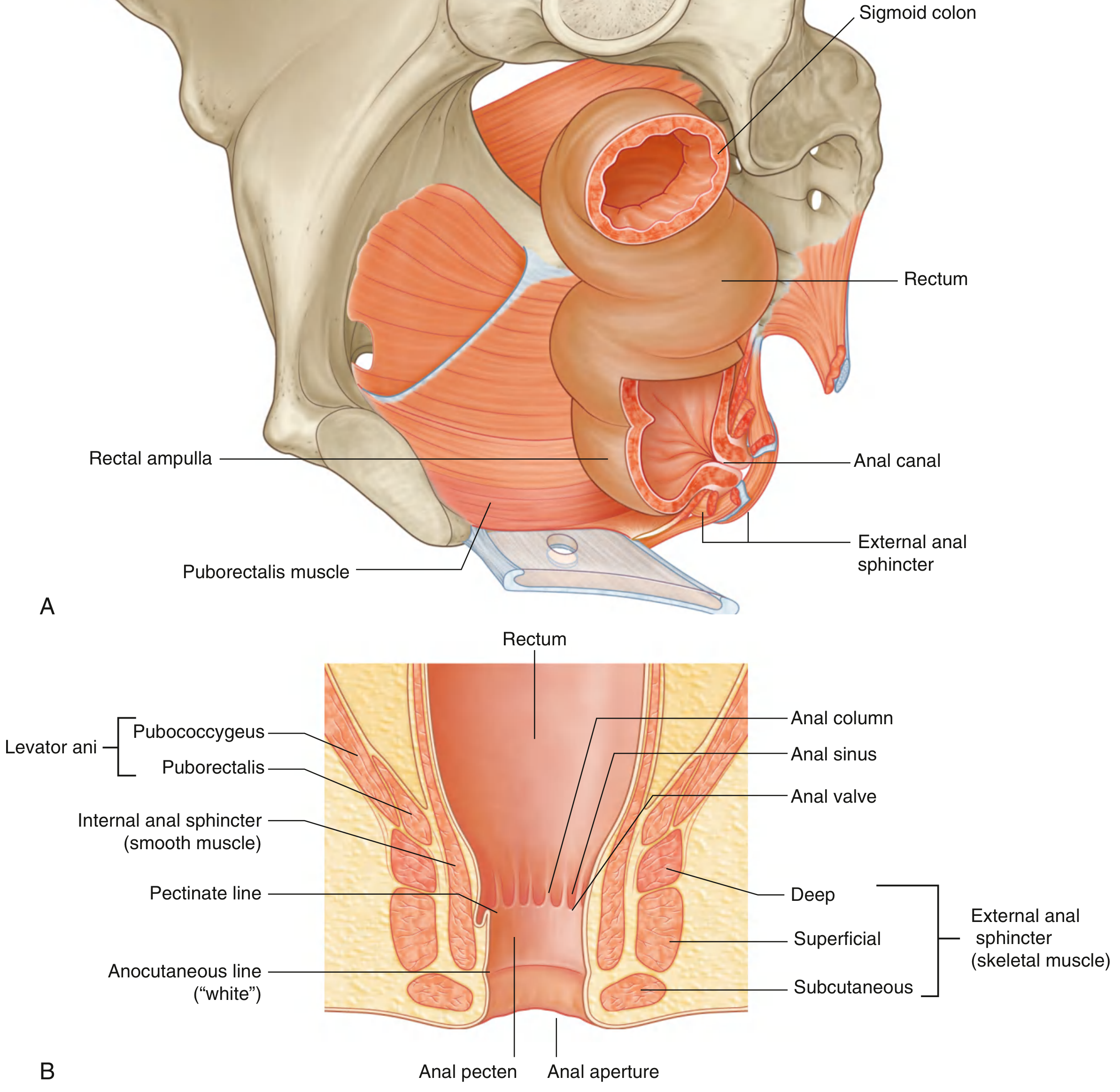
Formation of the Pectinate Line
- The upper part of the anal canal is lined by mucosa bearing longitudinal folds = anal columns (columns of Morgagni)
- The anal columns are connected inferiorly by crescentic folds = anal valves
- Superior to each valve is a pocket = anal sinus
- The bases of all anal valves together form a circle = the pectinate (dentate) line
- It marks the approximate position of the cloacal membrane in the fetus
- Embryologically: above = endoderm (hindgut); below = ectoderm (proctodeum)
Why It Is Called the "Watershed Line"
Everything changes at this line - it is the dividing line for 5 anatomical parameters:
1. Epithelium
| Above Pectinate Line | Below Pectinate Line |
|---|---|
| Columnar (simple columnar / transitional) mucosa | Non-keratinized stratified squamous - anoderm |
| Insensitive to pain | Highly pain-sensitive |
- Immediately below the pectinate line: anal pecten (transition zone lined by non-keratinized stratified squamous epithelium)
- The anal pecten ends at the anocutaneous line ("white line" / Hilton's line), where the lining becomes true skin
2. Arterial Supply
| Above | Below |
|---|---|
| Superior rectal artery (terminal branch of inferior mesenteric artery) | Inferior rectal artery (branch of internal pudendal artery) |
| + Middle rectal artery (from internal iliac artery) | - |
3. Venous Drainage
| Above | Below |
|---|---|
| Internal rectal (hemorrhoidal) plexus → superior rectal vein → inferior mesenteric vein → Portal system | External rectal plexus → inferior rectal vein → internal pudendal vein → internal iliac vein → Systemic (caval) system |
"Internal hemorrhoids originate above the pectinate line and are covered by colonic mucosa."
- Gray's Anatomy for Students
- This porto-systemic anastomosis at the pectinate line is an important portacaval shunt in portal hypertension
4. Lymphatic Drainage
| Above | Below |
|---|---|
| Internal iliac nodes (along internal iliac vessels) | Superficial inguinal lymph nodes |
Clinical note: Cancer of anal canal above the line spreads to pelvic (internal iliac) nodes; below the line spreads to inguinal nodes - changes staging and treatment completely.
5. Nerve Supply
| Above | Below |
|---|---|
| Autonomic - sympathetic (L1-L3, hypogastric plexus) + parasympathetic (S2-S4, nervi erigentes) | Somatic - inferior rectal nerve (branch of pudendal nerve, S2-S4) |
| Insensitive to pain - only senses pressure | Acutely pain-sensitive - touch, temperature, pain all perceived |
Summary Table (BDC Style)
| Feature | Above Pectinate Line | Below Pectinate Line |
|---|---|---|
| Epithelium | Columnar mucosa | Stratified squamous (anoderm) |
| Artery | Superior + middle rectal | Inferior rectal |
| Vein | Portal system | Systemic (caval) |
| Lymph nodes | Internal iliac | Superficial inguinal |
| Nerve | Autonomic (visceral) | Somatic (pudendal) |
| Pain | Insensitive | Highly sensitive |
| Hemorrhoids | Internal (painless) | External (painful) |
| Embryology | Endoderm (hindgut) | Ectoderm (proctodeum) |
Clinical Significance
- Hemorrhoids: Internal hemorrhoids (above - painless, bleed); External hemorrhoids (below - painful, thrombose). Rubber band ligation is done above the pectinate line to avoid pain.
- Anal fissure: Located below the pectinate line - extremely painful due to somatic innervation.
- Anal canal carcinoma: Spread to inguinal nodes if below the line; pelvic nodes if above.
- Portal hypertension: Porto-systemic anastomosis here dilates internal hemorrhoidal veins.
- Biopsy/procedures: No anesthesia needed above the line; local anesthesia mandatory below.
Reference: Gray's Anatomy for Students, Pelvis and Perineum chapter (Fig. 5.39)
Note: BD Chaurasia's Human Anatomy is not in the library's database. The content above is sourced from Gray's Anatomy for Students, which covers the same landmarks in the same manner as BDC, with the same watershed classification. The clinical detail and point-wise presentation match the BDC style.
This is a shared conversation. Sign in to Orris to start your own chat.