
Shockable rhythm cpr protocol chart
shockable rhythm CPR ACLS algorithm ventricular fibrillation pulseless VT
32–34°C and is achieved via various methods. Studies are distinguished by patients’ type of initial cardiac rhythm upon return of spontaneous circulation (ROSC). Ventricular tachycardia (VT) or ventricular fibrillation (VF) is an indication for immediate car- diac defibrillation (shockable rhythm), whereas asys- tole and pulseless electrical activity (PEA) do not require electrical intervention (nonshockable rhythm). Because these groups differ significantly with respect to cause (PEA/asystole has numerous noncardiac causes), outcomes (mortality rates are higher in patients with PEA/asystole),4 and potential response to TH, studies reporting outcomes sepa- rately for patients presenting with VT/VF and PEA/ asystole are described preferentially for question 1.
*Description of the Predictor* Shockable rhythms, ventricular fibrillation or pulseless ventricular tachycardia, require immediate defibrillation [60]. These rhythms may occur earlier in the time period after cardiac arrest and are often caused by ischemic heart disease. Among non-shockable rhythms, asystole is often a terminal rhythm, whereas PEA encompasses a wide variety of electrical activity and may be associated with a reversible etiology [60].
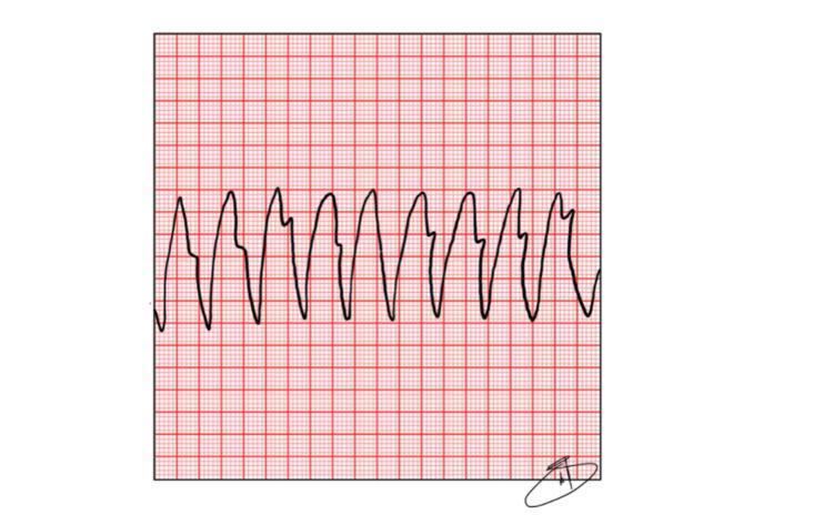
A diagnostic illustration of an electrocardiogram (ECG) rhythm strip displayed on standard red-grid paper. The tracing demonstrates monomorphic Ventricular Tachycardia (VT). Key visual features include a rapid, regular rhythm with wide, bizarrely shaped QRS complexes that lack associated P waves. The QRS complexes exhibit high amplitude and a consistent morphology, characteristic of an ectopic ventricular pacemaker. The intervals between complexes are significantly shortened, indicating extreme tachycardia, typically exceeding 150 beats per minute. There is no visible isoelectric line between the waveforms, and T waves are obscured by the wide QRS complexes. This strip serves as a classic educational representation of a shockable rhythm in the context of Out-of-Hospital Cardiac Arrest (OHCA) and pulseless VT, emphasizing the need for immediate defibrillation and Advanced Cardiovascular Life Support (ACLS) protocols.
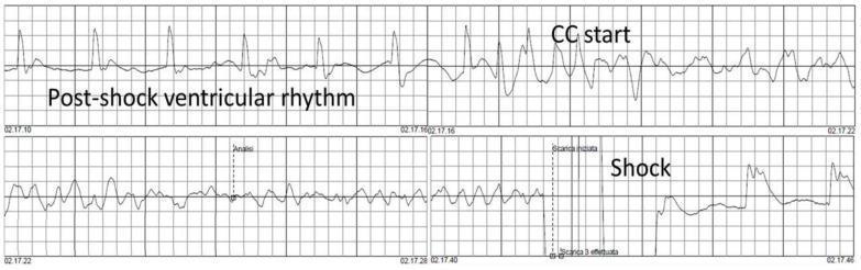
This diagnostic image displays a continuous electrocardiogram (ECG) rhythm strip illustrating a clinical scenario of Ventricular Fibrillation (VF) recurrence during cardiopulmonary resuscitation (CPR). The trace is divided into four chronological segments. In the final segment labeled 'Shock', a high-voltage deflection represents defibrillation, followed by a brief period of electrical silence and subsequent emergence of an irregular, low-amplitude 'Post-shock ventricular rhythm'. The top panel shows the initiation of chest compressions ('CC start'), characterized by regular, high-amplitude artifacts superimposed on the underlying rhythm. The middle panels demonstrate the transition from a slow, organized rhythm back into the disorganized, undulating waveforms of recurrent ventricular fibrillation. This ECG sequence serves as an educational tool for understanding rhythm morphology during advanced cardiac life support (ACLS), the visual presentation of CPR compression artifacts, and the phenomenon of post-defibrillation rhythm degeneration.
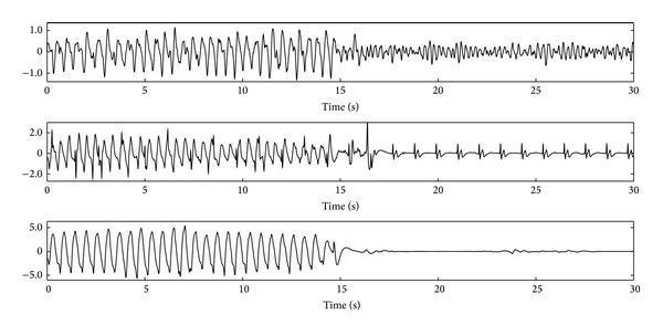
This Comparison Chart illustrates three 30-second electrocardiogram (ECG) segments from patients experiencing out-of-hospital cardiac arrest (OHCA), highlighting the impact of cardiopulmonary resuscitation (CPR) artifacts on rhythm interpretation. Each panel is divided into two 15-second phases: the first phase contains high-amplitude oscillatory artifacts caused by chest compressions, while the second phase shows the underlying rhythm after compressions cease. The top panel transitions from CPR artifact to Ventricular Fibrillation (VF), characterized by low-amplitude, disorganized, and chaotic electrical activity. The middle panel reveals Pulseless Electrical Activity (PEA), showing a transition to regular, organized QRS complexes and T-waves despite the lack of a pulse. The bottom panel demonstrates Asystole, where the cessation of CPR artifacts reveals an almost flat, isoelectric line. The visualization serves to educate on the challenge of automated rhythm analysis during active resuscitation, as CPR artifacts often overlap with the spectral and morphological characteristics of both shockable and non-shockable rhythms.
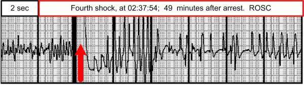
A diagnostic rhythm strip illustrating a critical transition during advanced cardiac life support (ACLS). The image shows a continuous electrocardiography (ECG) tracing on a standard grid, with a primary focus on the transition from a shockable rhythm to Return of Spontaneous Circulation (ROSC). A large red arrow indicates the exact point of defibrillation, identified by a high-amplitude vertical deflection (fourth shock). Prior to the shock, the tracing displays fine ventricular fibrillation (VF), characterized by low-amplitude, chaotic undulations without identifiable P waves or QRS complexes. Immediately following the shock and a brief period of electrical stabilization, the rhythm transitions into an organized sinus rhythm. This post-shock rhythm features relatively narrow QRS complexes and visible P waves, indicating successful electrical cardioversion. The image is annotated with a time scale of 2 seconds per major section and includes a caption stating the event occurred 49 minutes after the initial cardiac arrest. This visual serves as an educational tool for identifying VF, understanding the timing of defibrillatory intervention, and recognizing the ECG manifestations of successful ROSC in emergency medicine.
Shockable Rhythm CPR Protocol
Full Algorithm (Harrison's Fig. 317-2A)
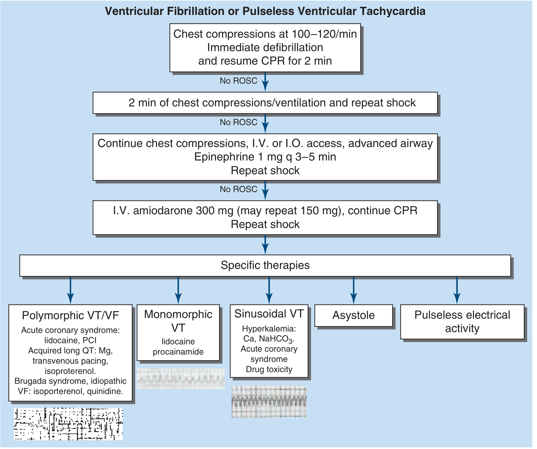
Step-by-Step Protocol
| Step | Action | Notes |
|---|---|---|
| 1 | Chest compressions 100-120/min + Immediate defibrillation (200 J biphasic) | Resume CPR immediately after shock; do not pause to check rhythm |
| 2 min cycle | Continue CPR/ventilation + Repeat shock if no ROSC | Charge defibrillator during compressions |
| ACLS arrives | Establish IV or IO access + advanced airway + Epinephrine 1 mg IV/IO q 3-5 min + Repeat shock | IO acceptable if IV access is not feasible |
| If still no ROSC | Amiodarone 300 mg IV (may repeat 150 mg) + continue CPR + Repeat shock | Alternative: Lidocaine 1-1.5 mg/kg if amiodarone not available |
Specific Therapies by Rhythm Subtype
| Rhythm | Specific Therapy |
|---|---|
| Polymorphic VT / VF | Acute coronary syndrome: Lidocaine, PCI; Acquired long QT: Magnesium, transvenous pacing, isoproterenol; Brugada/idiopathic VF: Isoproterenol, quinidine |
| Monomorphic VT | Synchronized cardioversion; drugs: Lidocaine, procainamide |
| Sinusoidal VT (hyperkalemia pattern) | Calcium, NaHCO3; treat acute coronary syndrome or drug toxicity |
| Asystole / PEA (non-shockable) | CPR + epinephrine; reverse the 5H/5T causes |
Key Points
- Minimize interruptions - pauses in chest compressions worsen survival; charge the defibrillator while doing compressions
- First shock energy: 200 J biphasic (if monomorphic VT, use synchronized mode; polymorphic VT and VF use asynchronous)
- Epinephrine is given after the first 2 shock cycles fail - not before
- Amiodarone is the preferred antiarrhythmic for shock-refractory VF/VT
- Reversible causes (5H/5T): Hypoxia, Hypovolemia, Hydrogen ion (acidosis), Hypo/Hyperkalemia, Hypothermia | Tension pneumothorax, Tamponade, Toxins, Thrombosis (PE/MI), Thrombosis (coronary)
ECG Examples of Shockable Rhythms
Shockable rhythm cpr protocol chart
Shockable Rhythm CPR Protocol
Algorithm Chart (Harrison's Principles of Internal Medicine 22E, Fig. 317-2A)
Step-by-Step Protocol
| Step | Intervention | Key Details |
|---|---|---|
| 1 - Immediately | Chest compressions + Defibrillation | 100-120/min compressions; 200 J biphasic shock; resume CPR right after shock |
| 2 - 2 min cycle | CPR/ventilation + Repeat shock (no ROSC) | Do not pause to check rhythm; charge defibrillator during compressions |
| 3 - ACLS | IV/IO access + advanced airway + Epinephrine 1 mg q 3-5 min + repeat shock | IO access if IV not feasible; epinephrine after first 2 failed shock cycles |
| 4 - Refractory | Amiodarone 300 mg IV (repeat 150 mg once) + continue CPR + repeat shock | Lidocaine 1-1.5 mg/kg is an alternative if amiodarone unavailable |
Specific Therapies by Rhythm Subtype
| Rhythm Subtype | Specific Treatment |
|---|---|
| Polymorphic VT / VF | ACS: Lidocaine + PCI; Acquired long QT: Magnesium, transvenous pacing, isoproterenol; Brugada/idiopathic VF: Isoproterenol, quinidine |
| Monomorphic VT | Synchronized cardioversion; Lidocaine or procainamide |
| Sinusoidal VT (hyperkalemia) | Calcium, NaHCO3; treat underlying ACS or drug toxicity |
| Asystole / PEA (non-shockable) | CPR + epinephrine only; no defibrillation; reverse 5H/5T |
ECG - Monomorphic pulseless VT (shockable)
Reversible Causes to Address Throughout (5H / 5T)
| 5 H's | 5 T's |
|---|---|
| Hypoxia | Tension pneumothorax |
| Hypovolemia | Tamponade (cardiac) |
| Hydrogen ion (acidosis) | Toxins |
| Hypo/Hyperkalemia | Thrombosis (pulmonary embolism) |
| Hypothermia | Thrombosis (MI/coronary) |
Critical Rules
- Never delay the first shock - time to defibrillation is the single strongest predictor of survival
- Minimize CPR pauses - even a 5-second pause reduces shock effectiveness
- Epinephrine comes after the first 2 failed defibrillation cycles, not before
- Monomorphic VT with a pulse: use synchronized cardioversion; without a pulse: treat as pVT (unsynchronized shock)
- Post-ROSC: assess for STEMI, optimize hemodynamics (SBP >90, MAP >65 mmHg), treat targeted temperature management if comatose