
Ent nose detailed notes
Reading File
Finding Sources
Finding Sources
Finding Sources
Finding Sources
Finding Sources
There's an ENT speciality with Cummings Otolaryngology. Let me get the full table of contents for the ENT books and read all key nose-related sections.
Finding Sources
Finding Sources
Finding Sources
Finding Sources
Excellent! Now let me read the key sections in parallel — nasal anatomy, sinonasal infection/sinusitis, nasal obstruction, and more from both textbooks.
Reading File
Reading File
Reading File
Reading File
Reading File
Reading File
Reading File
Finding Sources
Finding Sources
Reading File
Reading File
Reading File
Reading File
Reading File
Reading File
Searching Images
nasal anatomy turbinates septum Kiesselbach plexus
Searching Images
paranasal sinuses anatomy CT ostiomeatal complex
Here are comprehensive ENT nose notes compiled from Cummings Otolaryngology, K.J. Lee's Essential Otolaryngology, and Rosen's Emergency Medicine.
ENT — The Nose: Comprehensive Notes
1. ANATOMY
External Nose
- Inspected from frontal, profile, and base views for deformity or asymmetry
- Assess: nasal tip projection, dorsal width, alar base width, skin quality and thickness
- Nasolabial angle and height-to-length ratio change with age (tip droops due to weakening of lower lateral cartilage and caudal septum)
Nasal Cavity
Extends from the nares anteriorly → choana posteriorly, and from the hard palate inferiorly → cribriform plate superiorly.
| Structure | Description |
|---|---|
| Nasal septum | Midline divider; cartilaginous anteriorly, bony posteriorly |
| Inferior turbinate | Largest; most important for airflow resistance |
| Middle turbinate | Key surgical landmark; landmark for ostiomeatal complex |
| Superior turbinate | Overlies sphenoethmoidal recess |
| Nasal floor | Hard palate below |
| Cribriform plate | Roof; site of olfactory filaments (CN I) |
Ostiomeatal Complex (OMC)
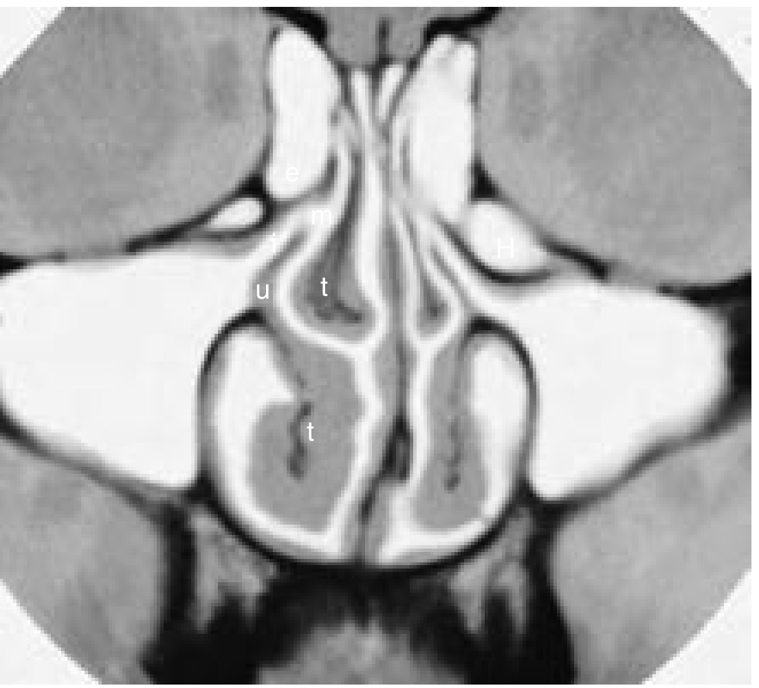
- Key drainage pathway for the frontal, maxillary, and anterior + middle ethmoid sinuses — all drain via the semilunar hiatus under the middle turbinate
- Posterior ethmoids + sphenoid drain through the sphenoethmoidal recess (under superior turbinate)
- A small lesion at the OMC can obstruct multiple sinuses simultaneously — critical concept for sinusitis pathogenesis
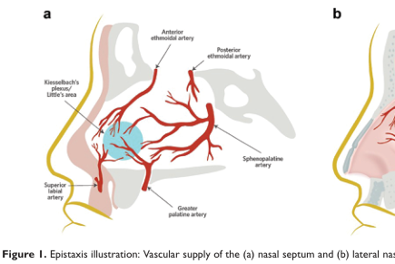
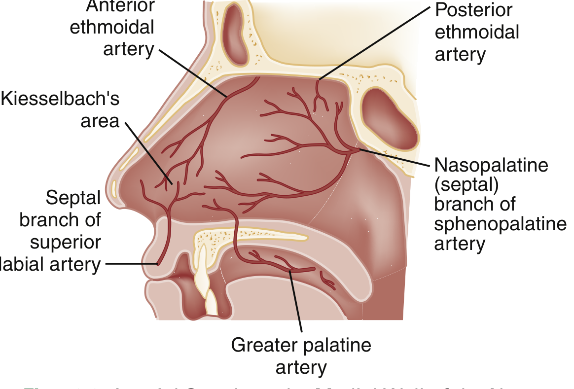
Blood Supply — Kiesselbach's Plexus (Little's Area)
The anterior septum is where branches of both external and internal carotid arteries anastomose:
| Artery | Source |
|---|---|
| Anterior ethmoidal a. | Internal carotid → ophthalmic a. |
| Posterior ethmoidal a. | Internal carotid → ophthalmic a. |
| Sphenopalatine a. | External carotid → maxillary a. → supplies turbinates and posterior septum |
| Greater palatine a. | External carotid → maxillary a. |
| Superior labial a. (facial a.) | External carotid → facial a. → anterior septal mucosa |
Kiesselbach's plexus = convergence of these vessels on the anteroinferior septum → most common site of anterior epistaxis (~90% of cases)
2. EXAMINATION OF THE NOSE
Anterior Rhinoscopy
- Uses headlamp + nasal speculum (directed laterally — never touch septum with metal edges)
- Assess: septum, inferior turbinate, drainage, polyps, foreign bodies, clots
- Mucosal appearance:
- Boggy, pale, edematous → allergic rhinitis
- Erythematous, edematous → sinusitis/infection
Nasal Endoscopy
- Rigid endoscope (0°) passed along nasal floor → visualises septum, inferior turbinate, eustachian tube orifice
- Reintroduced above inferior turbinate → middle turbinate, ostiomeatal complex, nasopharynx
- Accessory maxillary ostia may be visible (often mistaken for true ostium, which lies behind the uncinate process)
- Flexible fiberoptic scope = safer in children/uncooperative patients
Nasopharynx
- Extends from skull base → soft palate
- Inspect: adenoids, eustachian tube orifice, torus tubarius, fossae of Rosenmüller
- Adults should have minimal adenoid tissue; its presence suggests lymphoma or HIV
- All patients with unilateral otitis media → inspect nasopharynx for mass
3. PARANASAL SINUSES
Anatomy Summary
| Sinus | Drains Into |
|---|---|
| Frontal | Middle meatus (via frontonasal duct) |
| Maxillary | Middle meatus (via infundibulum) |
| Anterior + middle ethmoid | Middle meatus |
| Posterior ethmoid | Superior meatus |
| Sphenoid | Sphenoethmoidal recess |
Anatomic Variants (Clinical Significance)
- Haller cells (infraorbital ethmoid air cells) — can narrow the infundibulum
- Concha bullosa — pneumatized middle turbinate; can obstruct OMC
- Paradoxical middle turbinate — curves medially rather than laterally
- Deviated septum — very common; significant if causing obstruction or OMC narrowing
Imaging
- CT (non-contrast) is the gold standard for sinuses
- Screening: 5-mm axial slices
- Pre-operative (FESS): thin-slice coronal + axial + sagittal reconstructions
- Normal mucosa: not visible on CT
- Mucosal thickening → chronic sinusitis, polyps, retention cysts
- Air-fluid level → acute sinusitis, trauma
- MRI: superior for soft tissue characterisation, distinguishing tumour from mucus (different viscosity signals)

4. NASAL OBSTRUCTION
Causes (Nasal Level)
| Category | Specific Causes |
|---|---|
| Septal | Deviated septum, septal hematoma, perforation |
| Turbinate | Turbinate hypertrophy (allergic, vasomotor) |
| Valve | Nasal valve collapse (internal or external) |
| Adenoids | Adenoid hypertrophy (especially children) |
| Masses | Nasal polyps, tumours (benign/malignant) |
Nasal Valve
- Internal nasal valve (angle between upper lateral cartilage and septum, ~10–15°) = the narrowest segment of the airway
- Collapse → most common cause of nasal obstruction post-rhinoplasty
5. EPISTAXIS (NOSEBLEED)
Epidemiology
- Bimodal distribution: peaks in childhood and in elderly
- Higher incidence in winter (dry air desiccates mucosa, cold impairs coagulation)
- Anterior epistaxis: 90% of cases — Kiesselbach's plexus
- Posterior epistaxis: ~10% — more severe, older patients with comorbidities; sphenopalatine artery is most often identified
Risk Factors / Causes
| Category | Examples |
|---|---|
| Local | URI, nose picking, allergies, low humidity, foreign body, polyps, neoplasm |
| Systemic | Anticoagulants/antiplatelets, coagulopathy, thrombocytopenia, hereditary haemorrhagic telangiectasia (Rendu-Osler-Weber), hepatic disease, vitamin K deficiency, cocaine use |
| Post-procedure | Surgical epistaxis, nasal intubation (rate ~2–6%) |
Note: Hypertension is NOT causal, but persistent hypertension is associated with difficulty controlling bleeding.
Assessment
- Airway, perfusion, haemodynamics first
- History: timing, frequency, severity, medications (anticoagulants), comorbidities
- Examine: instruct patient to blow nose → compress cartilaginous nose for 10–15 minutes with 2 sprays of 0.05% oxymetazoline before pressure
- Floor of nose should be parallel to the room floor for examination
- Identify source → enables cautery (reduces recurrence)
Management — Step Ladder
| Step | Intervention |
|---|---|
| 1st | Direct pressure + oxymetazoline (10–15 min) |
| 2nd | Chemical cautery with silver nitrate (periphery → centre; do NOT apply bilaterally on septum simultaneously → risk of septal necrosis; <15 sec contact) |
| 3rd | Anterior nasal packing (absorbable or non-absorbable) |
| Adjunct | Topical tranexamic acid (500 mg IV solution on pledget) — moderate evidence; superior to packing in patients on antiplatelet therapy |
| Posterior bleed | Double balloon catheter (or Foley 12-Fr: inflate 5–7 mL in nasopharynx, pull anteriorly) |
| Refractory | Sphenopalatine artery ligation (surgical) OR endovascular embolisation (>80% success; preferred in anticoagulated patients) |
Prophylactic antibiotics after nasal packing: common practice for TSS prevention (Staph. aureus), though evidence is limited.
6. RHINOSINUSITIS
Definition
Inflammation of the nasal mucosa and paranasal sinuses. The maxillary and ethmoid sinuses are most commonly involved.
Classification
| Type | Duration | Key Features |
|---|---|---|
| Acute viral (common cold) | <4 weeks | Self-limiting; 98% of cases |
| Acute bacterial (ABRS) | <4 weeks | Bacterial superinfection; symptoms >10 days or worsening after 5–7 days |
| Chronic (CRS) | >12 weeks | Often polymicrobial biofilms; with or without nasal polyps |
| Subacute | 4–12 weeks | Between acute and chronic |
Symptoms
- Facial pain/pressure, nasal congestion, mucopurulent nasal discharge, postnasal drip
- Fever (more common in acute bacterial)
- Patients with CRS often relatively asymptomatic — mainly congestion and discharge
- Cough/bronchospasm from postnasal drainage (pulmonary presentation)
Bacteriology
| Setting | Organisms |
|---|---|
| Acute bacterial (adults & children) | H. influenzae, S. pneumoniae, M. catarrhalis; rarely S. pyogenes, S. aureus |
| Cystic fibrosis | Pseudomonas aeruginosa |
| Odontogenic sinusitis | Anaerobes |
| Chronic | Mixed: S. aureus, P. aeruginosa, polymicrobial anaerobes; biofilm formation |
| Immunocompromised | All of above + Aspergillus, Mucor/Rhizopus (invasive fungal sinusitis — high mortality) |
Treatment
Acute viral rhinosinusitis:
- Analgesics, nasal saline, topical intranasal corticosteroids
Acute bacterial rhinosinusitis — Adults:
- Watchful waiting (mild) OR amoxicillin ± clavulanate 5–10 days (first-line)
- Penicillin allergy: doxycycline, levofloxacin, or moxifloxacin
Acute bacterial rhinosinusitis — Children (AAP guidelines):
- Observation 72 hours if non-severe
- Amoxicillin ± clavulanate ≥10 days for severe/worsening/failing
- Penicillin allergy (non-severe): cefdinir, cefuroxime, or cefpodoxime
Chronic rhinosinusitis:
- Longer-duration antibiotics, nasal saline irrigation, intranasal steroids
- Polymicrobial biofilms → may need surgery (FESS) + antibiotic irrigations
- Do NOT use topical or systemic antifungals routinely in CRS without invasive fungal infection
Complications of Sinusitis
Adjacent compartments at risk: orbit, anterior cranial fossa, cavernous sinus, brain
- Orbital complications (from ethmoid sinusitis): preseptal cellulitis → orbital cellulitis → subperiosteal abscess → orbital abscess
- Cavernous sinus thrombosis (from sphenoid/ethmoid sinusitis): high morbidity
- Intracranial extension: meningitis, epidural/subdural/brain abscess
7. NASAL POLYPS
Characteristics
- Benign pedunculated outgrowths of nasal mucosa
- Pale, translucent, insensate (distinguish from turbinates which are pink and tender)
- More common with advancing age; associated with CRS, asthma, aspirin sensitivity (Samter's triad), cystic fibrosis, allergic rhinitis
Special Type: Antrochoanal Polyp
- Originates from maxillary sinus mucosa, prolapse through ostium into nasal cavity → choana → nasopharynx
- Usually unilateral; commoner in children/young adults
- Treatment: complete surgical excision (FESS)
Differential of Nasal Mass
- Nasal polyp (benign)
- Inverting papilloma (benign but locally aggressive; risk of malignant transformation)
- Angiofibroma (adolescent males — highly vascular)
- Esthesioneuroblastoma (olfactory neuroblastoma)
- SCC, adenocarcinoma, adenoid cystic carcinoma
- Lymphoma
- Meningoencephalocele (pulsatile, compressible — do NOT biopsy)
Imaging for Nasal Masses
- Coronal CECT or coronal/sagittal MRI to assess extent within nasal cavity, adjacent sinuses, orbits, cribriform plate
- "Cascading polyps" appearance on contrast CT
- MRI distinguishes tumour from different viscosity mucus — key for staging
8. DEVIATED NASAL SEPTUM
Anatomy
- Septum = quadrangular cartilage (anterior) + perpendicular plate of ethmoid (superior bony) + vomer (inferior bony)
- Caudal deviation: detected by palpation with gloved fingers anteriorly; most symptomatic type
- Bony spurs: at junction of cartilaginous and bony septum
Clinical Features
- Unilateral or bilateral nasal obstruction
- Predisposes to recurrent sinusitis (OMC narrowing), snoring, sleep-disordered breathing
Treatment
- Septoplasty: surgical correction of the septal cartilage/bone; often combined with turbinate reduction
- Combined with rhinoplasty = septorhinoplasty
9. OLFACTORY DYSFUNCTION
Physiology
- Olfactory neurons in the olfactory epithelium (roof of nasal cavity, cribriform plate) → CN I fibres → olfactory bulb
- Smell typically peaks in 3rd–4th decade, declines from 5th decade
- Higher rates of decline in men than women
Causes of Anosmia/Hyposmia
| Category | Examples |
|---|---|
| Conductive | Nasal polyps, CRS, septal deviation (blocking odorant access) |
| Sensorineural | Post-viral (most common), head trauma (cribriform plate shear), neurodegenerative |
| Neurodegenerative | Parkinson's disease (olfactory loss precedes motor signs by years), Alzheimer's disease, Lewy body disease |
| Medications | Lipid-lowering drugs, NSAIDs, antihypertensives, decongestants |
Clinical pearl: Olfactory dysfunction can be a screening marker for early Parkinson's disease — often present years before motor signs.
10. AGING NOSE
| Change | Mechanism |
|---|---|
| Drooping nasal tip | Weakening of lower lateral cartilage + loss of skin elasticity |
| Increased nasolabial angle | Upper lip lengthening, resorption of premaxillary fat |
| Increased nasal resistance | Mucosal atrophy, reduced ciliary function |
| Gustatory rhinitis | Loss of autonomic control of Bowman glands |
| Postnasal drip | Increased in elderly; loss of autonomic control |
| Epistaxis | Mucosal dryness, anticoagulant use |
| Nasal polyps | More common with advancing age |
11. SINONASAL MALIGNANCIES
| Tumour | Notes |
|---|---|
| Squamous cell carcinoma | Most common sinonasal malignancy |
| Adenocarcinoma | Associated with hardwood dust exposure (furniture workers) |
| Adenoid cystic carcinoma | Perineural spread; late recurrence |
| Inverting papilloma | Benign; locally aggressive; malignant transformation risk ~10% |
| Olfactory neuroblastoma (esthesioneuroblastoma) | Arises from olfactory epithelium; cribriform plate involvement |
| Angiofibroma | Juvenile nasopharyngeal; males; highly vascular; do NOT biopsy |
| Lymphoma | Consider in adults with enlarged adenoids or nasal mass; HIV-associated |
Warning Signs of Nasal Malignancy
- Unilateral nasal obstruction (progressive)
- Blood-stained unilateral discharge
- Facial swelling or pain
- Proptosis (orbital invasion)
- Loosening of teeth, trismus (palate/pterygoid invasion)
- Cervical lymphadenopathy
12. QUICK CLINICAL PEARLS
| Topic | Key Fact |
|---|---|
| Kiesselbach's plexus | Anteroinferior septum; 90% of anterior epistaxis |
| Sphenopalatine artery | Main vessel in posterior/severe epistaxis |
| Maxillary sinus | Most commonly affected in sinusitis |
| OMC | Key drainage bottleneck; small lesion → multi-sinus disease |
| ABRS first-line | Amoxicillin ± clavulanate |
| CRS antifungals | Do NOT use routinely |
| Silver nitrate cautery | Do NOT apply bilaterally on septum simultaneously |
| Foley for posterior bleed | 12-Fr, 5–7 mL balloon |
| Tranexamic acid | 500 mg IV solution topically; especially useful in antiplatelet patients |
| Antrochoanal polyp | Maxillary sinus origin; unilateral; children/young adults |
| Anosmia + Parkinson's | Smell loss precedes motor signs by years |
| Concha bullosa | Pneumatized middle turbinate; OMC obstruction |
| Haller cells | Infraorbital ethmoid cells; narrow infundibulum |
| Inverting papilloma | Benign but must excise fully; ~10% malignant transformation |
*Sources: Cummings Otolaryngology Head and Neck Surgery (9780323612173) | K.J. Lee's Essential Otolaryngology (9781260122237) | Rosen's Emergency Medicine (9780323757898)*These notes cover the full ENT nose syllabus across 12 sections. Here's what's included:
- Anatomy — external nose, nasal cavity, turbinates, ostiomeatal complex, blood supply (Kiesselbach's plexus with diagram)
- Examination — anterior rhinoscopy, nasal endoscopy, nasopharyngoscopy
- Paranasal Sinuses — anatomy, drainage pathways, anatomic variants (Haller cells, concha bullosa), CT/MRI imaging
- Nasal Obstruction — causes at each anatomical level
- Epistaxis — epidemiology, Kiesselbach's vs sphenopalatine, causes, step-ladder management (pressure → cautery → packing → tranexamic acid → balloon/Foley → embolisation/ligation)
- Rhinosinusitis — classification, bacteriology by type, treatment guidelines (AAP + adult), complications
- Nasal Polyps — features, antrochoanal polyp, differential of nasal mass
- Deviated Septum — anatomy, clinical features, septoplasty
- Olfactory Dysfunction — causes, Parkinson's connection
- Aging Nose — structural and functional changes
- Sinonasal Malignancies — tumour types, warning signs
- Quick Clinical Pearls table — high-yield exam facts
This is a shared conversation. Sign in to Orris to start your own chat.