
acute pancreatitis
"acute pancreatitis"[MeSH Terms]
acute pancreatitis management guideline diagnosis fluids nutrition 2024
acute pancreatitis contrast enhanced CT imaging pancreatic necrosis peripancreatic fluid collection
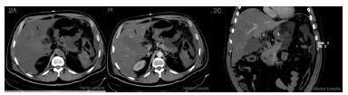
This diagnostic imaging series consists of axial and coronal abdominal Computed Tomography (CT) scans highlighting severe acute necrotizing pancreatitis and secondary gastric involvement. Figure 2A (axial unenhanced) and 2B (axial contrast-enhanced) demonstrate a slightly enlarged pancreatic body and tail with a notable absence of contrast enhancement, a hallmark of pancreatic necrosis. Peripancreatic fat stranding and inflammatory changes are evident. Figure 2C (coronal enhanced) reveals a markedly thickened and unenhanced gastric wall, consistent with gastric wall necrosis. A fluid collection is localized near the posterior gastric wall, representing a pancreatic-peripancreatic inflammatory collection. These findings are critical for diagnosing complications of severe acute pancreatitis, specifically necrotizing changes and secondary ischemia or necrosis of the adjacent stomach wall. The image is optimized for teaching radiological signs of abdominal organ necrosis and the spatial relationship between the pancreas and the posterior stomach.
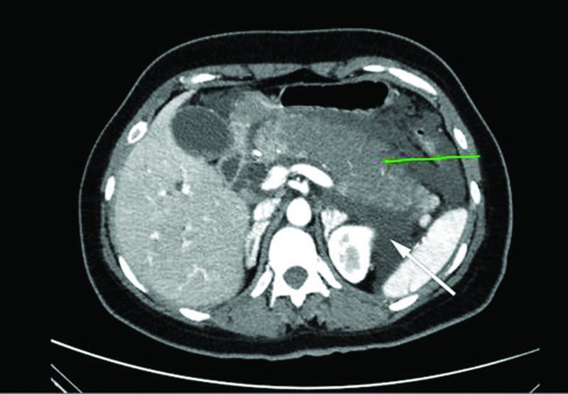
Axial contrast-enhanced computed tomography (CT) scan of the upper abdomen demonstrating severe acute necrotizing pancreatitis. The primary pathology involves the body and tail of the pancreas (indicated by a green arrow), which appear enlarged with heterogeneous parenchymal enhancement, suggestive of necrosis. Surrounding the pancreatic tissue is an extensive acute peripancreatic fluid collection (indicated by a white arrow), characterized by low-density, irregular margins. This collection occupies the peripancreatic space and extends into the pararenal spaces, causing displacement of the stomach and partial compression of the spleen and left kidney. Other visible anatomical structures include the liver, gallbladder, and abdominal aorta. This imaging study is characteristic of advanced acute pancreatitis complications and is used to assess the severity of pancreatic necrosis and the presence of localized fluid collections for clinical staging and intervention planning.
https://journals.lww.com/hbsn/_layouts/15/oaks.journals/down…
Acute pancreatitis
Diagnosis
- Typical acute, severe epigastric pain, often radiating to the back
- Serum lipase or amylase >3 times the upper limit of normal
- Characteristic imaging findings
Common causes
- Gallstones or biliary sludge
- Alcohol use
- Hypertriglyceridemia, particularly triglycerides >1,000 mg/dL
- Post-ERCP pancreatitis
- Drugs, trauma, hypercalcemia, infection, pancreatic tumor, or idiopathic disease
Severity
- Mild: no organ failure or local complications
- Moderately severe: transient organ failure, less than 48 hours, and/or local complications
- Severe: persistent organ failure for more than 48 hours
Initial hospital management
- Admit and assess severity frequently, especially during the first 24 hours.
- Give IV crystalloid fluids, usually lactated Ringer's, with reassessment to avoid both under-resuscitation and fluid overload.
- Provide adequate analgesia and antiemetics.
- Monitor urine output, vital signs, hematocrit, BUN/creatinine, electrolytes, glucose, calcium, and oxygenation.
- Start oral low-fat diet early once nausea/vomiting are controlled and pain improves. If oral feeding is not possible in severe disease, use enteral tube feeding rather than parenteral nutrition where feasible.
- Do not give prophylactic antibiotics for sterile pancreatic necrosis. Use antibiotics only for a documented or strongly suspected infection.
- ERCP is indicated urgently in biliary pancreatitis with cholangitis or persistent biliary obstruction, not routinely in all gallstone-related cases.
- In mild gallstone pancreatitis, cholecystectomy is usually performed during the same admission to prevent recurrence.
Imaging
- Ultrasound: first-line for gallstones.
- Contrast CT or MRI: not always needed at presentation. Consider if diagnosis is uncertain, complications are suspected, or the patient does not improve after about 48-72 hours.
- CT can identify pancreatic necrosis and fluid collections. - Schwartz's Principles of Surgery, p. 1470