
Arsenic poisoning
arsenic poisoning skin manifestations Mees lines hyperkeratosis
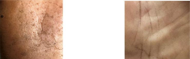
This clinical comparison photograph displays the plantar surfaces (soles) of two different individuals to illustrate skin manifestations of chronic arsenic exposure (arsenicosis). The image on the left shows an affected foot characterized by diffuse hyperpigmentation, mottled melanosis (darkened patches), and palmoplantar hyperkeratosis. Visible features include a rough, thickened skin texture with numerous small, raised, corn-like keratotic papules and irregular desquamation. In contrast, the image on the right depicts a healthy sole with a smooth, uniform skin texture, normal light pigmentation, and clearly defined dermatoglyphic lines without any evidence of lesions, nodules, or abnormal thickening. This comparison serves as an educational tool for identifying the dermatological signs of arsenic poisoning, specifically highlighting the diagnostic transition from healthy tissue to pathological keratotic and pigmentary changes characteristic of environmental or occupational toxin exposure.
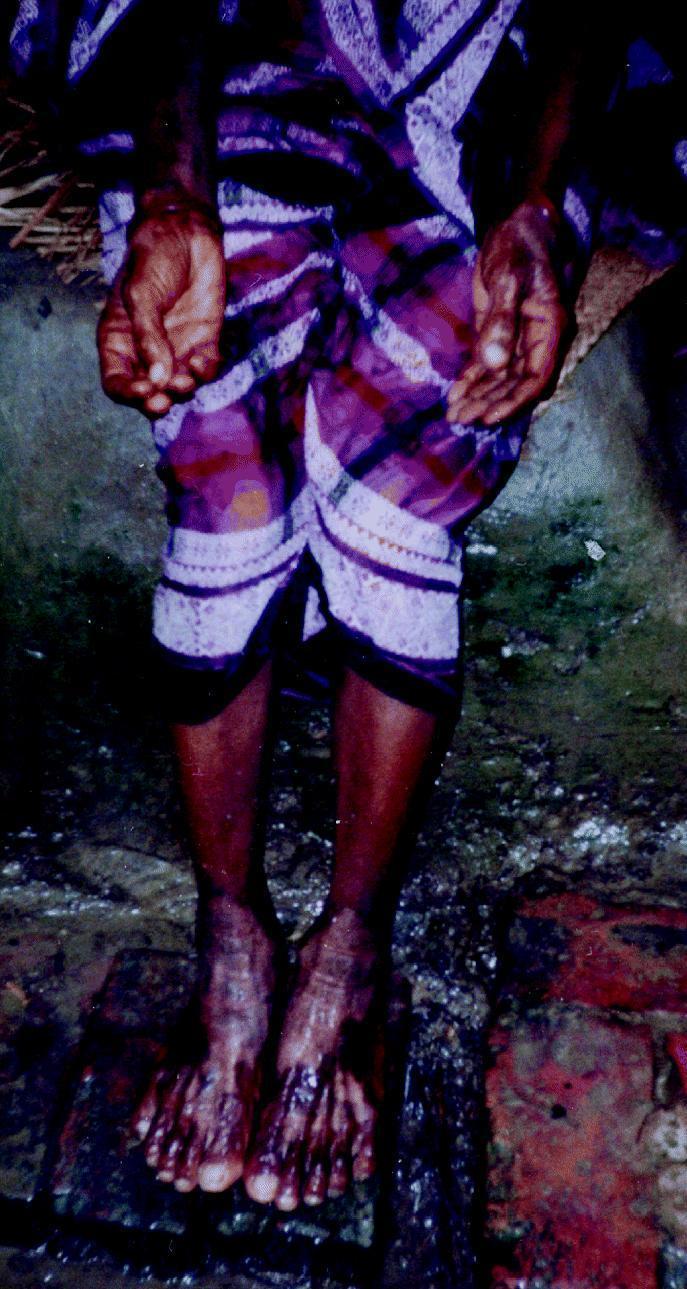
This clinical photograph illustrates the dermatological and vascular manifestations of chronic arsenic poisoning in a patient. The image displays the hands and feet of a female, highlighting two hallmark conditions: palmar keratosis and 'blackfoot disease.' The palmar surfaces of the hands show significant hyperkeratosis, characterized by thickened, roughened skin with exaggerated creases and hyperpigmentation. The feet and lower legs demonstrate advanced peripheral vascular changes. Specifically, the toes and distal portions of the feet exhibit severe dark discoloration ranging from deep mahogany to black, indicative of gangrenous changes or severe ischemia associated with blackfoot disease. The skin in these affected areas appears shiny or atrophic. This presentation is characteristic of long-term exposure to high levels of arsenic in drinking water, which leads to multisystemic toxicity, including hyperkeratosis and obliterative angiopathy. The photograph serves as an educational tool for identifying systemic signs of environmental toxicant exposure in endemic regions.
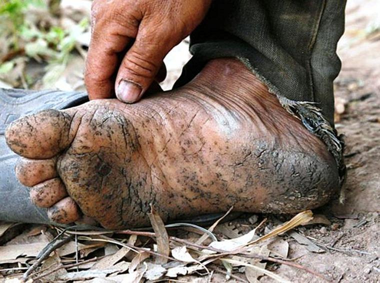
This clinical photograph displays characteristic cutaneous manifestations of chronic arsenicosis, specifically palmoplantar hyperkeratosis and dyspigmentation, on the foot of an individual. The plantar surface exhibits diffuse skin thickening (hyperkeratosis) with a rough, fissured, and cracked texture. Prominent hyperpigmented macules and patches are distributed across the sole, often described as a 'raindrops on a dusty road' pattern. The skin appears weathered and xerotic, with visible debris embedded in the epidermal cracks. Educational focus is placed on the recognition of chronic heavy metal toxicity signs, which often include these specific keratotic lesions and pigmentary changes. The surrounding context, including worn clothing and weathered skin on the hand, suggests prolonged environmental or occupational exposure to arsenic, typically through contaminated groundwater or industrial processes. This image serves as a diagnostic reference for dermatological signs of systemic arsenic poisoning.
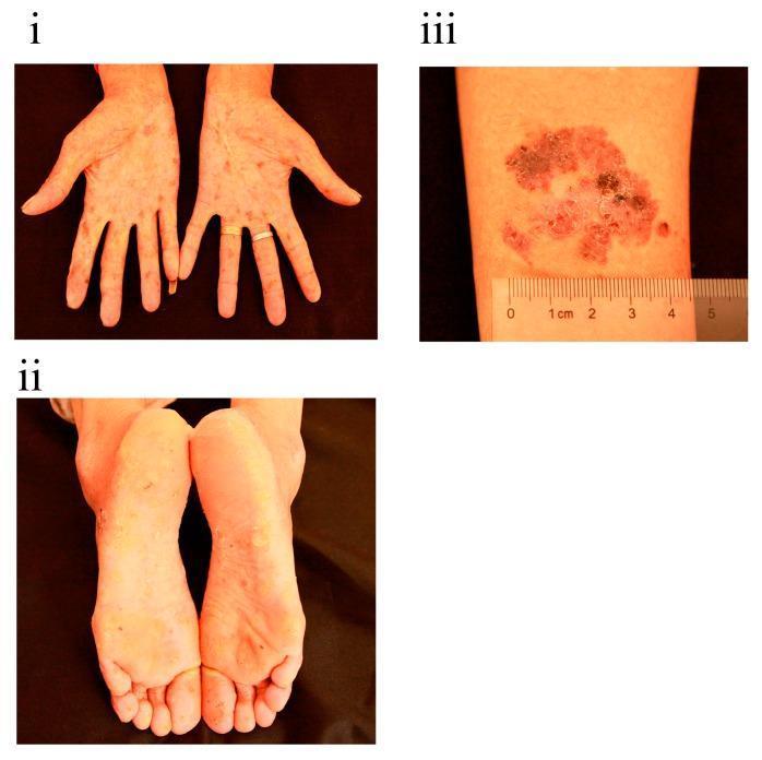
Clinical photographs showing dermatological manifestations of chronic arsenic poisoning in a single patient. Panel (i) displays the palms with diffuse, mottled pigmentation characterized by intermingled hyperpigmentation and de-pigmentation ('raindrop' appearance). Panel (ii) illustrates the soles of the feet exhibiting significant palmoplantar hyperkeratosis, appearing as thickened, yellowish, and rough skin localized primarily on weight-bearing areas. Panel (iii) presents a close-up of the right arm featuring a well-demarcated, erythematous, and scaly plaque approximately 3 cm in diameter, consistent with Bowen's disease (squamous cell carcinoma in situ). The plaque shows variegated color, including brownish-black pigmented areas and an irregular, rough surface texture. These visual signs are classic diagnostic indicators of long-term arsenic exposure, often leading to secondary cutaneous malignancies.
"arsenic poisoning" AND treatment
Arsenic Poisoning
Sources of Arsenic
- Pesticides/insecticides: Paris green (copper arsenite), calcium arsenate, lead arsenate
- Contaminated groundwater (major cause of chronic poisoning in South Asia, Bangladesh)
- Occupational exposure: smelting, mining, semiconductor industry, wood preservatives (CCA-treated timber)
- Homicidal use: historically common; tasteless and odorless in solution
- Traditional/alternative medicines (see Gerke & Seifert 2025 - men show greater sensitivity than women)
- Arsenic trioxide (ATO) used therapeutically in acute promyelocytic leukemia (APL)
Mechanism of Toxicity
- Sulfhydryl (SH) group inhibition - reversibly binds sulfhydryl groups in tissue proteins and enzymes, disrupting cellular metabolism (this is the primary mechanism; BAL/dimercaprol works by competing for these binding sites)
- Enzyme inhibition - interferes with pyruvate dehydrogenase and other enzymes of the TCA cycle, disrupting oxidative phosphorylation
- Arsenate substitutes for phosphate in ATP synthesis (arsenolysis), uncoupling oxidative phosphorylation
- Capillary dilation - causes vasodilation, transudation of fluid, and hemorrhage in the intestines
- Direct organ damage - fatty degeneration of liver, renal tubular necrosis
- Peripheral nerve damage - disintegration of the axon cylinder (axonal neuropathy) with fragmentation and resorption of myelin
Fatal Dose and Period
| Parameter | Value |
|---|---|
| Fatal dose (arsenic trioxide) | ~180 mg |
| Minimum lethal dose reported | 30 mg (arsenic trioxide) |
| Fatal period | 12-48 hours (can be as short as 2-3 hours) |
- P C Dikshit Textbook of Forensic Medicine and Toxicology
Acute Arsenic Poisoning
Gastrointestinal (Earliest)
- Metallic taste in mouth, garlicky odour on breath
- Xerostomia (dry mouth), dysphagia
- Severe nausea and vomiting
- Colicky abdominal pain
- Profuse diarrhoea with rice water stools (resembles cholera - see differential below)
- Bloody stools in some cases; tenesmus and anal irritation
Cardiovascular
- Diffuse capillary leak + GI fluid loss → hypotension and shock
- Congestive cardiomyopathy
- Cardiogenic or non-cardiogenic pulmonary edema
- QTc prolongation and ventricular arrhythmias (appears promptly or after days)
Hematologic (within 1 week)
- Pancytopenia
- Basophilic stippling of erythrocytes
Neurological
- Delirium, encephalopathy, coma (within first few days)
- Ascending sensorimotor peripheral neuropathy - delayed 2-6 weeks; may progress to proximal muscle involvement and neuromuscular respiratory failure
Late Finding
- Aldrich-Mees lines (transverse white striae in nails) - appear months after acute poisoning; each line = one episode of poisoning; multiple lines suggest repeated exposure
Differential Diagnosis: Arsenic vs. Cholera
| Feature | Arsenic Poisoning | Cholera |
|---|---|---|
| Pain in throat | Before vomiting | After vomiting |
| Purging | After vomiting | Before vomiting |
| Stools | Dark, bloody → later rice-watery | Rice-watery, non-bloody, involuntary jet |
| Tenesmus/anal irritation | Present | Absent |
| Vomited matter | Mucus, bile, blood | Watery, no mucus/bile/blood |
| Voice | Not affected | Rough and whistling |
| Conjunctivae | Inflamed | Not inflamed |
- The Essentials of Forensic Medicine and Toxicology, 36th ed (2026)
Chronic Arsenic Poisoning
Skin Changes (Most Characteristic)
- "Raindrop" hyperpigmentation - finely mottled brown spots, mainly on flexures, temples, eyelids, neck
- Palmoplantar hyperkeratosis - thickened, rough skin of palms and soles
- Mee's lines (Aldrich-Mees lines) - transverse white lines in fingernails; appear ~5 weeks after exposure, 1-2 mm width
- Alopecia - patchy or diffuse hair loss
- Peripheral/facial oedema
- Bowen's disease (squamous cell carcinoma in situ) - long-term complication, a marker of systemic neoplastic risk

Neurological
- Symmetrical sensorimotor polyneuropathy (resembles Guillain-Barré syndrome)
- Glove-and-stocking distribution of dysesthesia, paresthesia, numbness, pain
- Distal muscle weakness → wrist drop, inability to walk
- Decreased distal reflexes, muscular atrophy, possibly paralysis
- Encephalopathy: severe headache, personality changes, seizures, coma
Hematologic
- Normochromic normocytic anaemia (partly hemolytic)
- Leukopenia, thrombocytopenia, mild eosinophilia
- Karyorrhexis on bone marrow examination
- May mimic megaloblastic anaemia (arsenic interferes with folate metabolism)
Other Systems
- Cardiovascular: peripheral vascular disease, non-cirrhotic portal hypertension, possible hypertension/cardiovascular mortality
- Hepatic: hepatomegaly, jaundice, cirrhosis
- Renal: chronic nephritis
- Respiratory: cough, hemoptysis, dyspnea
- Endocrine/Metabolic: possible link to diabetes
Malignancies (major long-term risk)
- Lung cancer
- Skin cancer (Bowen's disease, squamous cell carcinoma, basal cell carcinoma)
- Bladder cancer
- Possibly kidney and liver cancer
- Leukemia
- Tobacco synergizes with arsenic to increase cancer risk
Postmortem Findings (Forensic)
- "Red velvet" appearance of gastric mucosa - lines of redness along the rugae
- Small intestine: flaccid, mucus flakes, pale-violet mucosa, sub-mucous hemorrhages
- Subendocardial petechial hemorrhages of the ventricle (characteristic finding)
- Liver/spleen/kidneys: congested, enlarged, cloudy swelling, fatty change
- Renal tubular necrosis
- Pulmonary congestion with subpleural ecchymoses
- Brain: edema with patchy necrosis or hemorrhagic encephalitis
- X-ray: arsenic may be visible in the GI tract (radiopaque)
- Chronic gastritis, patchy hemorrhagic erosions
- Small intestinal dilation, reddened thickened mucosa
- Hepatic fatty change or severe necrosis
Diagnosis
| Test | Finding |
|---|---|
| Urine arsenic (gold standard) | Thousands of mcg in first 2-3 days after acute poisoning |
| Blood arsenic | Unreliable (disappears rapidly); do not use except in anuric patients |
| Hair/nail arsenic | Useful for chronic exposure (arsenic deposits in keratin) |
| Reinsch test | Arsenic deposits as grey film on copper strip |
| Marsh test | Classic forensic chemical test - produces arsenic mirror |
| Bone marrow exam | Karyorrhexis, megaloblastic changes |
| ECG | QTc prolongation |
Treatment
Acute Poisoning
- Gut decontamination - gastric lavage if early presentation
- Intensive supportive care - IV fluids for hypotension, antiarrhythmics for QT prolongation
- Chelation therapy (start immediately if clinical suspicion is high - do NOT wait for lab confirmation):
| Agent | Route | Dose |
|---|---|---|
| Unithiol (DMPS) | IV (preferred) | 3-5 mg/kg every 4-6 hours |
| Dimercaprol (BAL) | IM | 3-5 mg/kg every 4-6 hours |
| Succimer (DMSA) | Oral (not preferred in acute - GI absorption unreliable due to gastroenteritis/splanchnic edema) | Adjunct |
- Katzung's Basic and Clinical Pharmacology, 16th ed
Chelation is most effective when given within minutes to hours of exposure. Efficacy drops significantly with delay.
Chronic Poisoning
- Remove from exposure source
- Oral DMSA (succimer) can be used
- Treat peripheral neuropathy symptomatically
- Monitor for malignant transformation
- Nutritional support (folate supplementation)
Key Forensic Points
- Arsenic is a protoplasmic poison and capillary poison
- Classic method of homicidal poisoning due to tastelessness and solubility
- Preservation in body: arsenic fixes in bone, hair, and nails - detectable years after death (resists putrefaction)
- A single episode of acute poisoning → one Mee's line; repeated exposures → multiple lines (width reflects duration of exposure)
- Arsenic is teratogenic
- P C Dikshit Textbook of Forensic Medicine and Toxicology
- The Essentials of Forensic Medicine and Toxicology, 36th ed (2026)
- Katzung's Basic and Clinical Pharmacology, 16th Edition
- Arsenical keratosis review (2024, PMID 39189802)