
Papilledema and bp
Reading File
Papilledema is swelling of the optic disc caused by raised intracranial pressure (ICP), and hypertension is one of its key causes. Let me pull the textbook content now.
Searching sources...
Finding Sources
Finding Sources
Searching PubMed
papilledema hypertension intracranial pressure
Good hits. Let me read the key sections now in parallel.
Reading File
Reading File
Reading File
Good content retrieved. Let me now read the malignant hypertension section and the treatment section from Wills Eye Manual.
Reading File
Reading File
Loading Image
I now have comprehensive content. Here is a full, synthesized answer.
Papilledema and Blood Pressure
What is Papilledema?
Papilledema refers specifically to optic disc edema secondary to raised intracranial pressure (ICP). It is not a generic term for any disc swelling - the cause must be elevated ICP. The mechanism: raised ICP blocks axoplasmic flow in optic nerve fibers, increasing axoplasm volume at the disc. - Bradley and Daroff's Neurology in Clinical Practice
The Retinal Fundus in Papilledema
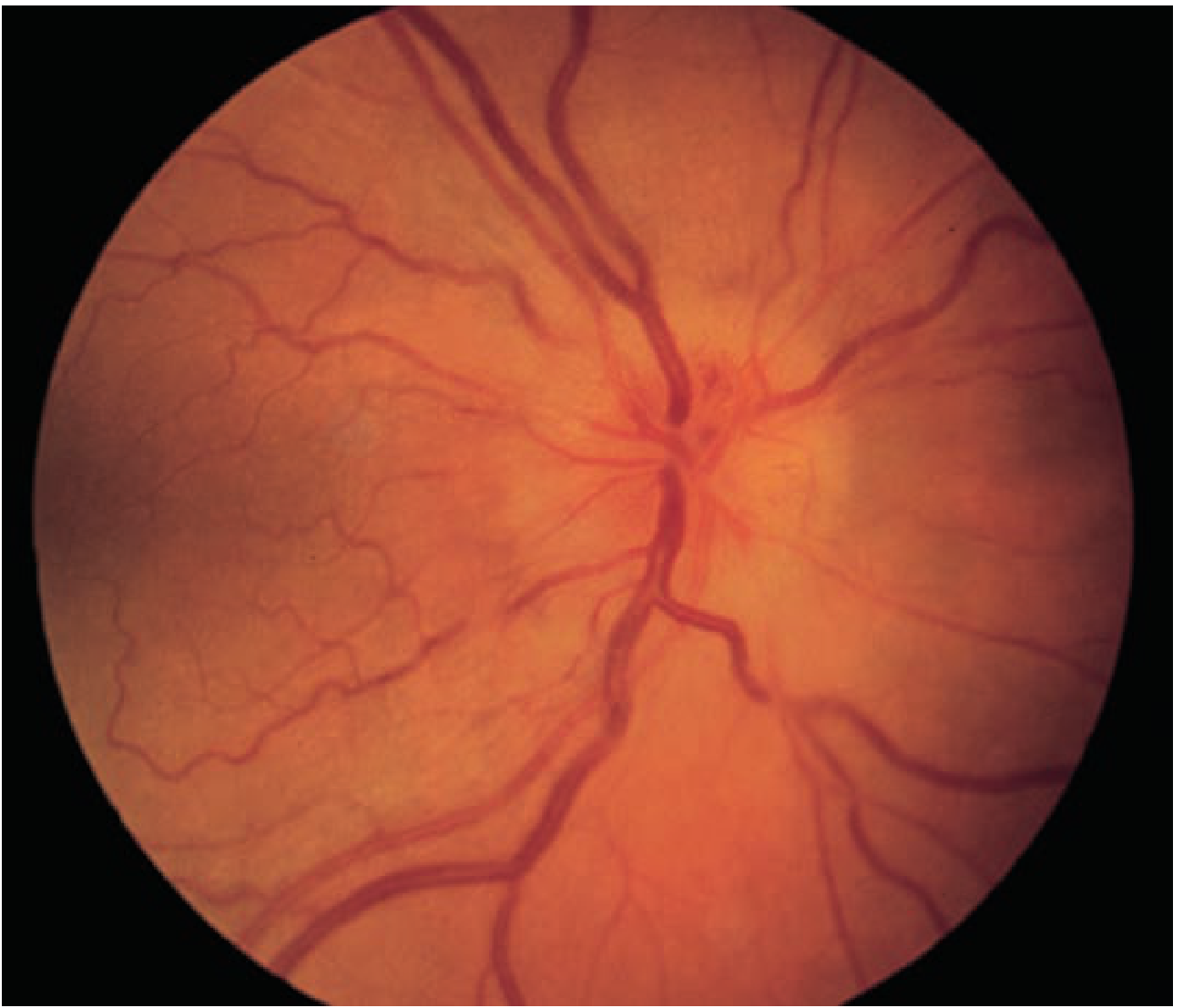
Fundus appearance showing vascular congestion, elevation of the nerve head, and blurred disc margins characteristic of papilledema - Tintinalli's Emergency Medicine
Stages of Papilledema
| Stage | Key Features |
|---|---|
| 1. Early | Minimal disc hyperemia, capillary dilation; mild nerve fiber layer opacification; absence of venous pulsations; peripapillary hemorrhage |
| 2. Fully developed | Engorged/tortuous veins; disc surface grossly elevated; cotton-wool spots; Paton lines (circumferential retinal folds); macular exudates or star |
| 3. Chronic (weeks-months) | Pale, gliotic disc; "champagne cork" appearance; pseudodrusen (extruded axoplasm); hemorrhages resolve; collateral optociliary vessels may appear |
| 4. Atrophic | Optic atrophy; severe irreversible visual loss |
- Localization in Clinical Neurology, 8e and Bradley and Daroff's Neurology
Clinical Features
- Usually bilateral (may be asymmetric due to variation in optic nerve subarachnoid septations)
- Visual acuity preserved early - a distinguishing hallmark; patients may be visually asymptomatic initially
- Transient visual obscurations (seconds-long visual loss)
- Visual field defects: enlarged blind spot (earliest), arcuate defects (typically inferonasal), concentric constriction
- No afferent pupillary defect unless disc edema is severe and asymmetric
- Absent spontaneous venous pulsations on fundoscopy (SVPs present = ICP likely normal)
- Fluorescein angiography: disc capillary dilation, dye leakage, microaneurysms
Causes - Including Hypertension
Primary causes of raised ICP (and thus papilledema):
- Idiopathic intracranial hypertension (IIH / pseudotumor cerebri)
- Mass lesions (tumor, hemorrhage, abscess, large infarction)
- Hydrocephalus
- Meningitis/encephalitis
- Subarachnoid hemorrhage
Hypertension and papilledema - two key relationships:
1. Malignant (Hypertensive Emergency) - BP causing disc edema
A marked elevation in blood pressure (typically >180/120 mmHg) can produce bilateral optic disc edema that is indistinguishable from papilledema. This is a hypertensive emergency. Associated findings include:
- Peripapillary cotton-wool spots
- Retinal hemorrhages
- Hypertensive encephalopathy / PRES (posterior reversible encephalopathy syndrome)
- Vision loss
"The changes associated with malignant hypertension can occur at lower blood pressures in patients with renal failure." - Bradley and Daroff's Neurology
The mechanism here involves both direct end-organ damage to retinal vasculature AND elevated ICP from PRES/encephalopathy.
2. Idiopathic Intracranial Hypertension (IIH) - raised ICP causing papilledema
This is a separate condition where ICP rises without a mass lesion or vascular cause. IIH is a primary cause of papilledema. Obesity and female sex are major risk factors. BP can be normal.
Papilledema vs. Pseudopapilledema
| Feature | Papilledema | Pseudopapilledema (e.g. drusen) |
|---|---|---|
| Disc color | Hyperemic | Pink/yellowish-pink |
| Disc margins | Indistinct (superior/inferior poles first) | Usually distinct, may be lumpy |
| Vessels | Normal distribution, SVPs absent | Emanate from center, anomalous pattern |
| Nerve fiber layer | Dull, edematous, obscures vessels | No edema; may glisten |
| Hemorrhages | Splinter type | Subretinal/retinal/vitreous |
Management
Treat the underlying cause:
- Hypertensive emergency: controlled reduction of BP (avoid over-rapid drop to prevent ischemia); IV agents in ICU setting
- IIH: weight loss, acetazolamide (reduces CSF production); in progressive visual loss - optic nerve sheath fenestration or CSF shunting
- Serial visual field monitoring for all cases with papilledema
Recent Evidence (2024-2026)
A 2025 meta-analysis (PMID: 40937960) found GLP-1 and dual GIP/GLP-1 receptor agonists (e.g., semaglutide) to be efficacious and safe in IIH - relevant given the obesity-IIH-hypertension overlap. A 2025 consensus statement (PMID: 38431253) from the Spanish Society of Neurology provides updated diagnostic and treatment criteria for disorders of intracranial pressure.
Key clinical pearl: In a hypertensive patient with bilateral disc edema, always consider whether the disc swelling reflects (a) direct hypertensive retinopathy/malignant hypertension, or (b) raised ICP from an underlying cause unrelated to BP. The distinction requires neuroimaging and BP measurement together.
This is a shared conversation. Sign in to Orris to start your own chat.