
Explain endocytosis and exocytosis
Endocytosis and exocytosis are the two main forms of vesicular transport - cells use them to move large molecules or particles across the plasma membrane that cannot pass through protein channels. Endocytosis brings material in, exocytosis sends material out. Both require energy (ATP) and involve membrane-bound vesicles.
Searching sources...
Reading File
Finding Sources
Reading File
Reading File
Searching Images
types of endocytosis phagocytosis pinocytosis receptor-mediated clathrin
Reading File
Reading File
Loading Image
Loading Image
Reading File
Reading File
Loading Image
Here is a thorough explanation drawn from Junqueira's Basic Histology and Harper's Illustrated Biochemistry:
Endocytosis and Exocytosis
Both are forms of vesicular (bulk) transport - they move large molecules, particles, or fluid that cannot cross the plasma membrane through channels. Both are energy-dependent (require ATP, Ca²⁺, and cytoskeletal elements) and involve membrane-bound vesicles.
The diagram below summarizes their relationship:
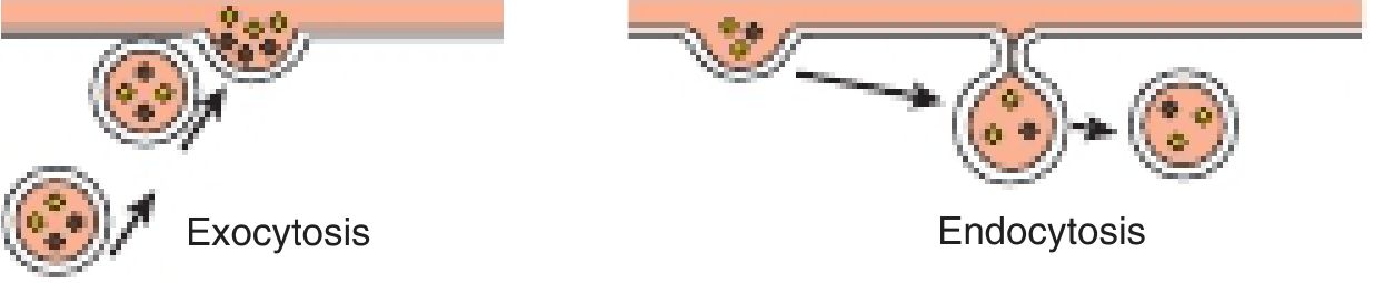
Comparison of exocytosis and endocytosis mechanisms - Harper's Illustrated Biochemistry, 32nd Ed.
Endocytosis ("bringing in")
Macromolecules enter the cell when plasma membrane folds inward, enclosing extracellular material into a vesicle that pinches off into the cytoplasm. There are three major types:
1. Phagocytosis ("cell eating")
- Ingestion of large particles - bacteria, dead cells, cellular debris, viruses
- Specialized cells only: macrophages, neutrophils, monocytes
- Cell extends pseudopodia that engulf the particle, forming an intracellular vacuole called a phagosome
- The phagosome fuses with a lysosome - hydrolytic enzymes degrade the contents
- Macrophages are extremely active; they can ingest 25% of their volume per hour and internalize 3% of their plasma membrane per minute
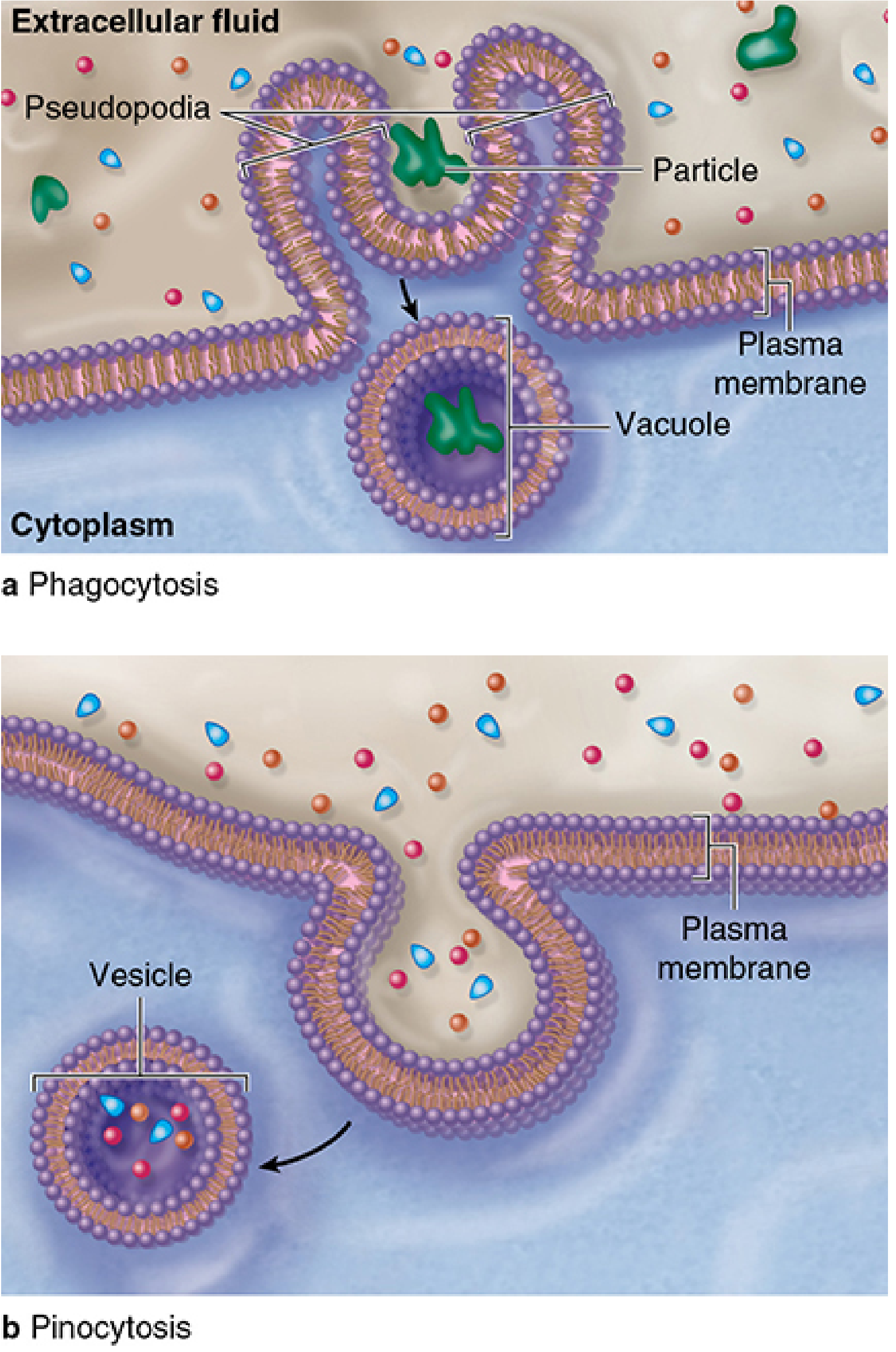
Phagocytosis (a) vs Pinocytosis (b) - Junqueira's Basic Histology, 17th Ed.
2. Pinocytosis ("cell drinking")
- Uptake of extracellular fluid and its dissolved contents in small vesicles (~80 nm diameter)
- Occurs in all eukaryotic cells continuously
- Two subtypes:
- Fluid-phase pinocytosis - nonselective; solute uptake is proportional to extracellular concentration; random and non-directed
- Absorptive (receptor-mediated) pinocytosis - see below
- Vesicles either fuse with lysosomes or travel to the opposite surface (transcytosis), enabling bulk transfer across a cell (e.g., across endothelial cells)
3. Receptor-Mediated Endocytosis (Absorptive Pinocytosis)
This is the most selective and clinically significant type:
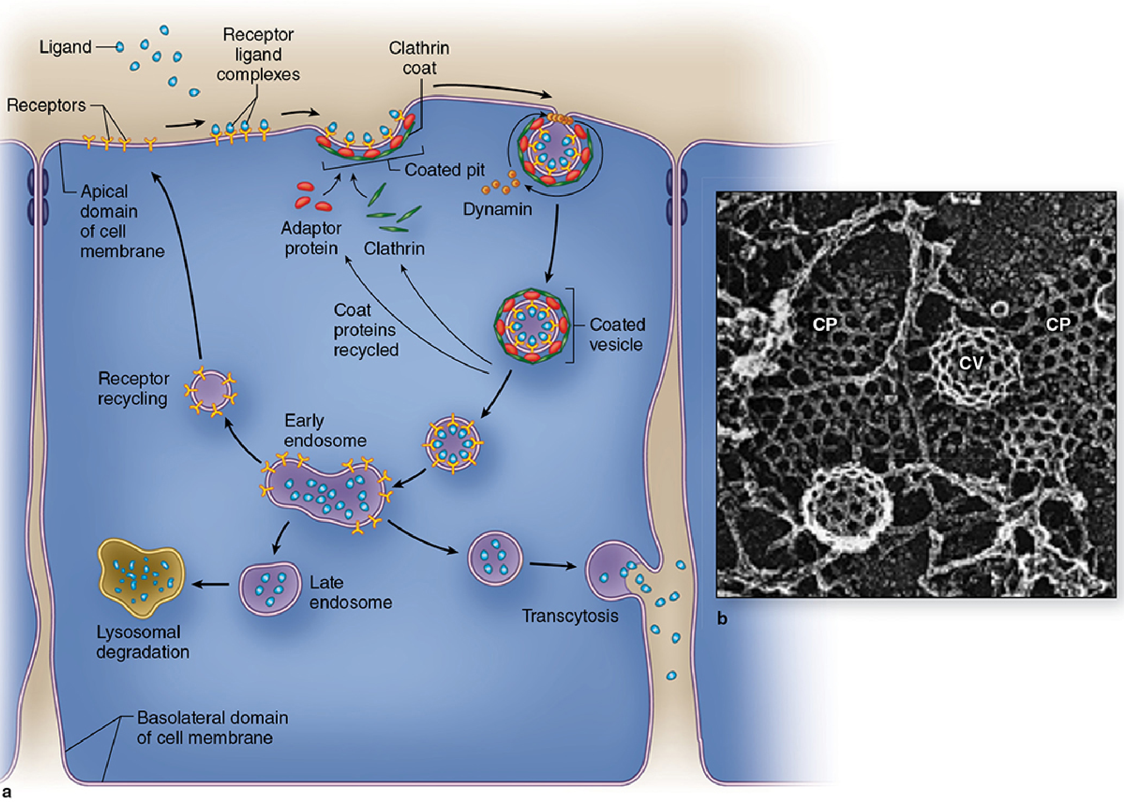
Receptor-mediated endocytosis pathway showing coated pits, vesicles, endosomes, and possible fates - Junqueira's Basic Histology, 17th Ed.
Steps:
- Specific ligands (e.g., LDL, hormones, transferrin) bind high-affinity receptors on the cell surface
- Receptor-ligand complexes aggregate in coated pits - membrane regions coated on the cytoplasmic side with clathrin (a three-limbed triskeleton protein) and adaptor proteins
- Dynamin (a GTPase) forms constricting rings around the pit neck and pinches it off as a clathrin-coated vesicle
- The clathrin coat is shed and recycled
- The vesicle fuses with an early endosome; the acidic interior uncouples ligand from receptor
Possible fates of internalized material:
- Receptor + ligand move to late endosome → lysosome for degradation
- Ligands released; empty receptors go to recycling endosomes and return to the cell surface (e.g., LDL receptor)
- Vesicles fuse with the opposite membrane surface → transcytosis (ligand released extracellularly)
Coated pits may constitute up to 2% of the surface area of some cells. Ca²⁺ is also required for endocytosis, and Ca²⁺ stimulates the process (which is why calcium phosphate is used in DNA transfection experiments).
Clinical relevance: Viruses causing hepatitis, poliomyelitis, AIDS, and COVID-19 exploit receptor-mediated endocytosis to enter cells. Iron toxicity begins with excessive endocytic uptake. LDL receptor defects underlie familial hypercholesterolemia.
Exocytosis ("sending out")
The reverse process: intracellular vesicles fuse with the plasma membrane and release their contents to the extracellular space. Also involved in membrane remodeling (replacing membrane removed during endocytosis).
Key trigger: Hormonal signals bind cell-surface receptors, causing a local, transient rise in intracellular Ca²⁺, which triggers vesicle-membrane fusion.
Two modes:
| Mode | Description | Examples |
|---|---|---|
| Constitutive secretion | Continuous release as soon as synthesis is complete; no regulatory signal needed | Collagen subunits, plasma proteins, extracellular matrix components |
| Regulated secretion | Products stored in granules; released only on specific stimuli | Insulin (glucose stimulus), digestive enzymes (pancreatic acinar cells, cholecystokinin), catecholamines, parathyroid hormone, neurotransmitters |
Fates of released molecules:
- Remain as membrane proteins on the cell surface
- Become part of the extracellular matrix (e.g., collagen, glycosaminoglycans)
- Enter extracellular fluid and signal other cells (endocrine/paracrine)
Membrane Trafficking - The Balance Between the Two
Since membrane is lost with each endocytotic event and added with each exocytotic one, the two processes must be balanced to maintain cell size and membrane composition. This continuous cycling of membrane components is called membrane trafficking. It is physiologically important for processes like reducing blood lipid levels and receptor regulation.
Related Concepts: Exosomes and Multivesicular Bodies
Some endosomal compartments accumulate small vesicles within their lumen by further inward invaginations, forming multivesicular bodies (MVBs). These can:
- Fuse with lysosomes for selective degradation
- Fuse with the plasma membrane and release their internal vesicles as exosomes (50-150 nm) - a form of cell-to-cell communication
Summary Comparison
| Feature | Endocytosis | Exocytosis |
|---|---|---|
| Direction | Extracellular → Intracellular | Intracellular → Extracellular |
| Membrane movement | Plasma membrane folds inward | Vesicle fuses with plasma membrane |
| Energy | ATP required | ATP required |
| Ca²⁺ requirement | Yes | Yes (triggers fusion) |
| Subtypes | Phagocytosis, Pinocytosis, Receptor-mediated | Constitutive, Regulated |
| Key proteins | Clathrin, Dynamin, Adaptor proteins | SNARE proteins, Ca²⁺-sensing proteins |
| Products | Endosomes, Phagosomes, Pinocytotic vesicles | Secreted proteins, hormones, neurotransmitters |
Sources: Junqueira's Basic Histology, 17th Ed., pp. 82-86 | Harper's Illustrated Biochemistry, 32nd Ed., pp. 493-494
This is a shared conversation. Sign in to Orris to start your own chat.