
Copd pathophysiology
Reading File
Now let me search the medical library for COPD pathophysiology content in parallel.
Finding Sources
Finding Sources
Searching PubMed
"COPD" AND "pathophysiology"
Good. Now let me read the key COPD sections from Fishman's Pulmonary and Harrison's, and also search for dedicated COPD pathophysiology headings.
Finding Sources
Reading File
Now let me read the main COPD chapters from Murray & Nadel's and Robbins Pathology:
Reading File
Reading File
Good content. Let me read more of Robbins Pathology for inflammatory mechanisms and the Harrison's chapter:
Reading File
Finding Sources
Excellent content from Robbins. Let me read more of this section and fetch the pathogenesis diagram:
Reading File
Reading File
Excellent content. Let me now fetch the key pathogenesis diagram from Robbins and read chronic bronchitis pathology:
Loading Image
Loading Image
Reading File
I now have comprehensive content from multiple authoritative textbooks. Let me compile a thorough, well-illustrated answer on COPD pathophysiology.
COPD Pathophysiology
Definition
COPD is defined by the WHO as "a common, preventable and treatable disease characterized by persistent respiratory symptoms and airflow limitation due to airway and/or alveolar abnormalities caused by exposure to noxious particles or gases." It affects >10% of adults over 40 years in the US and is the third leading cause of death worldwide.
COPD is an umbrella term that encompasses two major overlapping conditions - emphysema and chronic bronchitis - both resulting from chronic airway injury (predominantly smoking):
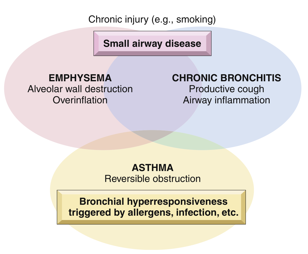
Risk Factors
- Cigarette smoking - the dominant cause; ~80% of COPD is attributable to it; 35-50% of heavy smokers develop COPD
- Poor lung development early in life
- Environmental/occupational pollutant exposure
- Airway hyperresponsiveness
- Genetic factors - particularly alpha-1 antitrypsin (A1AT) deficiency (Pi locus, chromosome 14)
- Women appear more susceptible than men
Core Pathogenic Mechanisms
The three pillars of COPD pathogenesis (especially emphysema) are shown below:
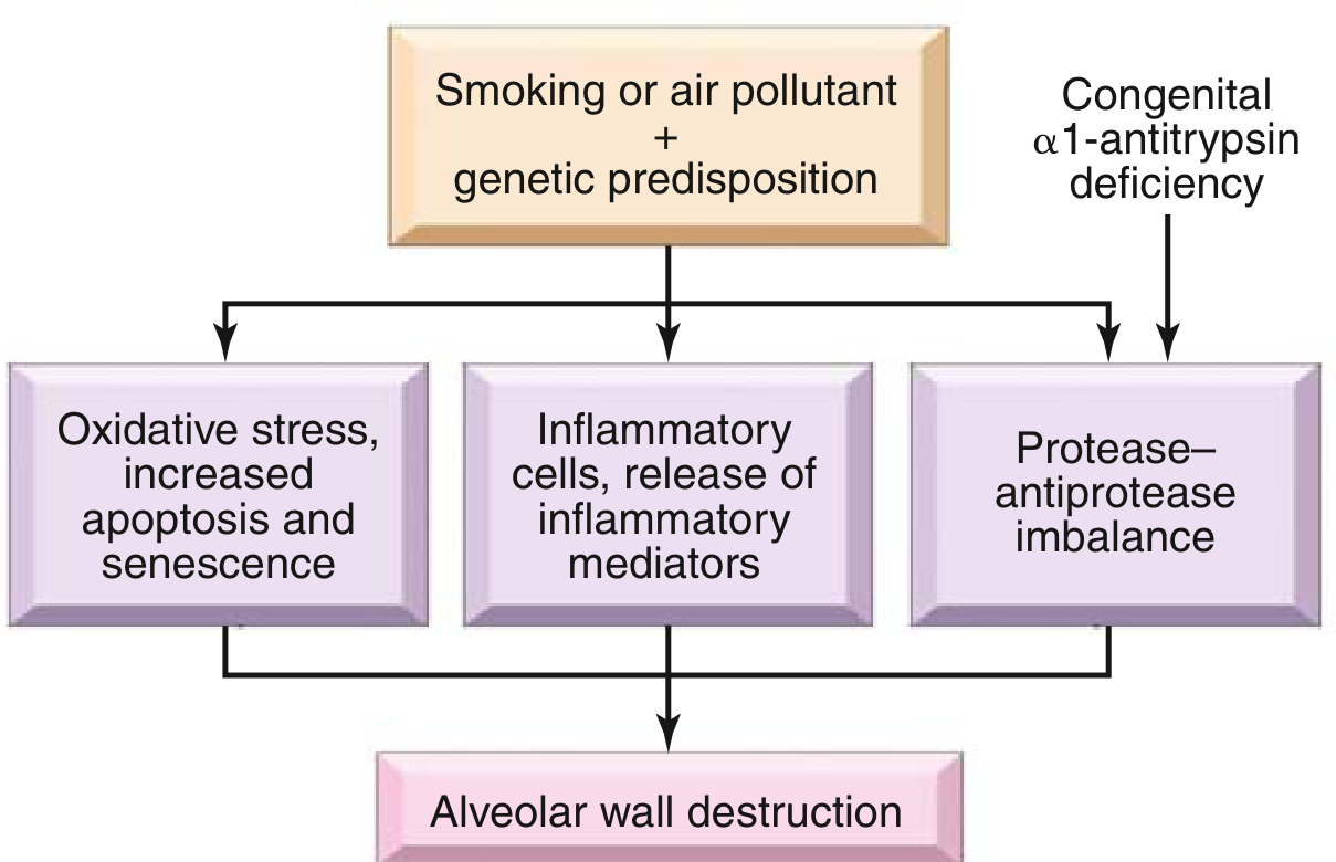
1. Inflammatory Cell Recruitment and Mediators
Inhaled cigarette smoke activates the innate and adaptive immune systems in the airways and lung parenchyma:
-
Key inflammatory cells: Neutrophils, alveolar macrophages, CD4+ and CD8+ T lymphocytes
-
Key mediators: Leukotriene B4 (LTB4), IL-8 (chemokine), TNF-alpha, IL-13
-
These mediators act as chemotactic factors (recruit more cells), proinflammatory cytokines (amplify the reaction), and growth factors (induce structural remodeling)
-
Notably, eosinophils are NOT prominent in COPD (this distinguishes it from asthma)
-
This inflammatory process persists even after smoking cessation
-
Robbins & Kumar Basic Pathology, p. 448
2. Protease-Antiprotease Imbalance
This is the central mechanism of alveolar wall destruction in emphysema:
- Inflammatory cells (especially neutrophils and macrophages) release proteases - notably neutrophil elastase and matrix metalloproteinases (MMPs)
- These enzymes degrade elastin and collagen in the alveolar walls and extracellular matrix
- Normally, antiproteases - chiefly alpha-1 antitrypsin (A1AT) - inhibit these proteases
- In smokers, oxidative stress directly inactivates A1AT, tipping the balance toward destruction
- In A1AT deficiency (homozygous Z allele, ~0.01% of US population): >80% develop symptomatic panacinar emphysema, earlier onset and more severe with smoking
- Loss of elastic tissue removes radial traction on small airways, causing airway collapse during expiration - this creates functional (dynamic) airflow obstruction without a fixed mechanical blockade
3. Oxidative Stress
-
Cigarette smoke directly contains reactive oxygen species (ROS): superoxide (O2-), hydroxyl radical (OH), and hydrogen peroxide (H2O2)
-
Activated macrophages and neutrophils generate additional ROS chronically
-
NOX4 (NADPH oxidase 4) expression is increased in airway smooth muscle and fibroblasts in COPD
-
ROS cause direct tissue injury and inflammation
-
A critical consequence: ROS activate NF-kB, a transcription factor that upregulates proinflammatory cytokines (IL-8, TNF), creating a self-amplifying loop
-
Cigarette smoke also inhibits histone deacetylase, further promoting proinflammatory gene expression
-
Oxidative stress also inactivates A1AT, compounding the protease-antiprotease imbalance
-
Fishman's Pulmonary Diseases and Disorders, p. 395
Emphysema - Anatomic Patterns
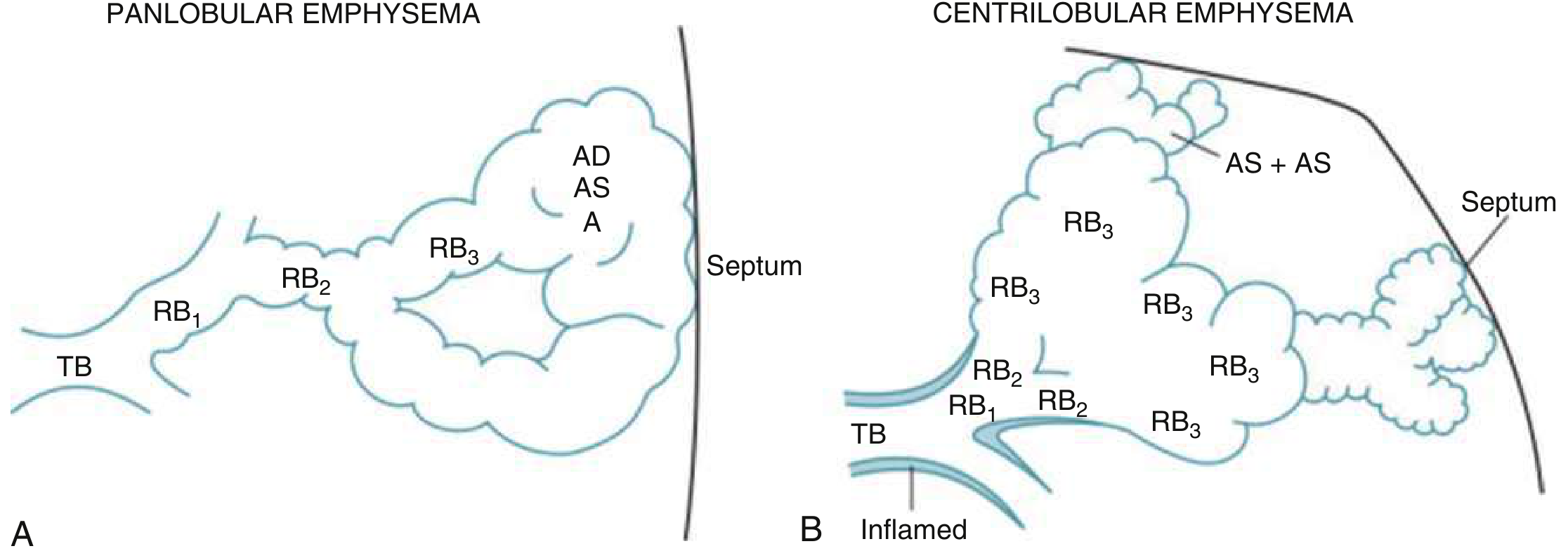
Centriacinar (Centrilobular) Emphysema
- Involves central/proximal acinus (respiratory bronchioles) with relative sparing of distal alveoli
- Both emphysematous and normal air spaces coexist within the same acinus
- More common and severe in upper lobes, apical segments
- Strongly associated with cigarette smoking - the predominant form
- About 20x more common than panacinar emphysema
Panacinar (Panlobular) Emphysema
- Uniform enlargement of the entire acinus, from respiratory bronchiole to terminal alveoli
- More common in lower lung zones
- Classically associated with alpha-1 antitrypsin deficiency
Distal Acinar (Paraseptal) Emphysema
- Affects the distal part of the acinus, near pleura and lobular septa
- Upper half of lungs more affected; can form bullae
- Classic association: spontaneous pneumothorax in young adults
Irregular Emphysema
- Irregular acinar involvement, almost always associated with scarring
- Usually clinically insignificant
Chronic Bronchitis - Pathogenesis
Defined clinically: productive cough for ≥3 months in ≥2 consecutive years.
The hallmark is mucus hypersecretion, driven by:
- Hypertrophy of mucous glands in trachea and large bronchi (assessed by the Reid Index - ratio of submucosal gland thickness to bronchial wall thickness; normally <0.4; elevated in chronic bronchitis)
- Goblet cell metaplasia - increase in goblet cells in smaller bronchi and bronchioles
- Inflammation - macrophages, neutrophils, lymphocytes (NOT eosinophils)
- Cytokine mediation - IL-13 from T cells drives mucin expression; neutrophil elastase (induced by tobacco smoke) further contributes
The airflow obstruction in chronic bronchitis results primarily from small airway disease (chronic bronchiolitis):
-
Mucous plugging of bronchiolar lumen
-
Luminal inflammation
-
Submucosal fibrosis causing luminal narrowing
-
In severe cases: bronchiolitis obliterans
-
Robbins & Kumar Basic Pathology, p. 449
Airflow Obstruction Mechanics
The final common pathway leading to obstructive physiology:
| Mechanism | How it causes obstruction |
|---|---|
| Loss of elastic recoil (emphysema) | Reduced radial traction on airways; collapse during expiration |
| Mucus hypersecretion (chronic bronchitis) | Plugging of bronchioles |
| Airway wall inflammation and fibrosis | Fixed narrowing of lumen |
| Dynamic hyperinflation | Air trapping; increased TLC and FRC |
Dynamic Hyperinflation
- Decreased elastic recoil + airflow limitation means insufficient time for passive exhalation to FRC
- Tidal breathing shifts to the upper portion of the flow-volume curve
- Results in progressive air trapping and increased TLC, FRC, and RV
- The diaphragm flattens, losing its domed shape - reducing its mechanical advantage
- In severe COPD, diaphragmatic contraction can paradoxically deflate the rib cage, making inspiration increasingly dependent on accessory muscles
Gas Exchange Abnormalities
Ventilation-Perfusion (V/Q) Mismatch
- Low V/Q areas (poorly ventilated but perfused): seen in airways disease/chronic bronchitis - cause hypoxemia
- High V/Q areas (alveolar dead space - ventilated but not perfused): seen in emphysema - waste ventilation
- Net result: inefficient gas exchange requiring higher minute ventilation to maintain normocapnia
Hypoxemia
- Results from low V/Q mismatch (the dominant mechanism in COPD, unlike diffusion limitation)
- Leads to hypoxic vasoconstriction, pulmonary hypertension, and eventually cor pulmonale
Hypercapnia (Type II Respiratory Failure)
-
PaCO2 typically does not rise until FEV1 falls to ~20-25% of predicted
-
Caused by:
- Increased physiologic dead space (V/Q mismatch) - requires increased ventilation to maintain eucapnia
- Mechanical disadvantage of the respiratory system (dynamic hyperinflation)
- Inspiratory muscle weakness (reduced force generation per cross-sectional area, reduced myosin heavy chain content, decreased Ca2+ sensitivity)
- Blunted ventilatory drive in some patients
-
Fishman's Pulmonary Diseases and Disorders, p. 2532
"Pink Puffer" vs. "Blue Bloater" Phenotypes
| Feature | Pink Puffer (Emphysema-predominant) | Blue Bloater (Chronic Bronchitis-predominant) |
|---|---|---|
| Body habitus | Thin, cachexic | Obese/stocky |
| Cyanosis | Absent (maintains oxygenation) | Present (cyanotic) |
| Cough/sputum | Minimal | Prominent productive cough |
| Breathing | Pursed-lip, increased WOB | Shallow, less labored |
| Hypoxemia | Mild-moderate | Marked |
| Hypercapnia | Absent/mild | Often present |
| PH/Cor pulmonale | Late | Earlier |
| OSA risk | Lower (hyperinflation may protect) | Higher (obesity, blunted drive) |
Downstream Systemic Consequences
- Pulmonary hypertension / Cor pulmonale: chronic hypoxia triggers hypoxic pulmonary vasoconstriction; vascular remodeling occurs even in mild COPD
- Polycythemia: secondary to chronic hypoxemia (EPO stimulation)
- Skeletal muscle dysfunction: reduced muscle fiber force generation, myosin heavy chain content, Ca2+ sensitivity - contributes to exercise limitation
- Nocturnal desaturation: physiologic changes of sleep are poorly tolerated; REM sleep causes most severe hypoventilation (especially with diaphragmatic dysfunction)
- Exacerbations: bacterial/viral infections trigger acute inflammatory flares; do not initiate, but perpetuate and worsen the disease
Summary Flow
Smoking / noxious particles + genetic predisposition
          ↓
Airway inflammation (neutrophils, macrophages, CD4+/CD8+ T cells)
          ↓
  ┌───────────────────┬───────────────────┐
  ↓                   ↓                   ↓
Oxidative stress   Protease-antiprotease   Mucus hypersecretion
(ROS, NF-kB)       imbalance               (goblet cell metaplasia,
                  (elastase > A1AT)         mucous gland hypertrophy)
  ↓                   ↓                   ↓
Alveolar wall     Alveolar wall         Small airway disease
apoptosis/        destruction           (bronchiolitis, fibrosis)
senescence        (EMPHYSEMA)           (CHRONIC BRONCHITIS)
  └───────────────────┴───────────────────┘
                        ↓
              Airflow obstruction (irreversible)
                        ↓
          V/Q mismatch → hypoxemia → PH/cor pulmonale
          Dead space → ↑ WOB → dynamic hyperinflation
          Hypercapnia (late, when FEV1 <25%)
Sources:
- Robbins & Kumar Basic Pathology - pp. 447-450
- Fishman's Pulmonary Diseases and Disorders, pp. 395, 2532
- Murray & Nadel's Textbook of Respiratory Medicine
Symptoms
Finding Sources
Finding Sources
Reading File
Reading File
Finding Sources
Reading File
Finding Sources
Reading File
Finding Sources
I have comprehensive content. Let me now compile the full symptoms answer.
COPD - Symptoms & Clinical Features
Key Point
COPD symptoms are insidious in onset - they develop gradually over years (typically after ~10 years of smoking exposure). By the time patients present, significant irreversible lung damage has usually already occurred. Physical examination findings only become apparent at advanced stages (FEV1 <50% predicted).
Cardinal Symptoms (The Triad)
1. Dyspnea
- The dominant and most disabling symptom
- Initially exertional - worse with hills, stairs, hurrying
- Progresses insidiously over years to dyspnea at rest in severe disease
- Patients often unconsciously reduce activity to avoid breathlessness, so they under-report it
- Graded using the mMRC (Modified Medical Research Council) Dyspnea Scale:
| Grade | Description |
|---|---|
| 0 | Breathless only with strenuous exercise |
| 1 | Short of breath when hurrying on level ground or walking up a slight hill |
| 2 | Walks slower than peers on level ground due to breathlessness, or stops for breath after ~100m |
| 3 | Stops for breath after a few minutes or ~100 yards on level ground |
| 4 | Too breathless to leave the house; breathless when dressing/undressing |
2. Chronic Cough
- Often the first symptom to appear
- Initially intermittent - the "smoker's cough" - easily dismissed
- Becomes persistent and daily as disease advances
- May be dry or productive
- Patients may not report it spontaneously as they consider it "normal" for a smoker
3. Sputum Production
- Typically mucoid (clear/white), produced in the morning ("morning sputum")
- Reflects mucous gland hypertrophy and goblet cell metaplasia of chronic bronchitis
- Heavy, daily sputum for ≥3 months per year for ≥2 consecutive years = chronic bronchitis by definition
- Becomes purulent (yellow/green) during exacerbations (bacterial colonization/infection)
4. Wheezing
- Due to airflow turbulence through narrowed airways
- May be present at rest or only on exertion
- Particularly prominent during exacerbations
- Not specific - can also occur in asthma, heart failure
Additional Symptoms (Advanced/Severe Disease)
| Symptom | Mechanism |
|---|---|
| Chest tightness | Bronchospasm, air trapping |
| Exercise intolerance | V/Q mismatch, dynamic hyperinflation, skeletal muscle dysfunction |
| Fatigue / weight loss | Systemic inflammation, increased work of breathing, cachexia |
| Poor sleep quality | Nocturnal hypoxemia, hyperinflation limiting comfortable positioning |
| Ankle swelling | Cor pulmonale / right heart failure from pulmonary hypertension |
| Morning headache | Nocturnal CO2 retention (hypercapnia) |
| Confusion / drowsiness | Severe hypercapnia (CO2 narcosis in acute exacerbations) |
| Depression / anxiety | Common comorbidities; impair quality of life significantly |
Comorbidities that are more common than expected in COPD: osteoporosis, depression, anxiety, cardiovascular disease, lung cancer, malnutrition, and diabetes - Washington Manual of Medical Therapeutics, p. 311
Physical Examination Signs
Physical signs appear late (usually FEV1 <50%):
Inspection
- Pursed-lip breathing - creates intrinsic PEEP to prevent airway collapse during expiration; characteristic of emphysema-predominant COPD
- Barrel chest - increased AP diameter due to chronic hyperinflation (increased TLC)
- Use of accessory muscles - sternocleidomastoid, scalenes recruited due to diaphragmatic disadvantage
- Hoover's sign - paradoxical inward movement of the lateral rib cage during inspiration (flattened diaphragm)
- Central and peripheral cyanosis - in hypoxemic patients (blue bloater phenotype)
- Cachexia / weight loss - in advanced emphysema-predominant disease
- Pursed-lip breathing posture ("tripod") - leaning forward with hands on knees
Percussion
- Hyperresonance - increased air in lungs (emphysema)
- Reduced diaphragmatic excursion - diaphragm flattened and fixed by hyperinflation
Auscultation
- Reduced breath sounds - loss of lung parenchyma, air trapping
- Prolonged expiratory phase - >6 seconds on forced exhalation (hallmark of obstruction)
- Expiratory wheeze and rhonchi - may or may not be present
- Muffled heart sounds - interposed hyperinflated lung attenuates cardiac sounds
Signs of Advanced Complications
- Raised JVP, peripheral edema, loud P2 - pulmonary hypertension / cor pulmonale
- Right ventricular heave - RV hypertrophy
Important: Clubbing is NOT a feature of COPD alone - its presence should prompt evaluation for other conditions, especially lung cancer. - Washington Manual of Medical Therapeutics, p. 312
Symptom Spectrum by Disease Severity
| Stage | Typical Symptoms |
|---|---|
| Mild (GOLD 1: FEV1 ≥80%) | Smoker's cough, minimal sputum; may have no dyspnea |
| Moderate (GOLD 2: FEV1 50-79%) | Dyspnea on exertion, chronic cough and sputum, possible exacerbations |
| Severe (GOLD 3: FEV1 30-49%) | Marked dyspnea, frequent exacerbations, reduced exercise tolerance, fatigue |
| Very Severe (GOLD 4: FEV1 <30%) | Severe dyspnea at rest, respiratory failure, cor pulmonale, life-threatening exacerbations |
Acute Exacerbation Symptoms
An exacerbation is defined as acute worsening of respiratory status beyond normal day-to-day variation, requiring a change in treatment. The classic Anthonisen triad (all three = antibiotic benefit):
- Increased dyspnea (beyond baseline)
- Increased sputum volume
- Increased sputum purulence (yellow/green)
Other exacerbation symptoms: increased cough, wheeze, chest tightness, tachypnea, tachycardia, decreased oxygen saturation.
Trigger: most commonly respiratory infections (bacterial or viral). Other triggers: air pollution, pulmonary embolism, pneumothorax, cardiac failure. - Rosen's Emergency Medicine, p. 2531
Symptom Assessment Tools
- CAT (COPD Assessment Test) - 8-item questionnaire scoring cough, sputum, chest tightness, breathlessness on stairs, home activity limitation, confidence leaving home, sleep quality, and energy (score 0-40; higher = worse)
- mMRC Dyspnea Scale - grades breathlessness 0-4
- BODE Index - multidimensional mortality predictor combining BMI, airflow Obstruction (FEV1), Dyspnea (mMRC), and Exercise capacity (6-minute walk test)
| BODE Score | 4-year survival |
|---|---|
| 1 point | ~80% |
| 4 points | ~67% |
| 7 points | ~18% |
Sources:
- Washington Manual of Medical Therapeutics, pp. 310-312
- Symptom to Diagnosis: An Evidence-Based Guide, 4th ed., p. 613
- Rosen's Emergency Medicine, p. 2531
Pathogenesis
COPD pathogenesis was covered in detail in the first response (Pathophysiology), but I'll now go deeper - focusing specifically on the molecular and cellular mechanisms, inflammatory pathways, and structural remodeling.
Searching sources...
Finding Sources
Finding Sources
Finding Sources
Finding Sources
Reading File
Finding Sources
Finding Sources
Finding Sources
Finding Sources
Reading File
Reading File
Loading Image
Reading File
I now have rich, detailed content from multiple sources. Let me compile a comprehensive, focused answer on COPD pathogenesis - going deeper than the pathophysiology overview.
COPD - Pathogenesis
COPD pathogenesis is a multi-hit process: a noxious stimulus (primarily cigarette smoke) acts on a genetically susceptible host, triggering a self-amplifying inflammatory response that drives structural remodeling across three compartments - the large airways, small airways, and alveolar parenchyma.
Step 1: Initiation - Noxious Stimulus Exposure
Cigarette Smoke (Primary Trigger)
Inhaled smoke delivers two simultaneous insults to the airway epithelium:
Direct chemical injury:
- Contains preformed ROS: superoxide (O₂⁻), hydroxyl radical (·OH), hydrogen peroxide (H₂O₂)
- Contains >4,000 toxic compounds including acrolein, aldehydes, and quinones
- Directly damages airway epithelial cells and mucociliary apparatus
- Disrupts tight junctions between epithelial cells
Epigenetic reprogramming:
- Inhibits histone deacetylase (HDAC) - normally represses proinflammatory genes
- HDAC inhibition → unopposed histone acetylation → sustained transcription of IL-8, TNF-α, and other cytokines
- This epigenetic change is partially irreversible, explaining why inflammation persists after smoking cessation
Step 2: Innate Immune Activation
Epithelial Cell Response
Airway epithelial cells are the first line sensors:
- Activated by cigarette smoke via pattern recognition receptors (TLRs)
- Release IL-8 (CXCL8) - the dominant neutrophil chemoattractant in COPD
- Release LTB4 (leukotriene B4) - another potent neutrophil and macrophage chemoattractant
- Upregulate ICAM-1 and other adhesion molecules on endothelium
Macrophage Activation
Alveolar and interstitial macrophages accumulate in large numbers:
- Activated by cigarette smoke components and damage-associated molecular patterns (DAMPs)
- Release: MMP-9, MMP-12 (macrophage metalloelastase), LTB4, IL-8, TNF-α
- MMP-12 is particularly critical - mice deficient in MMP-12 are protected from smoke-induced emphysema
- Also generate large quantities of ROS (via NOX2 and NOX4 enzymes)
Neutrophil Recruitment and Action
Neutrophils are the central effector cells of parenchymal destruction:
- Recruited to lung by IL-8, LTB4, and complement fragments
- Primary source of neutrophil elastase (NE) - the key destructive protease
- Also release: MMP-8, MMP-9, cathepsin G, proteinase 3
- NE degrades elastin, collagen, fibronectin, and laminin in alveolar walls
- NE also directly stimulates goblet cell mucus secretion and impairs mucociliary clearance
Step 3: Adaptive Immune Perpetuation
CD8+ T Lymphocytes (Cytotoxic T Cells)
- The hallmark lymphocyte of COPD (unlike asthma, which is CD4+/Th2 dominant)
- CD8+ T cells accumulate in airspaces and airway walls
- Release perforin and granzyme - directly induce alveolar cell apoptosis
- Also produce IFN-γ, which activates macrophages and amplifies the inflammatory loop
- Their antigen specificity is unknown - possibly reacting to smoke-modified self-antigens or cross-reacting with respiratory pathogens
CD4+ Th1 and Th17 Cells
- Th1 cells produce IFN-γ, activating macrophages
- Th17 cells produce IL-17, which further recruits neutrophils and contributes to mucus hypersecretion
- Regulatory T cell (Treg) dysfunction may prevent resolution of inflammation
Step 4: The Three Core Destructive Mechanisms
A. Protease-Antiprotease Imbalance
This is the central molecular mechanism of alveolar destruction:
NORMAL:                Neutrophil elastase  ←  INHIBITED by  →  α1-Antitrypsin (A1AT)

IN COPD:               ↑↑ Neutrophil elastase  +  ↓ A1AT activity  =  NET PROTEOLYSIS
Sources of excess protease activity:
- Massive neutrophil and macrophage recruitment → ↑↑ NE, MMP-9, MMP-12
- Oxidative stress directly inactivates A1AT (oxidizes its active-site methionine)
- ROS also activate latent MMPs
Genetic amplification - Alpha-1 Antitrypsin (A1AT) Deficiency:
- A1AT is a glycoprotein synthesized by hepatocytes; encoded at the Pi locus on chromosome 14
- It is the major inhibitor of neutrophil elastase in the lung
- ~0.01% of the population is homozygous for the Z allele (Pi*ZZ genotype)
- Pi*ZZ → markedly reduced serum A1AT levels
- Results in panacinar emphysema (lower lobe predominant), onset in 3rd-4th decade
-
80% develop symptomatic emphysema; smoking dramatically accelerates this
- Even in non-deficient smokers, ROS inactivate sufficient A1AT to tip the balance toward destruction
Consequence of unchecked proteolysis:
- Degradation of elastin, collagen, and proteoglycans in alveolar walls
- Loss of alveolar wall integrity → irreversible airspace enlargement (emphysema)
- Loss of the elastic fiber scaffold around small airways → reduced radial traction → dynamic airway collapse during expiration
B. Oxidative Stress
A self-amplifying cycle of oxidant injury:
Cigarette smoke ROS
       +
Activated macrophage/neutrophil ROS (NOX2, NOX4)
       ↓
Overwhelm antioxidant defenses (glutathione, SOD, catalase, Nrf2-regulated enzymes)
       ↓
   ┌────────────────────────────────────────┐
   ↓                                        ↓
Inactivate A1AT           Activate NF-κB transcription factor
(↑ proteolysis)           (↑ IL-8, TNF-α, IL-1β → more neutrophils/macrophages)
   ↓                                        ↓
Alveolar wall destruction       More ROS generated → self-perpetuating loop
Key molecules:
- NF-κB activation: the master switch for proinflammatory gene transcription - drives IL-8, TNF-α, IL-1β, MMP expression
- Nrf2 (transcription factor): the master antioxidant switch - activates superoxide dismutase (SOD), glutathione peroxidase, catalase. Impaired in COPD - its deficiency increases susceptibility to emphysema
- NOX4: a NADPH oxidase isoform highly expressed in airway smooth muscle and fibroblasts in COPD; generates H₂O₂, promoting inflammation and remodeling
- ROS also trigger alveolar epithelial cell apoptosis and mitochondrial dysfunction
C. Alveolar Cell Apoptosis and Impaired Repair
A more recently recognized mechanism:
- CD8+ T cells (perforin/granzyme) and ROS (via mitochondrial pathway) drive alveolar type I and type II pneumocyte apoptosis
- Normally, alveolar type II cells regenerate and repair alveolar walls
- In COPD, this repair capacity is exhausted - possibly through cellular senescence (accelerated aging)
- SOD mimetics (which scavenge ROS) prevent alveolar cell apoptosis and emphysema in animal models, confirming this link
- Result: net alveolar destruction exceeds repair → progressive airspace enlargement
Step 5: Structural Remodeling
Large Airway (Chronic Bronchitis) Remodeling
Triggered by IL-13 from T cells, tobacco smoke, and neutrophil elastase:
- Mucous gland hypertrophy - submucosal glands enlarge (Reid Index elevated >0.4)
- Goblet cell metaplasia - replacement of ciliated epithelium with mucus-secreting goblet cells in bronchi and bronchioles (normally absent in bronchioles)
- Mucociliary dysfunction - cilia damaged by cigarette smoke; reduced ciliary beat frequency
- Submucosal inflammation - macrophages, neutrophils, lymphocytes (not eosinophils)
Net effect: mucus hypersecretion + impaired clearance = mucus plugging of bronchioles
Small Airway (Bronchiolar) Disease - The "Quiet Zone"
This is where the earliest and most functionally significant obstruction begins:
- Small airways (<2 mm) contribute little to total airway resistance normally
- Inflammation → goblet cell metaplasia, mucus plugging, mural inflammation
- Fibroblast activation → submucosal fibrosis → luminal narrowing (irreversible)
- Loss of alveolar attachments reduces radial traction holding bronchioles open
- Severe: bronchiolitis obliterans - complete luminal obliteration by fibrous tissue
- This small airway narrowing is the primary fixed component of airflow obstruction in COPD
Pulmonary Vascular Remodeling
Occurs even in mild COPD and progresses independently:
- Chronic hypoxia → hypoxic pulmonary vasoconstriction (HPV) - mediated by inhibition of K⁺ channels in smooth muscle, causing depolarization and Ca²⁺ influx
- Hypercapnia potentiates HPV
- Sustained HPV → intimal hyperplasia and smooth muscle hypertrophy of pulmonary arteries
- Progressive loss of pulmonary capillary bed (from parenchymal destruction) reduces the vascular cross-sectional area
- Combined effect: pulmonary hypertension → RV pressure overload → cor pulmonale
Step 6: Systemic Effects of Lung Inflammation
COPD is not purely a lung disease - systemic inflammation "spills over":
| System | Mechanism | Effect |
|---|---|---|
| Skeletal muscle | Systemic TNF-α, IL-6; disuse; hypoxia | Myopathy, sarcopenia, exercise limitation |
| Cardiovascular | Systemic inflammation; shared smoking risk | ↑ MI, stroke, arrhythmia risk |
| Bone | Systemic inflammation; corticosteroid use | Osteoporosis |
| Metabolic | Chronic inflammation, disuse | Insulin resistance, diabetes |
| Brain | Hypoxia, systemic inflammation | Cognitive decline, depression, anxiety |
| Blood | Chronic hypoxemia → EPO | Secondary polycythemia (hyperviscosity) |
Master Summary Diagram
SMOKING / NOXIOUS PARTICLES  +  GENETIC SUSCEPTIBILITY (A1AT deficiency, etc.)
                    ↓
         Airway epithelial injury
                    ↓
    ┌───────────────────────────────────┐
    ↓                                   ↓
Innate immunity                    Epigenetic changes
(neutrophils, macrophages)         (HDAC inhibition → NF-κB sustained)
    ↓                                   ↓
Adaptive immunity                  Persistent inflammation
(CD8+ T cells, Th17)               even after smoking cessation
    ↓
Three converging pathways:
  1. PROTEASE EXCESS (NE, MMP-9, MMP-12) + A1AT inactivation
       → Alveolar wall & ECM destruction → EMPHYSEMA
  2. OXIDATIVE STRESS (NOX4, ROS) → NF-κB → proinflammatory amplification
       → Apoptosis, impaired repair → ACCELERATED AIRSPACE LOSS
  3. AIRWAY REMODELING (IL-13, NE, IL-17)
       → Goblet cell metaplasia, mucous gland hypertrophy, submucosal fibrosis
       → CHRONIC BRONCHITIS + SMALL AIRWAY OBSTRUCTION
                    ↓
       IRREVERSIBLE AIRFLOW OBSTRUCTION (FEV1/FVC < 0.7)
                    ↓
     V/Q mismatch → Hypoxia → HPV → Pulmonary Hypertension → Cor Pulmonale
     ↑Dead space → ↑WOB → Dynamic hyperinflation → Type II respiratory failure
                    ↓
              SYSTEMIC EFFECTS (muscle, cardiovascular, bone, metabolic)
Sources:
- Robbins & Kumar Basic Pathology, pp. 447-450
- Fishman's Pulmonary Diseases and Disorders, pp. 395-396
- Rosen's Emergency Medicine, pp. 942-944
- Fuster and Hurst's The Heart, 15th Ed.
- Murray & Nadel's Textbook of Respiratory Medicine
This is a shared conversation. Sign in to Orris to start your own chat.