
Explain everything and what’s the problem and give solution
cd /tmp/workspace/attachments/7732b0f9-725e-4966-a523-ac9a2660d785/ && python3 -c " import pdfplumber with pdfplumber.open('SHAILESH UMESH RUPWATE--CT Angio - Pulmonary --30.10--.pdf') as pdf: print(len(pdf.pages)) for p in pdf.pages: print(p.extract_text()) print('-----') "
"pulmonary embolism"[MeSH Terms] AND "treatment"
ESC 2019 acute pulmonary embolism guideline intermediate-high risk management anticoagulation thrombolysis RV dysfunction
saddle embolus CT pulmonary angiogram filling defect at bifurcation of pulmonary trunk
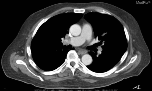
**Imaging Modality:** Contrast-enhanced Computed Tomography (CT) of the chest, axial view (CT Pulmonary Angiogram protocol).
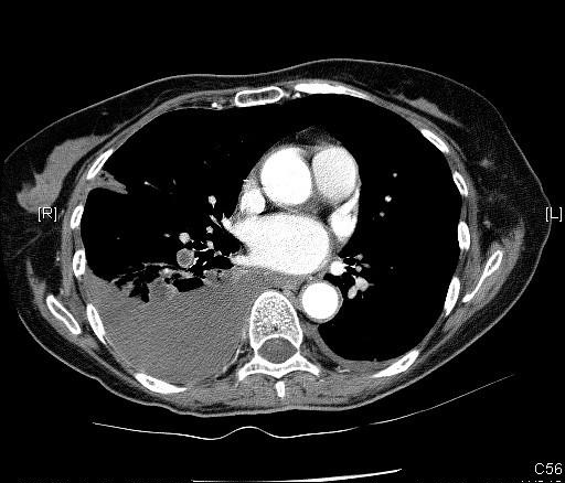
**Imaging Modality:** Contrast-enhanced Computed Tomography (CT) of the chest, axial section (Computed Tomography Pulmonary Angiogram - CTPA).
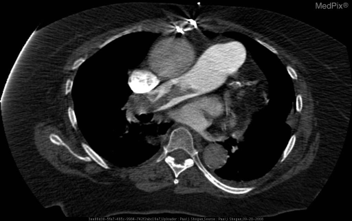
**Imaging Modality:** Contrast-enhanced Computed Tomography (CT) of the chest, specifically a CT Pulmonary Angiogram (CTPA).
CT Pulmonary Angiogram report: what it shows, what it means, and what to do
1. The problem in one line
2. What the report says, in plain language
| Report finding | What it means |
|---|---|
| Saddle thrombus at the bifurcation of the main pulmonary trunk, extending into the right and left main pulmonary arteries | A large clot sits across the point where the main lung artery splits into right and left branches. It blocks blood flow to both lungs. |
| A second saddle thrombus at the right/left main artery bifurcation, extending into lobar and segmental branches | The clot burden is extensive. It reaches deep into the smaller branches of the lung arteries. |
| CT pulmonary embolism index = 55 | This is a score for how much of the pulmonary artery tree is blocked. It is most likely the Qanadli obstruction index, shown as a percentage. If so, about 55% of the arterial tree is obstructed, which is a heavy clot load. The report doesn't state the unit, so confirm this with the radiologist. |
| Pulmonary trunk 2.8 cm, right and left pulmonary arteries 2.2 cm each; PA:aorta ratio maintained | The arteries are not enlarged. The upper limit of normal for the trunk is about 2.9 cm. This suggests the clot is acute or recent. Long-standing clots usually enlarge the artery because pressure has been high for a long time. |
| Mild prominence of right ventricle and right atrium | The right side of the heart is stretched because it is pumping against blockage. This is called right ventricular (RV) strain. |
| Dependent densities in both lower lobes | These are likely atelectasis (partial collapse) or gravity-related changes, which are common and usually not serious. |
| Mosaic attenuation in places | Some lung areas get less blood than others because of the blocked vessels. It supports the diagnosis of embolism. If it persists, doctors consider chronic thromboembolic disease. |
| No pleural effusion, consolidation or pulmonary infarct | There is no lung tissue death or fluid yet. This is good news. |
| Small hiatus hernia | A small part of the stomach sits through the diaphragm opening. It is an incidental finding and unrelated to the clots. |
| Trachea, bronchi, mediastinal nodes, ribs and bones normal | No other problems were found. |
3. Why this matters
4. What is needed: the solution
A. Immediate (today, in hospital)
- Admit to a monitored setting (HDU or ICU). This is not suitable for outpatient care given the clot burden and RV strain.
- Check these now:
- Blood pressure, heart rate, oxygen saturation, and syncope history.
- Troponin and BNP/NT-proBNP.
- Bedside echocardiogram to assess RV function.
- ECG.
- Baseline CBC, creatinine, coagulation profile and liver function.
- Oxygen to keep saturation above 90%.
- Start anticoagulation immediately if there is no bleeding contraindication:
- Preferred in the sicker patient is unfractionated heparin (IV infusion), because it can be stopped quickly if thrombolysis is needed.
- Alternatives are LMWH (enoxaparin) or fondaparinux.
- Rosen's states that apixaban and rivaroxaban are now first-line for most PE, but have not been extensively tested in severe PE. LMWH is preferred in that situation (Rosen's Emergency Medicine, Standard Anticoagulation).
- Avoid excess IV fluid. Too much fluid overdistends the right ventricle and can worsen collapse. Small boluses of 250-500 mL are acceptable if hypotensive. Norepinephrine is the first-choice vasopressor (Rosen's, Hemodynamic Resuscitation).
- Avoid intubation if possible. Positive-pressure ventilation can precipitate collapse in severe PE (Rosen's).
B. Escalation depends on the patient's status
| Status | Treatment |
|---|---|
| High-risk: low BP, shock or cardiac arrest | Systemic thrombolysis (for example alteplase) unless contraindicated. If thrombolysis is contraindicated or fails, use catheter-directed or surgical embolectomy. ECMO can bridge. |
| Intermediate-high risk: normal BP, but RV strain and raised troponin | Anticoagulation with close monitoring. Rescue reperfusion if the patient worsens. Catheter-directed therapy (mechanical thrombectomy or low-dose thrombolysis) is considered case by case. |
| Stable with only mild RV strain | Anticoagulation alone, usually with a short monitored stay first. |
C. After the acute phase
- Switch to an oral anticoagulant (apixaban or rivaroxaban, or warfarin or dabigatran/edoxaban after initial parenteral therapy) for at least 3 months.
- Look for the source and cause:
- Leg Doppler ultrasound for DVT.
- Review for recent surgery, immobility, long travel, cancer, hormonal therapy and family history.
- If there was no clear trigger, consider thrombophilia and cancer screening, and decide on extended therapy.
- Antiphospholipid syndrome needs warfarin rather than DOACs.
- Follow-up at 3 months: repeat echo. If breathlessness persists, check for chronic thromboembolic pulmonary hypertension (CTEPH) with V/Q scan. The mosaic attenuation makes this follow-up worthwhile.
- An IVC filter is only for patients who cannot take anticoagulants or who have recurrent clots despite treatment.
5. Recent evidence note
- Mechanical thrombectomy vs anticoagulation alone (PMID 41363677, 2026).
- Network meta-analysis of systemic thrombolysis (PMID 41076852, 2025).
6. Warning signs: go to the emergency room immediately
- Worsening breathlessness or chest pain
- Fainting or near-fainting
- Coughing blood
- Racing heart or low blood pressure
- Bluish lips
- Any bleeding on blood thinners