

Radiographic interpretation of this opg
OPG Radiographic Interpretation
Patient / Radiograph Details
- Adequate radiographic density and contrast; minor blurring due to screen photography but image is interpretable.
- Marker: R visible on lower left (patient's right side correctly labelled).
1. Teeth Present / Missing (FDI Notation)
| Quadrant | Teeth Present | Teeth Missing / Extracted |
|---|---|---|
| Upper Right (1) | 17 (tilted), possibly root of 16 | 11, 12, 13, 14, 15, 16 region |
| Upper Left (2) | 21, 22, 23 (anteriors partially visible), 27 | 24, 25, 26 |
| Lower Right (4) | 41, 42, 43, 44, 45, 46, 47 (tilted) | 48 (absent/extracted) |
| Lower Left (3) | 31, 32, 33, 34, 35, 36, 37 | 38 |
2. Key Pathological Findings
A. Tilted / Drifted / Malposed Teeth
- 17 (Upper Right 2nd Molar): Severely tilted mesioangularly and has drifted mesially into the edentulous space left by extraction of 16/15/14. This is a significant finding.
- 47 (Lower Right 2nd Molar): Also appears to be distally tilted, likely secondary to absence of 48 and lack of posterior support.
- 27 (Upper Left 2nd Molar): Appears to have supraerupted / drifted into the edentulous opposing space.
B. Alveolar Bone Loss
- Generalised moderate horizontal bone loss is noted across the mandible, consistent with the patient's age (65 yrs) and likely chronic periodontitis.
- Bone levels appear reduced around remaining mandibular premolars and molars - approximately 30-40% bone loss estimated in posterior regions.
- The edentulous ridges in the upper jaw show significant alveolar ridge resorption, indicating long-standing tooth loss.
C. Caries / Restorations
- No large radiopaque restorations (no obvious amalgam or metal crowns visible).
- Some radiolucent areas at the cervical regions of lower anteriors - possible cervical caries or root surface caries given patient age.
- Root surfaces of lower anteriors appear exposed (attrition/abrasion/gingival recession pattern).
D. Periapical Status
- No obvious large periapical radiolucencies are clearly identifiable, though the image resolution (photographed from screen) limits fine detail.
- Periapical areas of 17 and 47 should be assessed on dedicated periapical radiographs given their tilted positions and possible occlusal stress.
E. Lower Anterior Region
- Calculus deposits appear present (increased radioopacity along root surfaces of lower anteriors 31-42).
- Evidence of attrition on lower anterior incisal edges.
3. Maxillary Sinuses
- Bilateral maxillary sinuses are visible.
- The sinuses appear relatively clear with no obvious opacification or gross pathology noted.
- Given the missing upper posterior teeth, the sinus floor has likely pneumatised inferiorly - this is relevant if implant placement is considered.
4. TMJ / Condyles
- Both condylar heads are visible.
- No obvious gross condylar resorption, flattening, or asymmetry noted at this level of resolution.
- Condylar morphology appears within normal limits bilaterally.
5. Mandibular Structure
- The mandibular canal is partially visible bilaterally.
- No evidence of gross pathology (cysts, tumours) in the mandibular body or rami.
- Normal trabecular bone pattern in the symphysis region.
Summary / Differential Diagnoses & Clinical Implications
| Finding | Clinical Significance |
|---|---|
| Multiple missing upper teeth | Advanced tooth loss - likely long-standing, possibly combined aetiology (caries + periodontal disease) |
| Mesially tilted 17 | Overeruption risk, food trap, prosthetic challenge - consider uprighting or extraction before any prosthetic work |
| Generalised horizontal bone loss | Chronic generalised periodontitis (Stage III-IV, grade B-C) |
| Alveolar ridge resorption (upper) | Significant resorption - implants may require sinus lift / bone grafting |
| Calculus on lower anteriors | Periodontal treatment required |
| Sinus pneumatisation | Relevant for any implant planning in posterior maxilla |
Recommended Further Investigation
- Full mouth periapical radiographs - to assess individual tooth bone levels, periapical status, and caries more accurately.
- CBCT - if implant-based rehabilitation is planned, especially for upper posterior (sinus floor height) and lower posterior (inferior alveolar nerve proximity).
- Periodontal charting - to correlate with radiographic bone loss.
- Study models / clinical examination - for complete treatment planning.
Note: This interpretation is based on an OPG photographed from a screen, which reduces fine detail resolution. Formal interpretation should be performed on the original digital radiograph at full resolution.
What is that large radiolucency on right side of angle of mandible just below inferior alveolar nerve canal
Stafne bone cavity defect panoramic radiograph mandible below inferior alveolar canal
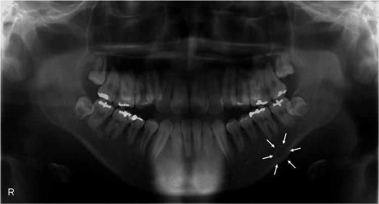
This diagnostic panoramic radiograph (orthopantomogram) displays the complete dentition, maxilla, and mandible. The primary clinical finding is a unilocular radiolucent lesion located in the left posterior mandible, specifically below the inferior alveolar nerve canal (mandibular canal) in the region apical to the first molar. The lesion is indicated by white arrows and exhibits a well-defined, corticated margin with a circular shape and internally homogeneous radiolucency. Measuring approximately 11x8 mm, the lower border of the lesion overlaps the mandibular inferior cortical bone. The rest of the mandible and maxilla show normal bone density and trabecular pattern. Several posterior teeth feature radiopaque restorations (fillings/crowns). This presentation is characteristic of a Stafne bone cavity or other odontogenic lesions such as ossifying fibroma, which require clinical correlation. The image is an essential educational tool for dental students and maxillofacial radiologists studying radiolucent mandibular pathologies and anatomical landmarks.
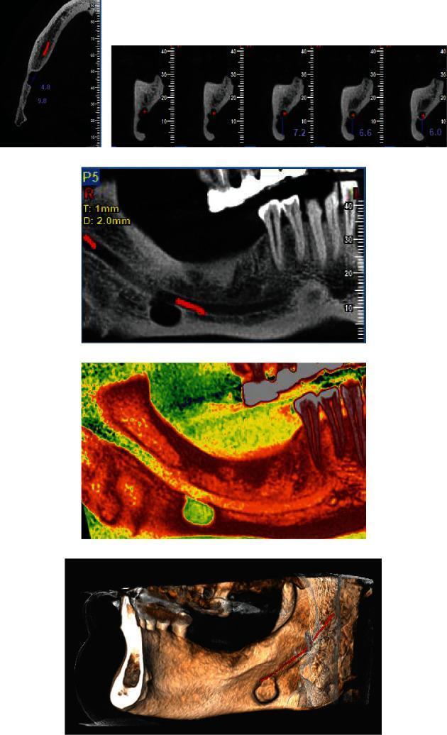
This composite diagnostic image features multiple Cone Beam Computed Tomography (CBCT) views of a Stafne bone cavity in the posterior mandible. (a) An axial cut identifies a well-defined lingual cortical defect measuring 9.8 x 4.8 mm in the right mandible. (b) Para-axial cross-sections demonstrate the monocystic nature of the cavity, showing its location on the lingual aspect of the mandible with a clear cortical margin situated approximately 7.2 mm below the mandibular canal. (c, d) Panoramic reconstructions, including one with color spectrum visualization, illustrate the ovoid, well-demarcated radiolucent lesion separated from the inferior alveolar nerve canal by a intact bone. (e) A 3D volumetric reconstruction provides a spatial overview of the defect, highlighting its relationship with the mandibular canal and the posterior mandibular anatomy. These views are characteristic of a submandibular gland depression, a developmental pseudocyst typically found incidentally below the mandibular canal in the molar-angle region.
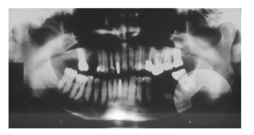
This diagnostic panoramic radiograph (orthopantomogram) of the maxilla and mandible demonstrates a large, well-circumscribed, oval-shaped radiolucent area in the left posterior mandible. The lesion is located below the mandibular canal, specifically in the submandibular fovea region, extending from the second molar area posteriorly toward the mandibular angle. It features a horizontal orientation and a distinct cortical margin. Key anatomical relationships include the apparent thinning of the inferior mandibular border and a close proximity to the mandibular canal; the upper border of the lesion appears to merge with the superior wall of the canal, while the lower canal wall remains visible within the radiolucency. The dental arch shows multiple teeth with radiopaque restorations and evidence of endodontic treatment. The clinical appearance and location are highly characteristic of a Stafne bone defect (static bone cavity), representing a developmental depression on the lingual surface of the mandible often containing salivary gland tissue. This image is an essential educational example for differentiating benign anatomical variants from cystic or neoplastic mandibular pathologies.
The Radiolucency at the Right Mandibular Angle is a Stafne Bone Cavity (Static Bone Cyst)
Radiographic Features Matching This Diagnosis
| Feature | This OPG | Stafne's Cavity (Classic) |
|---|---|---|
| Shape | Round to oval | Round/oval |
| Borders | Well-corticated, sclerotic | Well-demarcated, corticated |
| Location | Angle of mandible, below IAN canal | Premolar-angle region, beneath inferior dental canal |
| Internal structure | Unilocular, homogeneous radiolucency | Completely radiolucent, no septations |
| Association with teeth | No contact with roots | No root resorption or displacement |
| Cortical expansion | None | None |
| Size | ~1.5-2 cm | Usually 1-3 cm |
What Is It? (Pathophysiology)
- It is static - does not change in size over time (hence "static bone cyst")
- It is asymptomatic and discovered incidentally
- Most common in middle-aged to older males (fits this 65-year-old male perfectly)
- May be bilateral in some cases
Reference Images
Differential Diagnoses to Exclude (but unlikely here)
- Traumatic bone cyst (Simple bone cyst) - usually in young patients, typically above the IAN canal, scallops between roots
- Odontogenic keratocyst (OKC) - tends to grow along the medullary canal, often associated with impacted tooth, may displace IAN canal
- Ameloblastoma - multilocular ("soap bubble"), causes cortical expansion and root resorption
- Focal osteoporotic marrow defect - similar location but no well-defined corticated border
- Metastatic lesion - irregular, moth-eaten, no cortication - easily excluded here
Management
| Step | Action |
|---|---|
| No treatment required | It is a benign anatomical variant, not a pathological lesion |
| Confirm with CBCT | Shows the lingual cortical depression and confirms salivary tissue in the defect (no cystic lining) |
| MRI (optional) | Can confirm presence of salivary gland tissue within the defect if CBCT is inconclusive |
| Sialography | Historical method - now largely replaced by CBCT/MRI |
| Document and monitor | Radiographic follow-up in 1-2 years to confirm it is truly "static" |
| Biopsy NOT indicated | Unless the lesion shows progressive enlargement or atypical features |
As stated in Scott-Brown's Otorhinolaryngology Head & Neck Surgery: "Sialography is helpful in confirming the diagnosis and can prevent unnecessary surgical exploration."
Clinical Pearl
- Location below the IAN canal
- Well-corticated, sclerotic border
- 65-year-old male (typical demographic)