
Surface marking for sciatic nerve
The sciatic nerve is the largest nerve in the body. Its surface marking runs from the midpoint between the posterior superior iliac spine (PSIS) and the ischial tuberosity, down to the midpoint of the back of the thigh, then to the apex of the popliteal fossa where it divides into the tibial and common peroneal nerves.
Searching sources...
Reading File
Finding Sources
Finding Sources
The best sources are S Das (clinical surgery, most relevant for surface markings) and Gray's Anatomy for Students. Let me read both in parallel.
Reading File
Reading File
The S Das block didn't have the surface marking text at that offset. Let me read the correct section from S Das (line 7033 area) which is specifically about surface markings.
Reading File
Reading File
Good data from Gray's. Now let me search specifically for the surface marking description in S Das and also check the Gray's block at the posterior thigh section for the popliteal division point.
Finding Sources
Finding Sources
Excellent - I found a great snippet from Imaging Anatomy that has the exact surface marking text. Let me read that section fully, and also the Gray's "Avoiding the sciatic nerve" section and the Imaging Anatomy block.
Reading File
Reading File
I now have comprehensive information from authoritative sources. Let me also fetch the key diagram images to include in the answer.
Loading Image
Loading Image
I now have everything needed for a thorough, well-illustrated answer. Here is the complete surface marking of the sciatic nerve:
Surface Marking of the Sciatic Nerve
Overview
The sciatic nerve (L4, L5, S1, S2, S3) is the largest nerve in the body. Its surface marking is described in three segments: gluteal region, posterior thigh, and popliteal fossa.
1. Point of Entry into the Gluteal Region
The nerve exits the pelvis through the greater sciatic foramen, below the piriformis muscle.
Its surface marking in the gluteal region is defined by two reference points:
- Medially: within the middle third of the line joining the posterior superior iliac spine (PSIS) to the ischial tuberosity
- Laterally: halfway between the greater trochanter and the ischial tuberosity
It lies deep to the gluteus maximus at this midpoint between the ischial tuberosity and the greater trochanter.
(Imaging Anatomy: Bones, Joints, Vessels and Nerves, p. 417)

2. Course Through the Posterior Thigh
From the gluteal region, the nerve runs vertically downward along the midline of the posterior thigh, lying:
- On the posterior surface of adductor magnus
- Deep to the long head of biceps femoris
(Imaging Anatomy: Bones, Joints, Vessels and Nerves, p. 417)
3. Terminal Division at the Popliteal Fossa
The sciatic nerve divides into its two terminal branches at a variable level, usually near the apex of the popliteal fossa:
- Tibial nerve (medial, larger) - continues straight down
- Common fibular (peroneal) nerve (lateral, smaller) - winds around the neck of the fibula
(Imaging Anatomy: Bones, Joints, Vessels and Nerves, p. 417)
Summary: Surface Line
| Segment | Landmark |
|---|---|
| Upper point | Middle third of line from PSIS to ischial tuberosity; AND midpoint between greater trochanter and ischial tuberosity (gluteal region) |
| Course | Vertical line down the midline of the posterior thigh |
| Lower point | Apex of the popliteal fossa (division into tibial + common fibular nerves) |
Relations in the Gluteal Region
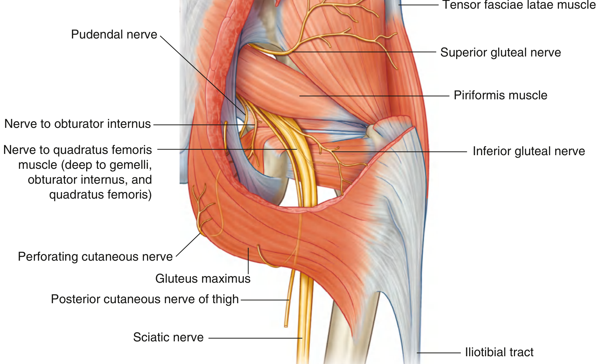
Key relations (posterior view, deep to gluteus maximus):
- Above: piriformis (nerve exits below it)
- Crosses posteriorly: obturator internus + gemelli, then quadratus femoris
- Medial to nerve: posterior cutaneous nerve of thigh and inferior gluteal artery
- In the thigh: deep to long head of biceps femoris, on the surface of adductor magnus
(Gray's Anatomy for Students, p. 682)
Clinical Relevance
Safe IM injection site: The gluteal region is divided into four quadrants by a vertical line from the highest point of the iliac crest and a horizontal line at the mid-point. The sciatic nerve courses through the upper lateral corner of the lower medial quadrant. Injections must be given in the upper lateral quadrant (anterosuperior quadrant) to avoid nerve injury. (Gray's Anatomy for Students, p. 770)
Vulnerability to injury: The nerve is commonly injured in:
- Posterior dislocation of the hip
- Subtrochanteric fractures of the femur
- Misplaced intramuscular injections
The common peroneal (fibular) division is preferentially affected in these injuries due to its more lateral, tethered position.
This is a shared conversation. Sign in to Orris to start your own chat.