
Microscopic feature of different types of renal stones Surgery
Reading File
Finding Sources
Finding Sources
Searching Images
urinary crystals microscopy calcium oxalate struvite uric acid cystine
Reading File
Reading File
Reading File
Searching PubMed
"kidney calculi"[MeSH Terms] AND "composition"
Reading File
Reading File
query_images_db
Reading File
Finding Sources
query_images_db
Renal stones are identified microscopically by the crystals in urine sediment and by polarized-light or crystallographic analysis of the stone. This matters in surgery because crystal type predicts stone type, which guides the choice of procedure and the work-up.
Microscopic features by stone type
| Stone type | Share of stones | Urine crystal appearance | Urine pH | Gross stone features |
|---|---|---|---|---|
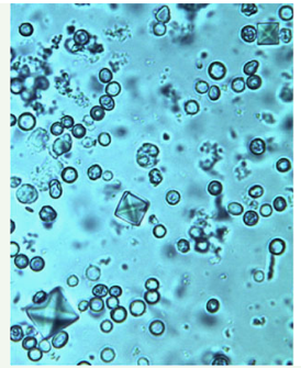
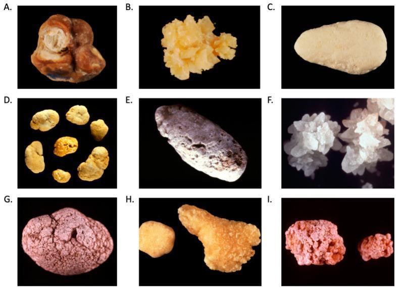
| Calcium oxalate (monohydrate or dihydrate) | About 80% (with calcium phosphate) | Dihydrate (weddellite): tetrahedral "envelope" crystals. Monohydrate (whewellite): oval, dumbbell or biconcave forms (my addition, not from the retrieved texts). Both are birefringent under polarized light. | Acidic or neutral | Dihydrate: sharp, spiculated, yellowish. Monohydrate: brown, multi-lobed, concentric layers. Radiopaque. |
| Calcium phosphate (hydroxyapatite, carbapatite, brushite) | Included in the 80% above | Brushite: needle-shaped. Apatite: amorphous granules (my addition). | Alkaline | Smooth, pale tan. Radiopaque. |
| Struvite (magnesium ammonium phosphate) | About 10% | Rectangular "coffin-lid" prisms | Alkaline (pH above 7) | White, branching, forms staghorn casts. Radiopaque. |
| Uric acid | About 6-7% | Amorphous shards, irregular plates, rhomboids and rosettes (my addition) | Acidic (pH below 5.5) | Small, rounded, yellow-orange. Radiolucent. |
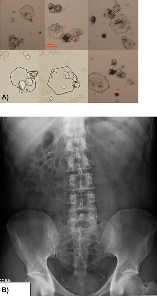
| Cystine | About 1-2% | Flat, colorless, hexagonal plates | Acidic | Yellow-brown, hard, waxy. Mildly radiopaque because of sulfur. |
The crystal descriptions come from Campbell-Walsh Urology, Table 92.2, and Henry's Clinical Diagnosis. Campbell-Walsh states: "Tetrahedral 'envelopes' are seen in calcium oxalate lithiasis, and rectangular, 'coffin-lid' crystals are often seen in patients with struvite calculi. Hexagonal crystals confirm cystinuria; uric acid crystals may be seen as amorphous fibers or as irregular plates."
Key points for each type
- Calcium stones
- Oxalate precipitates at acid or neutral pH. Calcium phosphate, as hydroxyapatite, forms at the normal urinary pH of 6.0 to 6.5 and in alkaline urine (Henry's Clinical Diagnosis, p. 576-577).
- About half of patients with calcium stones have hypercalciuria without hypercalcemia (Robbins Basic Pathology, p. 527-528).
- Struvite
- These form with urea-splitting organisms such as Proteus, Klebsiella and staphylococci, which make the urine alkaline.
- They are the usual cause of staghorn calculi. Robbins describes these as branching masses that cast the renal pelvis and calyces.
- Quick Compendium of Clinical Pathology notes that 75% of staghorn calculi are struvite.
- Infected stones contain far more endotoxin and often harbour organisms different from those in the preoperative bladder urine. In Campbell-Walsh, infected stones had 36 times more endotoxin.
- Uric acid
- These form in persistently acidic urine (pH below 5.5). They are linked to gout and high cell turnover, such as leukemia and its treatment.
- About half of patients have neither hyperuricemia nor hyperuricosuria (Robbins).
- They are radiolucent on plain film. This differs from the other types and is why CT is needed.
- Cystine
- These are due to a genetic defect in renal amino-acid transport. Hexagonal crystals confirm cystinuria.
- The stones are very hard, so they respond poorly to ESWL.
- Cystine crystals can be mistaken for hexagonal uric acid crystals. Henry's notes they are soluble in water at pH below 2 or above 8.
- Organic matrix: every stone contains mucoprotein matrix, about 2.5% by weight (Robbins).
Surgical relevance
- A urine pH above 7.0 suggests infection stones or renal tubular acidosis. A pH below 5.5 suggests uric acid stone risk (Campbell-Walsh).
- If cultures are positive, treat with antibiotics before any stone procedure. Operating during active infection risks bacteremia or sepsis (Campbell-Walsh).
- Gross stone morphology:
- Most stones are small (2-3 mm), smooth or jagged, and unilateral in about 80% of patients.
- Staghorn stones can fill the whole collecting system.
- Treatment options (Bailey & Love, p. 20):
- ESWL for stones under 2 cm.
- Ureteroscopy with fragmentation.
- PCNL for stones over 2 cm, ESWL-refractory stones and multiple stones.
I ran a PubMed search for recent (last 2 years) systematic reviews and meta-analyses on stone composition. It returned nothing that contradicts or updates the textbook material above.
This is a shared conversation. Sign in to Orris to start your own chat.