
Aneurysm
aortic aneurysm types diagram
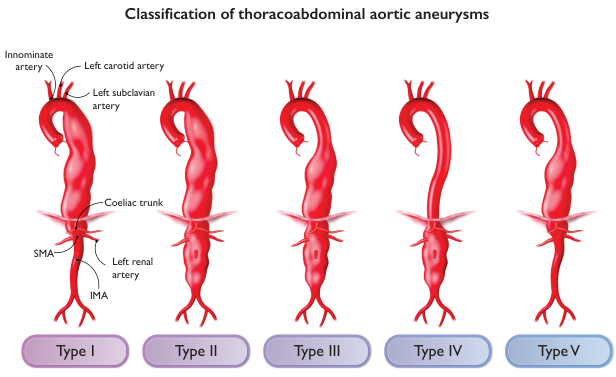
Summary : This figure illustrates the classification of thoracoabdominal aortic aneurysms into five types (I–V), showing the anatomical extent of each type along the aorta and its major branches.
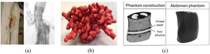
This composite image illustrates three types of 3D-printed medical phantoms used for clinical simulation and radiologic training. Figure (a) displays a 3D-printed abdominal aortic aneurysm (AAA) phantom designed for Fenestrated Endovascular Aortic Repair (FEVAR) simulation; it includes a photograph of the translucent physical model and a corresponding angiographic image showing a stent-graft with radiopaque markers within the vessel lumen. Figure (b) shows a 3D-printed biliary endoscopy phantom with a complex, branching coral-like morphology, fabricated in a uniform red material to simulate the biliary tree and distal cholangiocarcinoma anatomy. Figure (c) presents an abdominal phantom construction diagram and photograph; the diagram illustrates a layered assembly method using printed paper (containing CT image data) interleaved with polyethylene spacers to mimic tissue radiopacity, while the photograph shows the final phantom encapsulated in a black plastic film. These phantoms serve as high-fidelity tools for interventional radiology, surgical planning, and medical education in vascular and gastrointestinal procedures.
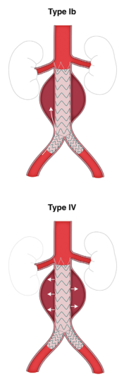
Summary : This figure illustrates two types of endoleaks (Type Ib and Type IV) following endovascular aneurysm repair (EVAR), showing the anatomical locations and flow patterns of each type in relation to the aortic stent graft and aneurysm sac.
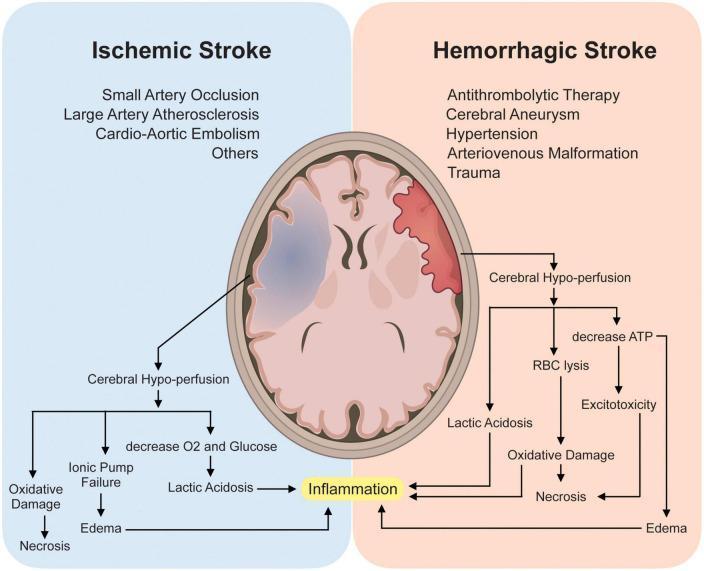
This medical infographic and pathophysiology flowchart compare Ischemic and Hemorrhagic stroke mechanisms. At the top, two axial brain illustrations highlight affected regions: a blue-shaded area in the left hemisphere represents ischemia, while a red-shaded area in the right hemisphere represents hemorrhage. Listed etiologies for Ischemic Stroke include small artery occlusion, large artery atherosclerosis, and cardio-aortic embolism. Hemorrhagic causes include antithrombolytic therapy, aneurysm, hypertension, arteriovenous malformation, and trauma. Below the illustrations, a logic flow depicts both conditions leading to cerebral hypo-perfusion. The ischemic pathway outlines oxygen/glucose deprivation, ionic pump failure, and lactic acidosis. The hemorrhagic pathway highlights red blood cell (RBC) lysis and ATP depletion. Both pathways converge on common secondary brain injury mechanisms: neuroinflammation, excitotoxicity, oxidative damage, and cerebral edema, all of which terminate in neuronal necrosis. This diagram serves as an educational tool for neurology students to understand the diverging initiating events and converging molecular outcomes of different stroke types.
intracranial berry aneurysm cerebral subarachnoid hemorrhage
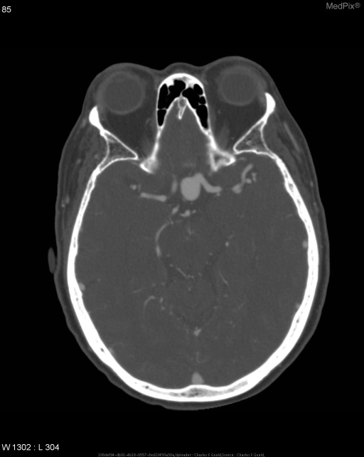
**Imaging Modality:** Computed Tomography Angiography (CTA), axial section.
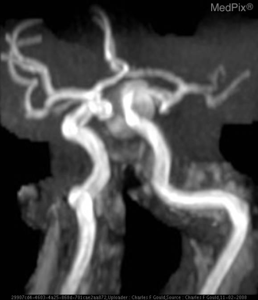
**Imaging Modality:** Magnetic Resonance Angiography (MRA) with 3D reconstruction (likely Time-of-Flight/TOF technique).
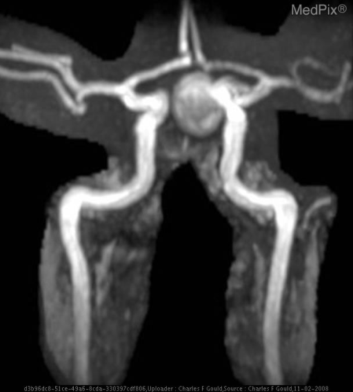
**Imaging Modality:** Time-of-Flight (TOF) Magnetic Resonance Angiography (MRA), Maximum Intensity Projection (MIP) reconstruction.
Aneurysm
Definition
Classification
1. By Wall Structure
| Type | Wall | Example |
|---|---|---|
| True | All three layers (intima, media, adventitia) | Atherosclerotic AAA |
| False (pseudoaneurysm) | Single layer of fibrous tissue | Post-traumatic, iatrogenic |
| Arteriovenous | Communication between artery and adjacent vein | Cirsoid aneurysm, AV fistula |
2. By Morphology (Shape)
- Fusiform - uniform, circumferential expansion of the entire arterial wall. Associated with atherosclerosis and causes symmetric widening.
- Saccular - expansion of only part of the arterial circumference; usually traumatic or congenital. In the cerebral circulation, saccular ("berry") aneurysms account for ~90% of intracranial aneurysms.
- Dissecting - blood forced through a ruptured tunica intima (often beneath an atheromatous plaque) splits the layers of the tunica media.
3. By Aetiology
- Berry aneurysm - congenital deficiency of the elastic lamina at branching points in the circle of Willis
- Cirsoid aneurysm (arteriovenous)
- Congenital AV fistula
- Marfan syndrome and Ehlers-Danlos syndrome
- Degenerative (most common): Atherosclerosis accounts for the vast majority. AAA has 95% association with atheromatous degeneration.
- Traumatic: Penetrating wounds, irradiation, indirect trauma (e.g. subclavian aneurysm distal to a cervical rib)
- Infective (Mycotic): Syphilis, bacterial seeding, polyarteritis nodosa; note "mycotic" is a misnomer - these are bacterial, not fungal.
Common Sites
- Abdominal aorta (most common large-vessel aneurysm; found in 2% of autopsy population; 95% occur below the renal arteries)
- Thoracoabdominal aorta
- Femoral and popliteal arteries (peripheral)
- Intracranial vessels (circle of Willis - especially MCA bifurcation, AComA, PComA)
- Splenic, renal, and carotid arteries
Clinical Features
- Pain - dull, aching; acute pain on sudden stretching; severe "bursting" pain on rupture
- Pulsatile mass - exhibits expansile pulsation (moves outward in all directions with each heartbeat); moves sideways but NOT along the arterial course
- Thrill on palpation; systolic bruit on auscultation
- Ischaemia - due to thrombosis or emboli from the aneurysm sac (claudication, rest pain, gangrene of toes)
- Compression of adjacent structures: venous obstruction, nerve compression (sciatica from AAA), dysphagia (aortic aneurysm compressing oesophagus)
- Haematemesis - from aorto-enteric fistula
Effects and Complications
- Veins → oedema of distal limb
- Nerves → pain, numbness, tingling, paraesthesia
- Bones → bone erosion
- Oesophagus → dysphagia
- Skin → redness, oedema (can be mistaken for abscess - dangerous if incised!)
Intracranial Aneurysms (Special Subtypes)
| Type | Features |
|---|---|
| Saccular (Berry) | ~90% of intracranial aneurysms; arise at bifurcations in the circle of Willis; responsible for most aneurysmal subarachnoid haemorrhage (aSAH) |
| Fusiform | Circumferential dilatation without a neck; caused by atherosclerosis; if elongated and tortuous = dolichoectasia (posterior circulation predilection); presents with brainstem compression or cranial nerve palsy |
| Dissecting | Intramural haematoma splits the vessel wall; can cause ischaemic stroke or SAH |
| Mycotic | Infected aneurysm from septic emboli, e.g. infective endocarditis |
Investigations
- Ultrasound - first-line for AAA (anteroposterior diameter ≥3 cm = aneurysm); UK National AAA Screening Programme offers USS to men aged 65
- CT angiography (CTA) - gold standard for surgical planning; defines extent and branch vessel involvement
- MR angiography (MRA) - used for intracranial aneurysms (TOF-MRA)
- Digital subtraction angiography (DSA) - most accurate for intracranial lesions; can miss AAA if laminated thrombus fills the sac
- AXR/CXR - may show calcified aortic wall or mediastinal widening
- Labs: FBC, electrolytes, LFTs, coagulation, lipids, cross-match
Management
Abdominal Aortic Aneurysm
| Scenario | Management |
|---|---|
| Asymptomatic, <55 mm | Regular USS surveillance (every 6-12 months) |
| Asymptomatic, ≥55 mm | Elective repair (open or EVAR) - annual rupture risk rises exponentially |
| 55-60 mm | ~5-10% annual rupture risk |
| ≥70 mm | ~25% annual rupture risk |
| Symptomatic / Ruptured | Emergency surgery (mortality ~50%) |
- Open repair: Resection of aneurysm sac + prosthetic graft; transabdominal approach; ~5% elective mortality
- EVAR (Endovascular Aneurysm Repair): Stent-graft deployed via femoral artery; lower short-term mortality; requires long-term surveillance for endoleaks
Intracranial Aneurysms
- Surgical clipping - open craniotomy with metal clip across the aneurysm neck
- Endovascular coiling (GDC coiling) - platinum coils deployed via catheter to thrombose the sac; preferred where anatomy allows (lower morbidity than clipping in most trials)
- Unruptured incidental aneurysms: management depends on size, location, patient age, and morphology
Peripheral Aneurysms (Femoral, Popliteal)
- Popliteal aneurysms carry high risk of limb-threatening thrombosis/embolism even without rupture
- Repair indicated: synthetic or autologous vein graft bypass + aneurysm exclusion
Differential Diagnosis of a Pulsatile Mass
- Transmitted pulsation through a cyst/mass overlying the artery (e.g. pseudopancreatic cyst)
- Swelling beneath the artery pushing it upward (e.g. cervical rib under subclavian)
- Pulsating tumours - telangiectatic osteosarcoma, vascular metastases (hypernephroma, follicular thyroid Ca), aneurysmal bone cyst
- Abscess overlying an artery - always palpate before incising to rule out aneurysm