
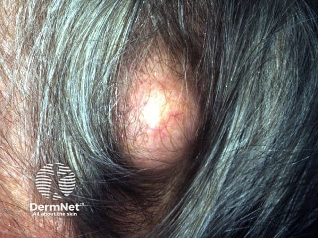
Thick skin on scalp but without itching no pain no redness elevated from normal skin Distinct lump No dandruff brittle hair on it 15 years 2*2 cm big Hard Not move under skin Rough surface It becomes less when I apply some tube on again back to shape after 3 yr 21 years old
Reading File
Finding Sources
Finding Sources
Finding Sources
Finding Sources
Searching the Web
hard fixed rough scalp lump 15 years brittle hair over it shrinks with topical cream recurs
Searching Images
pilomatricoma firm hard nodule scalp skin
Searching Images
nevus sebaceus scalp plaque alopecia rough surface
Searching PubMed
"pilomatricoma" diagnosis
Based on what you're describing, I looked through dermatology references (Andrews' Diseases of the Skin, Fitzpatrick's Dermatology) to match your description against known scalp lesions. I can't diagnose this remotely, but here's how your findings map onto the differential, and what actually needs to happen next.
Why this doesn't fit the most common scalp lump (pilar cyst)
The most frequent scalp lump in this age group is a pilar/trichilemmal cyst - but those are typically soft-to-firm, smooth-surfaced, and mobile under the skin. Your lesion is hard, fixed (not moving under the skin), and rough-surfaced - three features that point away from a simple cyst.
What actually fits better
Given hard consistency + rough surface + fixed to underlying tissue + present since childhood (roughly age 6, now 15 years), a few entities fit more closely:
-
Pilomatricoma (calcifying epithelioma of Malherbe) - a benign hair-matrix tumor that is classically firm-to-rock-hard (from calcification within it), slow-growing over years, and can feel fixed to skin though it usually moves with the skin, not deep tissue. It's one of the most common causes of a hard, longstanding scalp/head nodule in children and young adults. It's diagnosed clinically plus confirmed on excision - not something creams shrink permanently (Andrews' Diseases of the Skin, p. calcifying epithelioma section; Fitzpatrick's Dermatology).
-
Nevus sebaceus (organoid nevus) - a congenital hamartoma that presents as a rough, verrucous, slightly raised plaque on the scalp, often with sparse/absent or brittle hair over it (because hair follicles are replaced by sebaceous tissue), and can thicken further around puberty. This matches "rough surface," "brittle hair over it," and "since childhood" quite well (Andrews', p. Nevus Sebaceus section).
-
Osteoma cutis / dermoid cyst - both can present as hard, fixed, longstanding scalp nodules, though these are less common than the above two.
The detail that concerns me most
The part where it "becomes less when I apply some tube" and then "goes back to shape after 3 years" is unusual - true pilomatricomas, nevus sebaceus, and osteomas do not meaningfully shrink with topical creams. That pattern is more typical of an inflamed/infected cyst temporarily softened by a topical antibiotic or steroid, with the underlying cystic/solid lesion persisting and refilling. It does not rule out any of the above, but it tells me self-treatment with an unidentified "tube" has been masking rather than resolving whatever this is for years.
What you should actually do
A lesion that is hard, fixed, rough-surfaced, and has been present for 15 years should be examined in person by a dermatologist. They will likely:
- Do a clinical exam +/- dermoscopy
- Consider an ultrasound of the lesion (distinguishes cystic vs solid, checks for calcification, checks mobility relative to skull)
- Recommend excisional biopsy - this is both diagnostic and curative for pilomatricoma, pilar cysts, and nevus sebaceus-associated nodules
None of the entities discussed above are typically dangerous, but longstanding fixed lesions with an irregular/rough surface are exactly the kind of thing that should be biopsied rather than continually treated with an over-the-counter tube, partly to confirm it isn't something that mimics these (rare malignant transformation in longstanding nevus sebaceus or trichilemmal cysts is uncommon but reported).
I did not find any recent systematic reviews that change this general picture - the two recent pilomatricoma reviews I found (PMID 39825948, PMID 37787269) focus on the rare malignant variant (pilomatrix carcinoma) and reinforce that persistent, recurrent, or atypical lesions warrant biopsy rather than assumption of benignity.
Bottom line: this needs an in-person look and probably a biopsy/excision - not something I or any online source can safely pin down from a description alone.
This is a shared conversation. Sign in to Orris to start your own chat.