
. Necrosis. Reasons, mechanisms of development, morphological characteristics. Clinical and morphological forms of necrosis, pathological morphogenesis.
Necrosis: Causes, Mechanisms, Morphology, and Clinical-Morphological Forms
1. Definition
2. Causes (Etiological Factors)
| Category | Examples |
|---|---|
| Hypoxia / Ischemia | Arterial obstruction, thromboembolism, cardiac arrest |
| Physical agents | Extreme heat/cold, radiation, trauma, electrical injury |
| Chemical and toxic agents | Cyanide, carbon tetrachloride, heavy metals, strong acids/alkalis |
| Microbial toxins | Bacterial exo- and endotoxins, viral cytopathic effects, fungal toxins |
| Immune-mediated | Antigen-antibody complexes deposited in vessel walls, CTL-mediated killing |
| Enzymatic self-digestion | Trypsin leakage in acute pancreatitis - peripancreatic fat necrosis |
| Nutritional deficiencies | Severe protein or oxygen deprivation |
- Robbins & Kumar Basic Pathology, p. 21; Robbins, Cotran & Kumar Pathologic Basis of Disease, p. 53
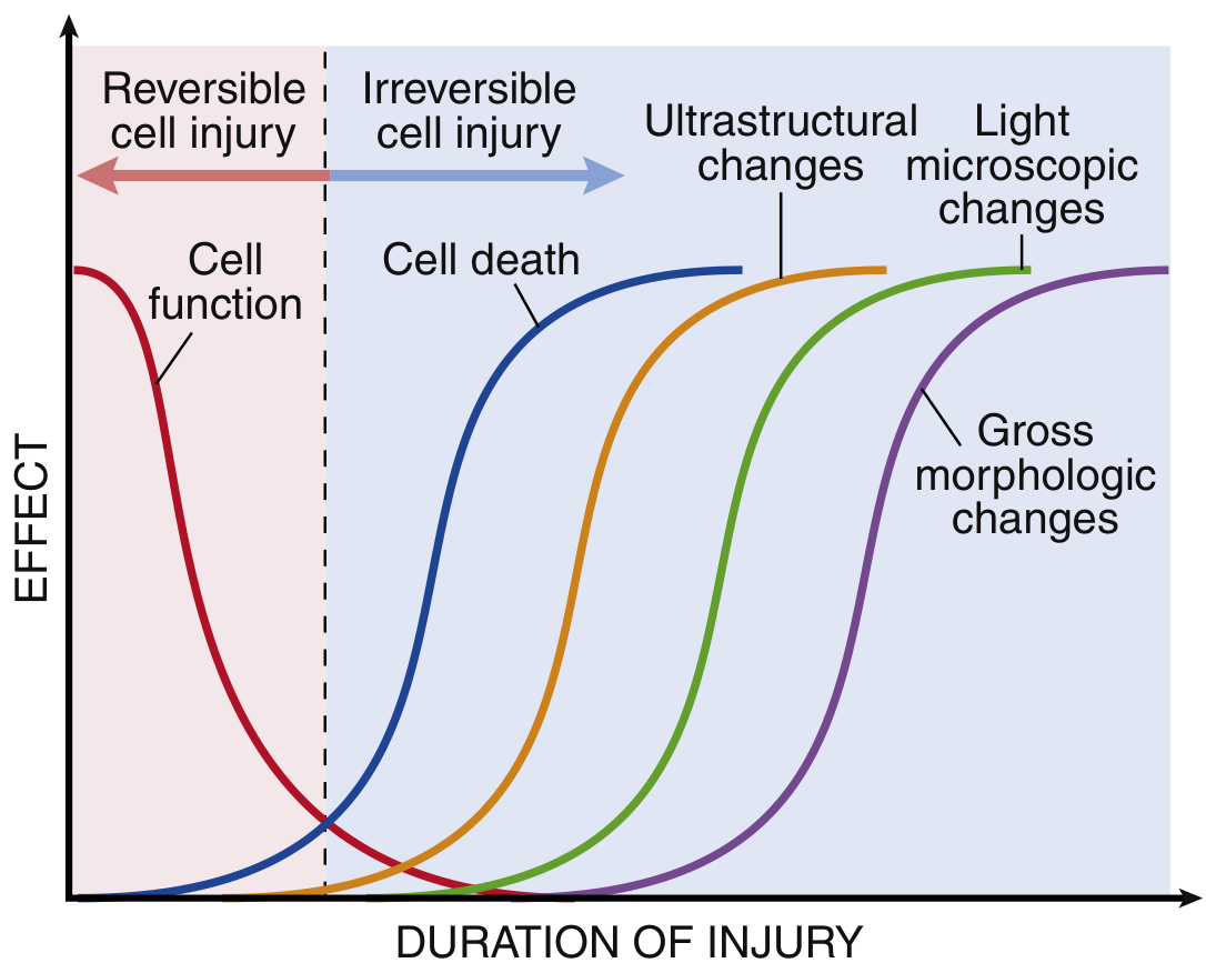
3. Pathogenesis - The "Point of No Return"
- Inability to reverse mitochondrial dysfunction - persistent failure of oxidative phosphorylation and ATP generation even after the original stimulus resolves.
- Profound, irreparable disturbances in membrane function - particularly lysosomal membrane rupture, which releases digestive enzymes into the cytoplasm.
Molecular mechanisms
- Reduction in Na⁺/K⁺-ATPase activity → cellular swelling (cell swells, ER dilates)
- Switch to anaerobic glycolysis → lactic acid accumulates → intracellular pH falls → clumping of nuclear chromatin
- Failure of Ca²⁺ pumps → intracellular Ca²⁺ rises → activates phospholipases, proteases, endonucleases, and ATPases
- Increased intracellular Ca²⁺, reactive oxygen species (ROS), and lipid breakdown products all damage mitochondrial membranes
- Mitochondrial permeability transition pore (MPTP) opens
- Cytochrome c and other pro-apoptotic signals leak out
- ROS cause lipid peroxidation of membranes
- Activated phospholipases degrade phospholipids in cell and organelle membranes
- Loss of plasma membrane integrity → cellular contents (troponins, transaminases, LDH, alkaline phosphatase) leak into blood - the basis for serum biomarker testing
- Robbins, Cotran & Kumar, p. 53-54
4. General Morphological Characteristics of Necrosis
4.1 Cytoplasmic changes
- Increased eosinophilia (bright pink on H&E): due to loss of cytoplasmic RNA (which normally binds hematoxylin/blue) and accumulation of denatured proteins (which bind eosin/red)
- Glassy, homogeneous appearance: loss of glycogen granules
- Vacuolated, "moth-eaten" cytoplasm: enzymatic digestion of organelles
- By electron microscopy: discontinuities in plasma and organelle membranes, marked mitochondrial dilation with large amorphous intramitochondrial densities, disrupted lysosomes, intracytoplasmic myelin figures (whorled phospholipid precipitates)
4.2 Nuclear changes (three patterns)
| Pattern | Description | Mechanism |
|---|---|---|
| Pyknosis | Nuclear shrinkage; increased basophilia; dark condensed mass | DNA condensation |
| Karyorrhexis | Fragmentation of the pyknotic nucleus | Further breakdown |
| Karyolysis | Fading of basophilia; nuclear dissolution in 1-2 days | Enzymatic DNase digestion of DNA |
4.3 Fate of necrotic cells
-
Persist for some time, then digested by lysosomal enzymes from recruited leukocytes and the dead cells themselves
-
Replaced by myelin figures, which are either phagocytosed or degraded into fatty acids
-
Fatty acids bind calcium salts → dystrophic calcification
-
Robbins & Kumar Basic Pathology, pp. 21-22; Robbins, Cotran & Kumar, pp. 54-55
5. Clinical-Morphological Forms of Necrosis
5.1 Coagulative Necrosis
- Tissue architecture is preserved for days to weeks (ghost outlines of cells remain)
- Firm texture
- Eosinophilic, anucleate ("ghost") cells with indistinct red nuclei
- Eventually cleared by leukocyte enzymes and phagocytosis
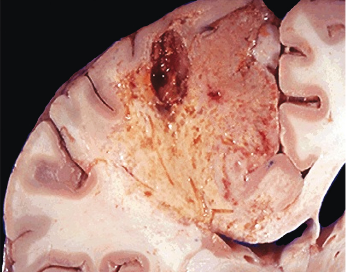
5.2 Liquefactive Necrosis
- Complete digestion of dead cells
- Viscous liquid, creamy yellow when pus-forming (due to neutrophils)
- No preserved architecture
- Bacterial (and occasionally fungal) infections - leukocyte enzymes digest the tissue; the yellow material = pus; a localized collection = abscess
- CNS ischemia/hypoxia (brain - for unknown reasons, hypoxic death manifests as liquefactive, not coagulative, necrosis)
5.3 Caseous Necrosis
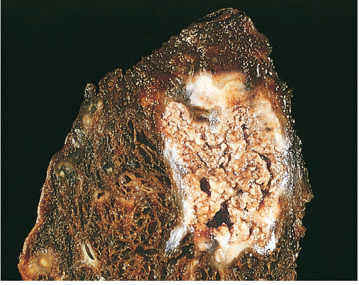
- Gross: Friable, white-yellow, "cheese-like" (caseous = Latin for cheese) material
- Microscopic: Structureless, amorphous granular pink debris; no preserved cell outlines; tissue architecture completely obliterated; surrounded by a rim of epithelioid macrophages and lymphocytes forming a granuloma
5.4 Fat Necrosis
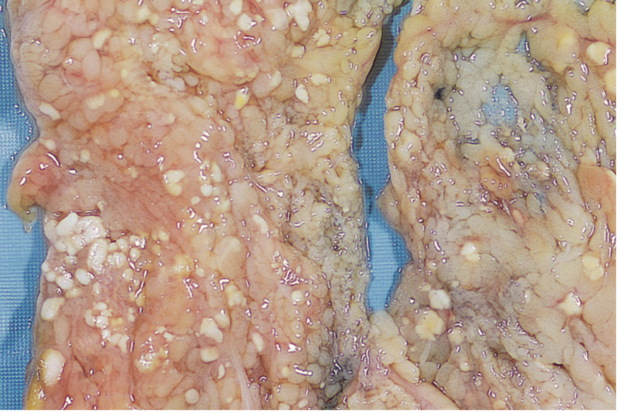
- Gross: Chalky white deposits (calcium soap foci) in adipose/mesentery
- Microscopic: Shadowy outlines of necrotic fat cells with granular basophilic calcium deposits and surrounding inflammatory reaction
- Acute pancreatitis - activated pancreatic enzymes (lipase) leak and digest peripancreatic fat
- Abdominal/breast trauma
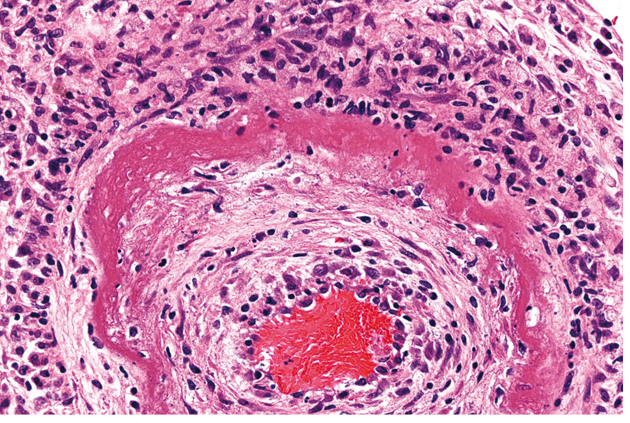
5.5 Fibrinoid Necrosis
- Detected only microscopically (no distinctive gross appearance)
- Bright pink (intensely eosinophilic), amorphous, homogeneous material in vessel walls on H&E = "fibrinoid" appearance
- Circumferential involvement of the arterial wall with associated inflammation (neutrophils)
- Immune vasculitis (antigen-antibody complex deposition)
- Severe/malignant hypertension
- Organ transplant rejection
5.6 Gangrenous Necrosis
| Subtype | Mechanism | Appearance |
|---|---|---|
| Dry gangrene | Coagulative necrosis from ischemia, without infection | Dry, dark, mummified tissue with clear demarcation |
| Wet gangrene | Ischemic coagulative necrosis + superimposed bacterial infection | Liquefactive component; swollen, moist, malodorous; less distinct boundary |
| Gas gangrene | Clostridium perfringens infection; gas-producing enzymes | Crepitus (gas in tissues), rapidly spreading, life-threatening |
6. Summary Table
| Form | Dominant mechanism | Preserved architecture? | Prototype condition | Typical organ/site |
|---|---|---|---|---|
| Coagulative | Protein denaturation | Yes (days-weeks) | Myocardial/renal infarct | All solid organs except brain |
| Liquefactive | Enzymatic digestion | No | Abscess, brain infarct | Brain, bacterial infection sites |
| Caseous | Mixed; granulomatous | No | Tuberculosis | Lung, lymph nodes |
| Fat | Lipase activation | Partial | Pancreatitis | Peripancreatic fat, breast |
| Fibrinoid | Immune complex deposition | N/A (vessel wall) | Vasculitis, malignant HTN | Blood vessel walls |
| Gangrenous | Coagulative ± liquefactive | Varies | Peripheral vascular disease | Lower limb |
7. Clinical Significance - Serum Biomarkers
-
Cardiac troponins (cardiac-specific troponin I and T): detectable as early as 2 hours after myocardial cell necrosis - gold standard for MI diagnosis
-
Transaminases (ALT, AST): liver cell necrosis (hepatitis, ischemic hepatitis)
-
Alkaline phosphatase isoforms: bile duct epithelium injury
-
LDH, CK-MB: tissue-specific markers of cell death
-
Robbins, Cotran & Kumar Pathologic Basis of Disease, p. 53
- Robbins & Kumar Basic Pathology (10th ed.), pp. 19-22
- Robbins, Cotran & Kumar Pathologic Basis of Disease (10th ed.), pp. 53-57
Show
What would you like me to show?
A clean, professional medical education summary diagram titled "Clinical-Morphological Forms of Necrosis". The diagram is organized as a structured visual chart with 6 rows (one per necrosis type) and columns for: Type, Dominant Mechanism, Architecture Preserved, Prototype Cause, Typical Site/Organ. Types listed: 1) Coagulative - protein denaturation - Yes (days-weeks) - Myocardial/renal infarct - All solid organs except brain. 2) Liquefactive - enzymatic digestion - No - Abscess / Brain infarct - Brain, bacterial infections. 3) Caseous - mixed granulomatous - No - Tuberculosis - Lung, lymph nodes. 4) Fat - lipase activation - Partial - Pancreatitis - Peripancreatic fat, breast. 5) Fibrinoid - immune complex deposition - N/A vessel wall - Vasculitis / malignant hypertension - Blood vessel walls. 6) Gangrenous - coagulative +/- liquefactive - Varies - Peripheral vascular disease - Lower limb. Below the table, a smaller box shows "Nuclear changes in necrosis": Pyknosis (shrinkage), Karyorrhexis (fragmentation), Karyolysis (dissolution). At the bottom, a box shows "Causes of necrosis": ischemia, toxins, infections, physical agents, immune reactions, enzymatic (pancreatitis). Use a white background, color-coded rows with soft medical colors (light blue, light green alternating), bold headers, clear legible sans-serif font. Medical textbook quality.
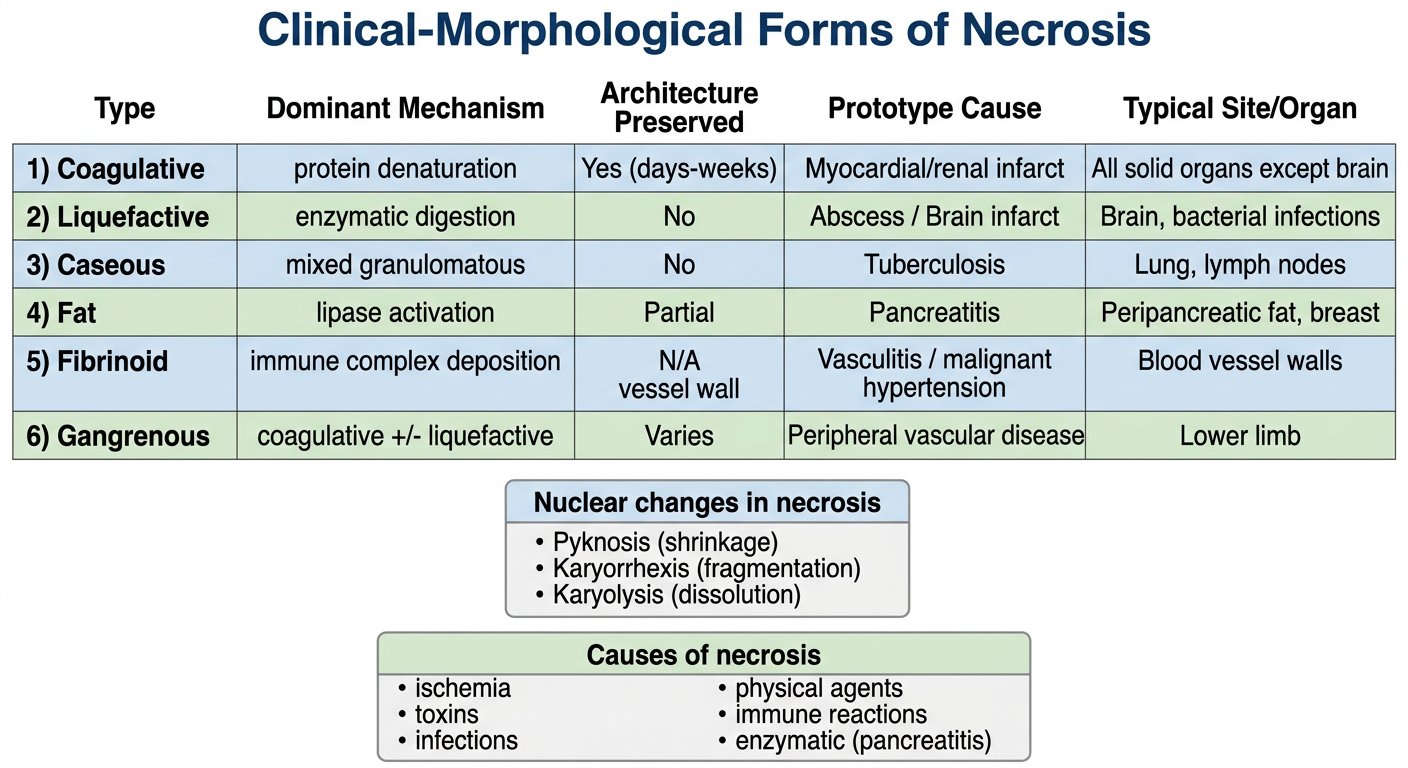
- All 6 forms - coagulative, liquefactive, caseous, fat, fibrinoid, gangrenous
- Dominant mechanism for each form
- Architecture preservation status
- Prototype cause and typical site
- Nuclear changes (pyknosis → karyorrhexis → karyolysis)
- Main causes of necrosis at a glance
Show
What would you like me to show?
Isse hinglish me explain kro
Necrosis - Hinglish mein Samjho! 🧬
Necrosis kya hota hai?
Necrosis Kyun Hota Hai? (Causes)
| Cause | Example |
|---|---|
| Ischemia (Blood supply band) | Heart attack, thrombosis |
| Physical injury | Burns, radiation, thanda/garmi |
| Chemical/Toxic | Zeher, heavy metals, strong acids |
| Bacteria/Virus ke toxins | Infections |
| Immune reaction | Antigen-antibody complex vessel wall mein |
| Enzymes leak ho jayein | Pancreatitis mein lipase leak |
Mechanism kaise hota hai? (Pathogenesis)
Step-by-step samjho:
- Na⁺/K⁺ pump band → cell swell karta hai
- Anaerobic glycolysis shuru → lactic acid banta hai → pH girta hai → chromatin clump hota hai
- Phospholipases activate → membrane digest hone lagti hai
- Proteases activate → proteins toot jaate hain
- Endonucleases activate → DNA toot jaata hai
- Mitochondrial permeability pore (MPTP) khul jaata hai
- Cytochrome c leak hota hai
- ATP generation permanently band → point of no return
- Digestive enzymes cytoplasm mein aa jaate hain
- Cell apne aap ko digest kar leta hai (autolysis)
- Cellular contents bahar aa jaate hain (blood mein)
- Troponin, transaminases, LDH → biomarkers ka basis yahi hai!
- Bahar aaye molecules (DAMPs) → inflammation trigger karte hain
Morphology - Microscope mein Kya Dikhta Hai?
Cytoplasm mein changes:
- Eosinophilia badhti hai → cell bright pink dikhta hai H&E mein
- (RNA khatam hoti hai jo blue color deti thi, denatured proteins eosin/red se bind karte hain)
- Glassy appearance → glycogen khatam
- Moth-eaten look → organelles digest ho gayi
Nucleus mein 3 changes (yaad karo teen K):
Pyknosis → Karyorrhexis → Karyolysis
(Shrink) (Fragment) (Dissolve)
- Pyknosis - nucleus sikar jaata hai, bahut dark ho jaata hai
- Karyorrhexis - pyknotic nucleus toot ke fragments ban jaata hai
- Karyolysis - basophilia fade ho jaati hai, nucleus dissolve ho jaata hai (1-2 din mein)
6 Clinical-Morphological Forms - Ek Ek Karke!
1. 🔴 Coagulative Necrosis
- Heart attack (myocardial infarct)
- Kidney infarct
- Spleen infarct
2. 💛 Liquefactive Necrosis
- Bacterial/fungal infections → pus banta hai → localized collection = abscess
- Brain ischemia → brain mein coagulative nahi, liquefactive hoti hai (reason unclear)
3. 🟡 Caseous Necrosis
- Amorphous, granular pink debris - koi cell outline nahi
- Architecture completely khatam
- Surrounded by epithelioid macrophages = granuloma banta hai
4. ⬜ Fat Necrosis
- Necrotic fat cells ke shadowy outlines
- Basophilic (blue) calcium deposits granular form mein
- Surrounding inflammatory reaction
- Acute pancreatitis - pancreatic lipase leak karke peripancreatic fat digest karta hai
- Breast/abdominal trauma
5. 🩷 Fibrinoid Necrosis
- Bright pink (intensely eosinophilic), amorphous, homogeneous material vessel wall mein
- Circumferential involvement
- Neutrophils surrounding area mein
- Vasculitis (immune complex deposition)
- Malignant/severe hypertension
- Organ transplant rejection
6. 🖤 Gangrenous Necrosis
| Type | Kya hota hai | Appearance |
|---|---|---|
| Dry gangrene | Sirf coagulative, koi infection nahi | Dry, dark, mummified, clear boundary |
| Wet gangrene | Coagulative + bacterial infection | Swollen, moist, badbu, unclear boundary |
| Gas gangrene | Clostridium perfringens | Crepitus (gas feel hota hai), rapidly spreading |
Ek Line Summary (Exam ke liye!)
| Form | Cause | Key Feature |
|---|---|---|
| Coagulative | Ischemia (brain chhod ke) | Ghost cells, architecture preserved |
| Liquefactive | Bacteria, brain ischemia | Pus, liquid tissue |
| Caseous | TB | Cheese jaisi, granuloma |
| Fat | Pancreatitis | Chalky white, saponification |
| Fibrinoid | Vasculitis, HTN | Bright pink vessel wall |
| Gangrenous | Limb ischemia ± infection | Clinical term, coagulative base |
Clinical Importance - Serum Biomarkers
- Troponin I/T → Heart cell necrosis (MI) - 2 ghante mein detect hota hai!
- ALT/AST (transaminases) → Liver cell necrosis
- LDH, CK-MB → Various tissue necrosis
- Alkaline phosphatase → Bile duct injury