
hey
help me with anatomy i want finish every thing in 2 weeks i already have idea about everything its just like a revision
What level is this anatomy for? · Which regions/systems do you want to cover? · How do you want me to help you?
2-Week Anatomy Revision Plan (MBBS 1st Year)
Ground Rules
- 2 sessions/day - morning (2 hrs) + evening (1 hr review/quiz)
- Each day = 1 main topic + evening recall/questions
- You tell me when you're on a topic and I'll quiz you or explain anything
WEEK 1 - Gross Anatomy (Regional)
| Day | Morning - Study | Evening - Review |
|---|---|---|
| Day 1 | Upper Limb - bones, joints (shoulder, elbow, wrist) | Quiz: brachial plexus roots & branches |
| Day 2 | Upper Limb - muscles, nerve supply, dermatomes | Quiz: muscle actions + clinical (wrist drop, claw hand, etc.) |
| Day 3 | Lower Limb - bones, joints (hip, knee, ankle) | Quiz: femoral triangle, popliteal fossa contents |
| Day 4 | Lower Limb - muscles, lumbosacral plexus, dermatomes | Quiz: common nerve injuries (foot drop, etc.) |
| Day 5 | Thorax - heart, great vessels, mediastinum | Quiz: heart borders, surface anatomy, coronary arteries |
| Day 6 | Thorax - lungs, pleura + Abdomen - peritoneum, GI tract | Quiz: lung segments, peritoneal vs retroperitoneal |
| Day 7 | Rest / Weak spots review | Light revision of Days 1-6 |
WEEK 2 - Head & Neck + Neuro + Histology/Embryology
| Day | Morning - Study | Evening - Review |
|---|---|---|
| Day 8 | Abdomen - liver, pancreas, spleen, kidneys | Quiz: porta hepatis, pancreatic ducts, suprarenal glands |
| Day 9 | Pelvis & Perineum - pelvic floor, bladder, uterus/prostate | Quiz: inguinal canal, femoral hernia vs inguinal |
| Day 10 | Head & Neck - skull, scalp, face, parotid, TMJ | Quiz: CN branches, dangerous area of face |
| Day 11 | Head & Neck - pharynx, larynx, thyroid, triangles of neck | Quiz: recurrent laryngeal nerve, carotid sheath |
| Day 12 | Neuroanatomy - spinal cord, brainstem, cerebellum | Quiz: tracts (DCML, spinothalamic), cerebellar signs |
| Day 13 | Neuroanatomy - cerebrum, cranial nerves, ventricles/CSF | Quiz: cranial nerve exam points, circle of Willis |
| Day 14 | Full mock revision - High-yield clinicals + past Qs | Cover all weak areas flagged during the week |
How I Can Help Each Day
- Study with you - paste a topic and I'll give you a tight, high-yield summary from anatomy textbooks
- Quiz you - I'll ask 5-10 MCQs or short-answer questions on the day's topic
- Explain on demand - stuck on a nerve plexus, a structure, a clinical correlation? Just ask
Generate a quick reference PDF on high-yield anatomy
mkdir -p /home/daytona/workspace/anatomy-pdf && ls /home/daytona/workspace/anatomy-pdf/
~/anatomy-pdf/generate_anatomy_pdf.py
from reportlab.lib.pagesizes import A4
from reportlab.lib import colors
from reportlab.lib.styles import getSampleStyleSheet, ParagraphStyle
from reportlab.lib.units import mm, cm
from reportlab.platypus import (
    SimpleDocTemplate, Paragraph, Spacer, Table, TableStyle,
    HRFlowable, PageBreak, KeepTogether
)
from reportlab.lib.enums import TA_CENTER, TA_LEFT
from reportlab.pdfgen import canvas
from reportlab.platypus.doctemplate import PageTemplate, BaseDocTemplate
from reportlab.platypus.frames import Frame

OUTPUT = "/home/daytona/workspace/anatomy-pdf/HighYield_Anatomy_QuickRef.pdf"

# ── Colour palette ──────────────────────────────────────────────────────────
NAVY   = colors.HexColor("#1a2c5b")
TEAL   = colors.HexColor("#0d7a8a")
AMBER  = colors.HexColor("#e07b1a")
LGREY  = colors.HexColor("#f0f4f8")
WHITE  = colors.white
BLACK  = colors.black
DKGREY = colors.HexColor("#333333")

# ── Custom document with header/footer ──────────────────────────────────────
class AnatomyDoc(BaseDocTemplate):
    def __init__(self, filename, **kwargs):
        super().__init__(filename, **kwargs)
        frame = Frame(1.5*cm, 2*cm, 18*cm, 25.5*cm, id='normal')
        template = PageTemplate(id='main', frames=[frame], onPage=self._draw_page)
        self.addPageTemplates([template])

    def _draw_page(self, canvas_obj, doc):
        canvas_obj.saveState()
        # Header bar
        canvas_obj.setFillColor(NAVY)
        canvas_obj.rect(0, A4[1]-1.5*cm, A4[0], 1.5*cm, fill=1, stroke=0)
        canvas_obj.setFillColor(WHITE)
        canvas_obj.setFont("Helvetica-Bold", 10)
        canvas_obj.drawString(1.5*cm, A4[1]-1.0*cm, "HIGH-YIELD ANATOMY  |  MBBS Quick Reference")
        canvas_obj.setFont("Helvetica", 8)
        canvas_obj.drawRightString(A4[0]-1.5*cm, A4[1]-1.0*cm, f"Page {doc.page}")
        # Footer bar
        canvas_obj.setFillColor(NAVY)
        canvas_obj.rect(0, 0, A4[0], 1.2*cm, fill=1, stroke=0)
        canvas_obj.setFillColor(WHITE)
        canvas_obj.setFont("Helvetica-Oblique", 7)
        canvas_obj.drawCentredString(A4[0]/2, 0.45*cm, "For revision use only  |  July 2026")
        canvas_obj.restoreState()

# ── Styles ───────────────────────────────────────────────────────────────────
styles = getSampleStyleSheet()

TITLE_STYLE = ParagraphStyle("title", fontName="Helvetica-Bold", fontSize=26,
    textColor=WHITE, alignment=TA_CENTER, spaceAfter=4)
SUBTITLE_STYLE = ParagraphStyle("subtitle", fontName="Helvetica-Oblique", fontSize=12,
    textColor=colors.HexColor("#cce0ff"), alignment=TA_CENTER, spaceAfter=0)
H1 = ParagraphStyle("h1", fontName="Helvetica-Bold", fontSize=13,
    textColor=WHITE, backColor=NAVY, leftIndent=-4, rightIndent=-4,
    spaceAfter=6, spaceBefore=14, leading=18,
    borderPadding=(4,6,4,6))
H2 = ParagraphStyle("h2", fontName="Helvetica-Bold", fontSize=10,
    textColor=TEAL, spaceBefore=8, spaceAfter=3)
BODY = ParagraphStyle("body", fontName="Helvetica", fontSize=8.5,
    textColor=DKGREY, leading=13, spaceAfter=2)
BULLET = ParagraphStyle("bullet", fontName="Helvetica", fontSize=8.5,
    textColor=DKGREY, leading=13, leftIndent=12, bulletIndent=2, spaceAfter=1)
CLINICAL = ParagraphStyle("clinical", fontName="Helvetica-Oblique", fontSize=8,
    textColor=colors.HexColor("#8b0000"), leftIndent=8, leading=12, spaceAfter=2)
BOLD_CELL = ParagraphStyle("boldcell", fontName="Helvetica-Bold", fontSize=8, textColor=WHITE)
REG_CELL  = ParagraphStyle("regcell",  fontName="Helvetica", fontSize=8, textColor=DKGREY, leading=11)

def h1(text):
    return Paragraph(f"  {text}", H1)

def h2(text):
    return Paragraph(text, H2)

def body(text):
    return Paragraph(text, BODY)

def bullet(text):
    return Paragraph(f"• {text}", BULLET)

def clinical(text):
    return Paragraph(f"★ Clinical: {text}", CLINICAL)

def space(n=4):
    return Spacer(1, n*mm)

def hr():
    return HRFlowable(width="100%", thickness=0.5, color=TEAL, spaceAfter=4, spaceBefore=2)

def table(headers, rows, col_widths=None):
    """Generic shaded table."""
    header_row = [Paragraph(h, BOLD_CELL) for h in headers]
    data = [header_row]
    for i, row in enumerate(rows):
        data.append([Paragraph(str(c), REG_CELL) for c in row])
    if col_widths is None:
        col_widths = [18*cm / len(headers)] * len(headers)
    t = Table(data, colWidths=col_widths, repeatRows=1)
    style = TableStyle([
        ("BACKGROUND", (0,0), (-1,0), TEAL),
        ("ROWBACKGROUNDS", (0,1), (-1,-1), [LGREY, WHITE]),
        ("GRID", (0,0), (-1,-1), 0.3, colors.HexColor("#bbccd8")),
        ("VALIGN", (0,0), (-1,-1), "TOP"),
        ("TOPPADDING", (0,0), (-1,-1), 4),
        ("BOTTOMPADDING", (0,0), (-1,-1), 4),
        ("LEFTPADDING", (0,0), (-1,-1), 5),
    ])
    t.setStyle(style)
    return t

# ══════════════════════════════════════════════════════════════════════════════
# CONTENT
# ══════════════════════════════════════════════════════════════════════════════
story = []

# ── COVER ────────────────────────────────────────────────────────────────────
def cover_page(canvas_obj, doc):
    """Full-bleed cover - called via onFirstPage."""
    pass  # handled inline with a coloured table block

# Title block as a styled table
cover_data = [[Paragraph("HIGH-YIELD ANATOMY", TITLE_STYLE)],
              [Paragraph("MBBS First Year  |  Quick Reference Guide", SUBTITLE_STYLE)],
              [Paragraph("All Regions  •  Clinical Pearls  •  Key Tables", SUBTITLE_STYLE)]]
cover_table = Table(cover_data, colWidths=[18*cm])
cover_table.setStyle(TableStyle([
    ("BACKGROUND", (0,0), (-1,-1), NAVY),
    ("TOPPADDING", (0,0), (-1,-1), 12),
    ("BOTTOMPADDING", (0,0), (-1,-1), 8),
    ("LEFTPADDING", (0,0), (-1,-1), 8),
    ("RIGHTPADDING", (0,0), (-1,-1), 8),
    ("LINEBELOW", (0,-1), (-1,-1), 3, AMBER),
]))
story += [space(8), cover_table, space(6)]
story.append(body("This reference card summarises the highest-yield anatomy topics for MBBS "
                  "first-year examinations. Use it alongside your 2-week revision plan. "
                  "Each section includes key facts, nerve supplies, clinical correlations, "
                  "and memory aids."))
story.append(PageBreak())

# ════════════════════════════════════════════════════════════════════════════
# 1. UPPER LIMB
# ════════════════════════════════════════════════════════════════════════════
story.append(h1("1. UPPER LIMB"))
story.append(space(2))

story.append(h2("Brachial Plexus - Roots to Branches"))
story.append(table(
    ["Component", "Roots", "Key Branches / Nerves"],
    [
        ["Roots", "C5-C8, T1", "Dorsal scapular (C5), Long thoracic (C5-7)"],
        ["Trunks", "Upper (C5-6), Middle (C7), Lower (C8-T1)", "Suprascapular (upper trunk)"],
        ["Divisions", "3 Anterior + 3 Posterior", "—"],
        ["Cords", "Lateral, Posterior, Medial", "Named by relation to axillary artery"],
        ["Terminal Branches", "5 nerves", "Musculocutaneous, Axillary, Radial, Median, Ulnar"],
    ],
    col_widths=[4*cm, 6*cm, 8*cm]
))
story.append(space(2))

story.append(h2("Key Nerve Injury Patterns"))
story.append(table(
    ["Nerve", "Root", "Injury Cause", "Deformity / Deficit"],
    [
        ["Axillary (C5-6)", "C5,6", "Surgical neck humerus fracture / dislocation", "Flattened deltoid; loss of shoulder abduction >15°"],
        ["Radial (C5-T1)", "C5-T1", "Midshaft humerus fracture / Saturday night palsy", "Wrist drop; loss of extension fingers & wrist"],
        ["Median (C6-T1)", "C6-T1", "Supracondylar fracture (kids); carpal tunnel", "Ape hand; thenar wasting; Pope's blessing sign"],
        ["Ulnar (C8-T1)", "C8,T1", "Medial epicondyle fracture; cubital tunnel", "Claw hand (ring & little); froment sign"],
        ["Musculocutaneous (C5-7)", "C5-7", "Coracobrachialis piercing injury", "Weak elbow flexion; loss of lat forearm sensation"],
        ["Long Thoracic (C5-7)", "C5-7", "Mastectomy / axillary dissection", "Winged scapula"],
    ],
    col_widths=[3.5*cm, 1.8*cm, 5*cm, 7.7*cm]
))
story.append(clinical("Erb's Palsy (C5-C6 injury): 'Waiter's tip' position - arm adducted, medially rotated, forearm pronated."))
story.append(clinical("Klumpke's Palsy (C8-T1): Claw hand + Horner syndrome (if T1 ramus communicans involved)."))

story.append(h2("Rotator Cuff - SITS"))
story.append(table(
    ["Muscle", "Nerve", "Action", "Common Pathology"],
    [
        ["Supraspinatus", "Suprascapular (C4-6)", "Initiates abduction (0-15°)", "Most commonly torn (supraspinatus tendon)"],
        ["Infraspinatus", "Suprascapular (C4-6)", "Lateral rotation", "Torn in shoulder dislocation"],
        ["Teres Minor", "Axillary (C5-6)", "Lateral rotation", "—"],
        ["Subscapularis", "Upper/Lower subscapular", "Medial rotation", "Least commonly torn"],
    ],
    col_widths=[3.5*cm, 4.5*cm, 4*cm, 6*cm]
))
story.append(PageBreak())

# ════════════════════════════════════════════════════════════════════════════
# 2. LOWER LIMB
# ════════════════════════════════════════════════════════════════════════════
story.append(h1("2. LOWER LIMB"))
story.append(space(2))

story.append(h2("Lumbar & Sacral Plexus Key Nerves"))
story.append(table(
    ["Nerve", "Root", "Motor", "Sensory", "Injury Cause"],
    [
        ["Femoral", "L2-4", "Quadriceps (knee extension)", "Medial leg/foot", "Psoas haematoma; hip arthroplasty"],
        ["Obturator", "L2-4", "Hip adductors", "Medial thigh", "Pelvic surgery; obturator hernia"],
        ["Sciatic", "L4-S3", "Hamstrings + all below knee", "Posterior thigh + leg/foot", "Posterior hip dislocation; IM injection"],
        ["Common Fibular", "L4-S2", "Dorsiflexors, evertors", "Lateral leg, dorsum foot", "Fibular neck fracture"],
        ["Tibial", "L4-S3", "Plantar flexors, invertors", "Plantar foot", "Tarsal tunnel syndrome"],
        ["Superior Gluteal", "L4-S1", "Gluteus medius & minimus", "—", "Hip arthroplasty → Trendelenburg gait"],
        ["Inferior Gluteal", "L5-S2", "Gluteus maximus", "—", "Posterior hip surgery"],
    ],
    col_widths=[3.2*cm, 1.6*cm, 4.5*cm, 3.8*cm, 4.9*cm]
))
story.append(clinical("Foot drop = common fibular nerve palsy; patient high-steps gait, cannot dorsiflex or evert."))
story.append(clinical("Trendelenburg sign: pelvis drops on unsupported side → superior gluteal nerve lesion ipsilateral."))

story.append(h2("Femoral Triangle & Femoral Canal"))
story.append(body("Boundaries: Inguinal ligament (superior) | Sartorius (lateral) | Adductor longus (medial) | Floor: iliopsoas + pectineus."))
story.append(body("Contents (lateral→medial): Nerve - Artery - Vein - Empty space - Lymphatics  →  mnemonic NAVEL"))
story.append(body("Femoral canal: most medial compartment of femoral sheath; contains lymphatics + fat; ring = site of femoral hernia."))
story.append(clinical("Femoral hernia: lateral to pubic tubercle, below inguinal ligament. More common in women. High risk of strangulation."))

story.append(h2("Hip Joint"))
story.append(body("Type: Synovial ball-and-socket. Head of femur (2/3 sphere) in acetabulum deepened by labrum."))
story.append(body("Blood supply to femoral head: Medial circumflex femoral artery (main) → at risk in subcapital fracture → avascular necrosis."))
story.append(clinical("Posterior hip dislocation: leg shortened, adducted, internally rotated. Sciatic nerve at risk."))
story.append(PageBreak())

# ════════════════════════════════════════════════════════════════════════════
# 3. THORAX
# ════════════════════════════════════════════════════════════════════════════
story.append(h1("3. THORAX"))
story.append(space(2))

story.append(h2("Heart - Surface Anatomy & Borders"))
story.append(table(
    ["Border", "Structure", "Surface Landmark"],
    [
        ["Right border", "Right atrium", "Right sternal edge, 3rd-6th rib"],
        ["Left border", "Left ventricle (mostly) + left auricle", "Left sternal edge 2nd rib to apex"],
        ["Apex", "Left ventricle", "5th intercostal space, midclavicular line"],
        ["Base (posterior)", "Left atrium (mainly)", "Behind sternum, T5-T8 level"],
        ["Inferior (diaphragmatic)", "Right + left ventricles", "Rests on central tendon of diaphragm"],
    ],
    col_widths=[4*cm, 7*cm, 7*cm]
))

story.append(h2("Coronary Arteries"))
story.append(table(
    ["Artery", "Origin", "Main Supply", "Occlusion Effect"],
    [
        ["Left anterior descending (LAD)", "Left coronary artery", "Anterior LV, anterior 2/3 IVS, apex", "Anterior MI - most common ('widow maker')"],
        ["Left circumflex (LCx)", "Left coronary artery", "Lateral & posterior LV, LA", "Lateral/posterior MI"],
        ["Right coronary (RCA)", "Right aortic sinus", "RV, SA node (60%), AV node (80%), posterior IVS", "Inferior MI; heart block"],
    ],
    col_widths=[4*cm, 4*cm, 5.5*cm, 4.5*cm]
))
story.append(clinical("Right dominant circulation (most people): RCA gives posterior interventricular artery."))

story.append(h2("Mediastinum Divisions"))
story.append(table(
    ["Division", "Key Contents"],
    [
        ["Superior", "Thymus, SVC, arch of aorta & branches, trachea, oesophagus, thoracic duct, vagus, phrenic, left recurrent laryngeal"],
        ["Anterior (inf. ant.)", "Thymus remnant, fat, lymph nodes"],
        ["Middle", "Heart + pericardium, ascending aorta, SVC, bifurcation of trachea, phrenic nerves"],
        ["Posterior", "Descending aorta, oesophagus, thoracic duct, azygos vein, sympathetic trunk, vagus"],
    ],
    col_widths=[4*cm, 14*cm]
))
story.append(clinical("Posterior mediastinal mass: think neurogenic tumour (commonest), oesophageal pathology, or descending aortic aneurysm."))

story.append(h2("Lung Lobes & Bronchopulmonary Segments"))
story.append(body("Right lung: 3 lobes (upper, middle, lower) | 10 segments | Oblique + horizontal fissures"))
story.append(body("Left lung: 2 lobes (upper, lower) | 8-10 segments | Oblique fissure only | Lingula = homologue of right middle lobe"))
story.append(clinical("Inhaled foreign body most likely in right lower lobe (wider, more vertical right main bronchus)."))
story.append(PageBreak())

# ════════════════════════════════════════════════════════════════════════════
# 4. ABDOMEN
# ════════════════════════════════════════════════════════════════════════════
story.append(h1("4. ABDOMEN"))
story.append(space(2))

story.append(h2("Peritoneal vs Retroperitoneal Structures"))
story.append(table(
    ["Retroperitoneal (SAD PUCKER)", "Intraperitoneal"],
    [
        ["Suprarenal (adrenal) glands", "Stomach"],
        ["Aorta & IVC", "Liver, spleen, gallbladder"],
        ["Duodenum (2nd-4th parts)", "Jejunum & ileum"],
        ["Pancreas (body & tail; head retroperitoneal too)", "Transverse & sigmoid colon"],
        ["Ureters", "Ovaries / testes (intraperitoneal origin)"],
        ["Colon (ascending & descending)", "Appendix"],
        ["Kidneys", "—"],
        ["Rectum (lower 2/3)", "—"],
    ],
    col_widths=[9*cm, 9*cm]
))

story.append(h2("Porta Hepatis Contents"))
story.append(body("Contents (anterior→posterior): Bile duct (right) | Hepatic artery (left) | Portal vein (posterior)  →  mnemonic: BD-HA-PV or 'Butter Has Protein'"))
story.append(clinical("Pringle's manoeuvre: compress hepatoduodenal ligament (contents of porta hepatis) to control liver haemorrhage."))

story.append(h2("Inguinal Canal"))
story.append(table(
    ["Feature", "Detail"],
    [
        ["Length", "~4 cm; above medial half of inguinal ligament"],
        ["Deep ring", "Lateral to inferior epigastric vessels; in transversalis fascia"],
        ["Superficial ring", "Above pubic tubercle; in external oblique aponeurosis"],
        ["Roof", "Internal oblique + transversus abdominis (arching fibres)"],
        ["Floor", "Inguinal ligament + lacunar ligament (medially)"],
        ["Anterior wall", "External oblique (full length) + internal oblique (lateral 1/3)"],
        ["Posterior wall", "Transversalis fascia + conjoint tendon (medially)"],
        ["Male contents", "Spermatic cord: vas deferens, testicular artery, pampiniform plexus, cremasteric + genital branch of genitofemoral nerve"],
        ["Female contents", "Round ligament of uterus + ilioinguinal nerve"],
    ],
    col_widths=[5*cm, 13*cm]
))
story.append(clinical("Indirect inguinal hernia: enters deep ring (lateral to inferior epigastric vessels), travels through canal. Congenital patent processus vaginalis."))
story.append(clinical("Direct inguinal hernia: pushes through Hesselbach's triangle (medial to inferior epigastric vessels). Acquired weakness."))
story.append(PageBreak())

# ════════════════════════════════════════════════════════════════════════════
# 5. HEAD & NECK
# ════════════════════════════════════════════════════════════════════════════
story.append(h1("5. HEAD & NECK"))
story.append(space(2))

story.append(h2("Cranial Nerves - High-Yield Summary"))
story.append(table(
    ["CN", "Name", "Type", "Key Function", "Test / Clinical"],
    [
        ["I", "Olfactory", "S", "Smell", "Unilateral anosmia → anterior fossa fracture"],
        ["II", "Optic", "S", "Vision", "RAPD, visual field defects; optic disc oedema"],
        ["III", "Oculomotor", "M+Para", "Eye mvmt (except LR, SO); pupil constriction; lid", "'Down & out' pupil; ptosis; posterior communicating artery aneurysm"],
        ["IV", "Trochlear", "M", "SO → intorsion, depression of adducted eye", "Vertical diplopia, head tilt; longest intracranial course"],
        ["V", "Trigeminal", "S+M", "Face sensation (V1/V2/V3); mastication", "Corneal reflex afferent (V1); trigeminal neuralgia"],
        ["VI", "Abducens", "M", "Lateral rectus → abduction", "Medial squint; raised ICP (false localising sign)"],
        ["VII", "Facial", "M+S+Para", "Facial expression; taste ant. 2/3; lacrimal/salivary", "UMN (forehead spared) vs LMN (Bell's palsy, all face)"],
        ["VIII", "Vestibulocochlear", "S", "Hearing + balance", "SNHL, tinnitus, vertigo; acoustic neuroma"],
        ["IX", "Glossopharyngeal", "M+S+Para", "Stylopharyngeus; taste post. 1/3; parotid", "Gag reflex afferent; carotid sinus innervation"],
        ["X", "Vagus", "M+S+Para", "Larynx, pharynx, thoracoabdominal viscera", "Hoarseness (recurrent laryngeal); uvula deviates away"],
        ["XI", "Accessory", "M", "SCM + trapezius", "Can't shrug or turn head against resistance"],
        ["XII", "Hypoglossal", "M", "Tongue movements", "Tongue deviates TOWARD side of LMN lesion"],
    ],
    col_widths=[0.8*cm, 3.2*cm, 1.2*cm, 5.5*cm, 7.3*cm]
))

story.append(h2("Triangles of the Neck"))
story.append(table(
    ["Triangle", "Boundaries", "Key Contents"],
    [
        ["Anterior triangle", "Midline, SCM, lower border of mandible", "Carotid arteries, IJV, vagus, submandibular gland, thyroid"],
        ["Posterior triangle", "SCM (anterior), trapezius (posterior), clavicle (base)", "Accessory nerve (XI), brachial plexus trunks, subclavian vessels"],
        ["Carotid triangle", "Sub-triangle of anterior", "Common/internal/external carotid, IJV, CN X, XII"],
        ["Muscular triangle", "Sub-triangle of anterior", "Strap muscles, thyroid, trachea, oesophagus"],
    ],
    col_widths=[4*cm, 7*cm, 7*cm]
))
story.append(clinical("Accessory nerve (XI) crosses posterior triangle - vulnerable in cervical lymph node biopsy → trapezius palsy, shoulder drop."))
story.append(clinical("Recurrent laryngeal nerve (branch of X): loops under aortic arch (left) / subclavian artery (right) → hooks up to larynx. Damaged in thyroid surgery, apical lung tumour → hoarseness."))
story.append(PageBreak())

# ════════════════════════════════════════════════════════════════════════════
# 6. NEUROANATOMY
# ════════════════════════════════════════════════════════════════════════════
story.append(h1("6. NEUROANATOMY"))
story.append(space(2))

story.append(h2("Spinal Cord Tracts"))
story.append(table(
    ["Tract", "Location in Cord", "Carries", "Features"],
    [
        ["Dorsal columns (DCML)", "Posterior", "Fine touch, vibration, proprioception, 2-point discrimination", "Ipsilateral ascent; cross at medulla (decussation of medial lemniscus)"],
        ["Spinothalamic (ALS)", "Anterolateral", "Pain, temperature (anterior), crude touch", "Cross within 1-2 segments of entry; contralateral loss below lesion"],
        ["Corticospinal (lateral)", "Posterior lateral", "Voluntary motor", "Cross at pyramidal decussation (medulla); UMN signs below lesion"],
    ],
    col_widths=[4*cm, 3*cm, 5.5*cm, 5.5*cm]
))
story.append(clinical("Brown-Séquard syndrome (hemisection): ipsilateral motor + DCML loss; contralateral pain & temperature loss below level."))

story.append(h2("Circle of Willis"))
story.append(body("Formed by: ICAs + basilar artery + communicating arteries."))
story.append(body("Anterior: ACA (x2) joined by anterior communicating artery."))
story.append(body("Posterior: PCA (x2) joined to ICA by posterior communicating artery (PComm)."))
story.append(clinical("PComm aneurysm: CN III palsy (first sign - fixed dilated pupil, 'down and out' eye)."))
story.append(clinical("ACA territory stroke: contralateral leg weakness > arm weakness (homunculus - leg area medial)."))
story.append(clinical("MCA territory stroke (most common): contralateral face & arm > leg; aphasia if dominant hemisphere."))

story.append(h2("Ventricular System & CSF"))
story.append(table(
    ["Structure", "Details"],
    [
        ["Lateral ventricles (x2)", "Largest; in cerebral hemispheres; communicate via interventricular foramina (Monroe) with 3rd ventricle"],
        ["3rd ventricle", "Diencephalon (between thalami); CSF flows via cerebral aqueduct (Sylvius) to 4th ventricle"],
        ["4th ventricle", "Between pons/medulla and cerebellum; foramina of Luschka (x2 lateral) + Magendie (1 medial) → subarachnoid space"],
        ["CSF production", "Choroid plexus (mainly lateral ventricles); ~500 mL/day; total volume ~150 mL"],
        ["Absorption", "Arachnoid granulations → dural venous sinuses → IJV"],
    ],
    col_widths=[4.5*cm, 13.5*cm]
))
story.append(clinical("Hydrocephalus: obstructive (non-communicating) vs communicating. Bulging fontanelle, 'sunset sign' eyes in infants."))
story.append(PageBreak())

# ════════════════════════════════════════════════════════════════════════════
# 7. EMBRYOLOGY HIGH-YIELD
# ════════════════════════════════════════════════════════════════════════════
story.append(h1("7. HIGH-YIELD EMBRYOLOGY"))
story.append(space(2))
story.append(table(
    ["Week", "Key Events"],
    [
        ["Week 1", "Fertilisation → zygote → morula → blastocyst → implantation (day 6-7)"],
        ["Week 2", "Bilaminar disc (epiblast + hypoblast); primitive streak appears end of week 2"],
        ["Week 3", "Gastrulation → trilaminar disc (ectoderm, mesoderm, endoderm); notochord forms"],
        ["Week 3-4", "Neurulation: neural plate → neural tube (closes by day 28); somites form"],
        ["Week 4", "Heart begins to beat; limb buds; embryonic folding; pharyngeal arches appear"],
        ["Week 4-8", "Organogenesis - most sensitive to teratogens; all major organs formed"],
        ["Week 9+", "Fetal period: growth and maturation"],
    ],
    col_widths=[3*cm, 15*cm]
))
story.append(space(2))
story.append(table(
    ["Defect", "Embryological Basis", "Presentation"],
    [
        ["Spina bifida", "Failure of neural tube closure (posterior)", "Meningocele / myelomeningocele; associated with folate deficiency"],
        ["Anencephaly", "Failure of cranial neural tube closure", "Absent cerebral hemispheres; AFP elevated; incompatible with life"],
        ["Cleft lip", "Failure of fusion of maxillary + medial nasal process", "Unilateral or bilateral; may be isolated or with cleft palate"],
        ["Meckel's diverticulum", "Persistent vitello-intestinal (omphalomesenteric) duct", "2 inches long, 2 feet from ileocaecal valve, 2% population; may contain gastric/pancreatic tissue"],
        ["Patent ductus arteriosus", "Ductus arteriosus fails to close (kept open by PGE2)", "Continuous 'machinery' murmur; treat with indomethacin (NSAIDs)"],
        ["VSD", "Failure of interventricular septum formation", "Most common congenital heart defect"],
    ],
    col_widths=[4*cm, 6*cm, 8*cm]
))
story.append(PageBreak())

# ════════════════════════════════════════════════════════════════════════════
# 8. MNEMONICS & QUICK-FIRE FACTS
# ════════════════════════════════════════════════════════════════════════════
story.append(h1("8. MNEMONICS & QUICK-FIRE FACTS"))
story.append(space(2))

story.append(h2("Mnemonics"))
mnemonics = [
    ("Brachial plexus", "Roots Trunks Divisions Cords Branches  →  'Real Teens Drink Cold Beer'"),
    ("Carpal bones (lateral→medial, proximal row)", "Scaphoid, Lunate, Triquetrum, Pisiform  →  'She Likes To Party'"),
    ("Carpal bones (distal row)", "Trapezium, Trapezoid, Capitate, Hamate  →  'Then The Carpal Hurts'"),
    ("Structures under inguinal ligament (lateral→medial)", "Nerve, Artery, Vein, Y-fronts (empty), Lymphatics  →  NAVEL (Y = empty space)"),
    ("Cranial nerves", "I II III IV V VI VII VIII IX X XI XII  →  'Oh Oh Oh To Touch And Feel Very Good Velvet AH!'"),
    ("Cranial nerve types (S/M/B)", "Some Say Marry Money But My Brother Says Big Brains Matter More  →  S/S/M/M/B/M/B/S/B/B/M/M"),
    ("Rotator cuff", "SITS: Supraspinatus, Infraspinatus, Teres minor, Subscapularis"),
    ("Femoral triangle contents", "NAVEL: Nerve, Artery, Vein, Empty canal, Lymphatics (lateral → medial)"),
    ("Layers of scalp", "SCALP: Skin, Connective tissue (dense), Aponeurosis (epicranial), Loose areolar tissue, Pericranium"),
    ("Thoracic duct drainage", "Drains everything EXCEPT right thorax, right upper limb, right head/neck (→ right lymphatic duct)"),
]
for title, content in mnemonics:
    story.append(body(f"<b>{title}:</b> {content}"))

story.append(space(3))
story.append(h2("Quick-Fire High-Yield Facts"))
facts = [
    "Longest nerve in body: Sciatic nerve (L4-S3)",
    "Smallest bone: Stapes (ear ossicle)",
    "Only bone not articulating with another bone: Hyoid",
    "Kidney relations: Right kidney lower than left (displaced by liver). Left kidney related to tail of pancreas.",
    "Most common site of Berry aneurysm: Anterior communicating artery (AComm) - ~35%",
    "Foramen ovale closes at birth due to reversal of pressure gradient (LA > RA)",
    "Diaphragm openings: T8 = IVC; T10 = oesophagus + vagus; T12 = aorta + thoracic duct + azygos (mnemonic: I 8 (ate) 10 eggs at 12)",
    "Portal-systemic anastomoses: oesophageal, para-umbilical, rectal/anal, retroperitoneal - dilate in portal hypertension",
    "Nerve to skin of medial thigh: Obturator (L2-4) - tested in obturator hernia (Howship-Romberg sign)",
    "McBurney's point: 1/3 from ASIS to umbilicus - maximum tenderness in appendicitis",
    "Commonest cause of primary amenorrhoea: Turner syndrome (45, XO) - streak gonads, short stature",
    "Spermatic cord layers (outside-in): External spermatic fascia, cremasteric muscle/fascia, internal spermatic fascia",
]
for f in facts:
    story.append(bullet(f))

story.append(PageBreak())

# ════════════════════════════════════════════════════════════════════════════
# 9. PELVIS & PERINEUM
# ════════════════════════════════════════════════════════════════════════════
story.append(h1("9. PELVIS & PERINEUM"))
story.append(space(2))
story.append(h2("Pelvic Floor Muscles"))
story.append(table(
    ["Muscle", "Origin → Insertion", "Nerve Supply", "Function"],
    [
        ["Levator ani", "Body of pubis + ischial spine → perineal body, coccyx, anococcygeal raphe", "S3-4 + inferior rectal nerve", "Main pelvic floor support; urinary/faecal continence"],
        ["Pubococcygeus (part of LA)", "Pubis → coccyx", "S3-4", "Most important part; 'puborectalis sling' maintains anorectal angle"],
        ["Iliococcygeus (part of LA)", "Ischial spine + arcus tendineus → coccyx", "S3-4", "—"],
        ["Coccygeus", "Ischial spine → coccyx/sacrum", "S4-5", "Supports coccyx"],
    ],
    col_widths=[3.5*cm, 6*cm, 4*cm, 4.5*cm]
))
story.append(clinical("Pelvic floor damage in childbirth → stress urinary incontinence; uterine prolapse."))

story.append(h2("Ureters - Key Relations"))
story.append(body("In females: ureter passes under uterine artery ('water under the bridge') - at risk in hysterectomy."))
story.append(body("In males: vas deferens crosses over ureter near bladder."))
story.append(body("Narrowings (sites of renal stone lodgement): (1) Pelviureteric junction (PUJ) | (2) Pelvic brim (crossing iliac vessels) | (3) Vesicoureteric junction (VUJ) - narrowest."))
story.append(clinical("Renal colic: loin-to-groin pain; haematuria. Stones most commonly lodge at VUJ."))

story.append(PageBreak())

# ════════════════════════════════════════════════════════════════════════════
# FINAL PAGE - REVISION TIPS
# ════════════════════════════════════════════════════════════════════════════
story.append(h1("10. REVISION STRATEGY TIPS"))
story.append(space(2))
tips = [
    ("Active recall over re-reading", "Close the book and write out the brachial plexus or cranial nerve table from memory. Check after. Repeat."),
    ("Draw diagrams", "The cross-section of spinal cord, circle of Willis, and brachial plexus are best understood visually. Draw them daily."),
    ("Teach it back", "Explain a nerve injury (e.g. radial nerve palsy) out loud as if teaching a classmate. Gaps become obvious."),
    ("Link structure to clinical", "Every muscle/nerve/vessel should be tied to at least one clinical scenario (fracture, surgery complication, hernia)."),
    ("Use the quiz feature", "Tell me 'quiz me on the lower limb' after each day's study. I'll fire questions at you."),
    ("Spaced repetition", "Day 1 → review Day 1 again on Day 3, then Day 7. Stops forgetting."),
]
for title, tip in tips:
    story.append(body(f"<b>{title}:</b> {tip}"))

story.append(space(6))
story.append(hr())
footer_data = [[Paragraph(
    "This quick reference was generated for your 2-week MBBS anatomy revision. "
    "Combine with atlas diagrams (Netter / Gray's), past exam questions, and active recall quizzes for best results.",
    ParagraphStyle("footer_note", fontName="Helvetica-Oblique", fontSize=8, textColor=TEAL, alignment=TA_CENTER)
)]]
footer_table = Table(footer_data, colWidths=[18*cm])
footer_table.setStyle(TableStyle([
    ("BACKGROUND", (0,0), (-1,-1), LGREY),
    ("TOPPADDING", (0,0), (-1,-1), 8),
    ("BOTTOMPADDING", (0,0), (-1,-1), 8),
    ("LEFTPADDING", (0,0), (-1,-1), 10),
    ("RIGHTPADDING", (0,0), (-1,-1), 10),
]))
story.append(footer_table)

# ── BUILD ─────────────────────────────────────────────────────────────────
doc = AnatomyDoc(OUTPUT, pagesize=A4,
                 topMargin=1.8*cm, bottomMargin=1.5*cm,
                 leftMargin=1.5*cm, rightMargin=1.5*cm,
                 title="High-Yield Anatomy Quick Reference",
                 author="Orris AI")
doc.build(story)
print(f"PDF created: {OUTPUT}")
cd /home/daytona/workspace/anatomy-pdf && python generate_anatomy_pdf.py
ls -lh /home/daytona/workspace/anatomy-pdf/HighYield_Anatomy_QuickRef.pdf
HighYield Anatomy QuickRef
PDF Document · PDF
| Section | Coverage |
|---|---|
| 1. Upper Limb | Brachial plexus table, nerve injury patterns (wrist drop, claw hand, ape hand), rotator cuff SITS |
| 2. Lower Limb | Lumbar/sacral plexus, femoral triangle NAVEL, hip joint blood supply, nerve injuries |
| 3. Thorax | Heart borders/apex, coronary arteries & MI territories, mediastinum divisions, lung lobes |
| 4. Abdomen | Retroperitoneal structures, porta hepatis, inguinal canal full table, hernia types |
| 5. Head & Neck | All 12 cranial nerves table, neck triangles, recurrent laryngeal nerve, accessory nerve |
| 6. Neuroanatomy | Spinal cord tracts (DCML vs spinothalamic), Brown-Sequard, Circle of Willis, ventricular system |
| 7. Embryology | Weekly development timeline, neural tube defects, common congenital anomalies |
| 8. Mnemonics | SITS, NAVEL, SCALP, diaphragm openings (I 8 10 eggs at 12), CN mnemonics + 12 quick-fire facts |
| 9. Pelvis | Pelvic floor muscles, ureter relations ('water under bridge'), stone lodgement sites |
| 10. Revision Tips | Active recall, spaced repetition, teaching back strategy |
i can't download the pdf
ls -lh /home/daytona/workspace/anatomy-pdf/
HIGH-YIELD ANATOMY - MBBS Quick Reference
1. UPPER LIMB
Brachial Plexus
| Component | Roots | Key Nerves |
|---|---|---|
| Roots | C5-C8, T1 | Dorsal scapular (C5), Long thoracic (C5-7) |
| Trunks | Upper (C5-6), Middle (C7), Lower (C8-T1) | Suprascapular (upper trunk) |
| Cords | Lateral, Posterior, Medial | Named by relation to axillary artery |
| Terminal Branches | 5 nerves | Musculocutaneous, Axillary, Radial, Median, Ulnar |
Nerve Injury Patterns
| Nerve | Root | Cause | Deformity |
|---|---|---|---|
| Axillary | C5,6 | Surgical neck humerus fracture | Flattened deltoid; loss shoulder abduction >15° |
| Radial | C5-T1 | Midshaft humerus / Saturday night palsy | Wrist drop |
| Median | C6-T1 | Supracondylar fracture / carpal tunnel | Ape hand; Pope's blessing sign |
| Ulnar | C8-T1 | Medial epicondyle fracture | Claw hand (ring & little); Froment sign |
| Long thoracic | C5-7 | Mastectomy / axillary dissection | Winged scapula |
- Erb's palsy (C5-C6): "Waiter's tip" - arm adducted, medially rotated, forearm pronated
- Klumpke's palsy (C8-T1): Claw hand + Horner syndrome
Rotator Cuff - SITS
| Muscle | Nerve | Action |
|---|---|---|
| Supraspinatus | Suprascapular | Initiates abduction (0-15°) - most commonly torn |
| Infraspinatus | Suprascapular | Lateral rotation |
| Teres Minor | Axillary | Lateral rotation |
| Subscapularis | Subscapular | Medial rotation |
2. LOWER LIMB
Key Nerve Injuries
| Nerve | Root | Cause | Deficit |
|---|---|---|---|
| Femoral | L2-4 | Psoas haematoma | Weak knee extension |
| Obturator | L2-4 | Pelvic surgery | Weak hip adduction |
| Sciatic | L4-S3 | Posterior hip dislocation | Everything below knee |
| Common Fibular | L4-S2 | Fibular neck fracture | Foot drop; high-step gait |
| Superior Gluteal | L4-S1 | Hip arthroplasty | Trendelenburg gait |
- Femoral triangle contents (lateral→medial): NAVEL - Nerve, Artery, Vein, Empty space, Lymphatics
- Hip blood supply: Medial circumflex femoral artery → damaged in subcapital fracture → avascular necrosis
- Posterior hip dislocation: leg shortened, adducted, internally rotated; sciatic nerve at risk
3. THORAX
Heart Borders
| Border | Structure | Landmark |
|---|---|---|
| Right | Right atrium | Right sternal edge, 3rd-6th rib |
| Left | Left ventricle + auricle | Left sternal edge, 2nd rib to apex |
| Apex | Left ventricle | 5th ICS, midclavicular line |
| Base (posterior) | Left atrium | T5-T8 |
Coronary Arteries
| Artery | Supplies | Occlusion |
|---|---|---|
| LAD | Anterior LV, anterior 2/3 IVS | Anterior MI ("widow maker") |
| LCx | Lateral/posterior LV | Lateral/posterior MI |
| RCA | RV, SA node (60%), AV node (80%) | Inferior MI; heart block |
Mediastinum
| Division | Key Contents |
|---|---|
| Superior | Aortic arch, trachea, oesophagus, thymus, SVC, thoracic duct, vagus, left RLN |
| Middle | Heart, pericardium, ascending aorta, tracheal bifurcation, phrenic nerves |
| Posterior | Descending aorta, oesophagus, thoracic duct, azygos, sympathetic trunk |
- Right lung: 3 lobes, 10 segments | Left lung: 2 lobes, 8-10 segments
- Inhaled foreign body: most likely right lower lobe (wider, more vertical right main bronchus)
4. ABDOMEN
Retroperitoneal Structures - SAD PUCKER
Porta Hepatis
Inguinal Canal
| Feature | Detail |
|---|---|
| Deep ring | Lateral to inferior epigastric vessels |
| Superficial ring | Above pubic tubercle |
| Roof | Internal oblique + transversus abdominis arching fibres |
| Floor | Inguinal ligament + lacunar ligament (medially) |
| Posterior wall | Transversalis fascia + conjoint tendon (medially) |
| Male contents | Spermatic cord: vas deferens, testicular artery, pampiniform plexus |
| Female contents | Round ligament of uterus |
- Indirect hernia: lateral to inferior epigastric vessels; congenital
- Direct hernia: medial (through Hesselbach's triangle); acquired
- Femoral hernia: lateral to pubic tubercle, below inguinal ligament; more common in women; high strangulation risk
5. HEAD & NECK
12 Cranial Nerves
| CN | Name | Type | Key Function | Clinical |
|---|---|---|---|---|
| I | Olfactory | S | Smell | Anosmia → anterior fossa fracture |
| II | Optic | S | Vision | RAPD; visual field defects |
| III | Oculomotor | M+Para | Eye movement; pupil; lid | "Down & out"; ptosis; PComm aneurysm |
| IV | Trochlear | M | Superior oblique (intorsion) | Vertical diplopia; head tilt |
| V | Trigeminal | S+M | Face sensation; mastication | Corneal reflex afferent (V1); trigeminal neuralgia |
| VI | Abducens | M | Lateral rectus | Medial squint; false localising sign |
| VII | Facial | M+S+Para | Facial expression; taste ant. 2/3 | UMN (forehead spared) vs LMN (Bell's palsy) |
| VIII | Vestibulocochlear | S | Hearing + balance | SNHL; acoustic neuroma |
| IX | Glossopharyngeal | M+S+Para | Taste post. 1/3; parotid | Gag reflex afferent |
| X | Vagus | M+S+Para | Larynx; viscera | Hoarseness (RLN); uvula away from lesion |
| XI | Accessory | M | SCM + trapezius | Can't shrug or turn head |
| XII | Hypoglossal | M | Tongue movements | Tongue deviates TOWARD LMN lesion |
Neck Triangles
| Triangle | Key Contents |
|---|---|
| Anterior | Carotid arteries, IJV, vagus, thyroid, submandibular gland |
| Posterior | CN XI (accessory nerve), brachial plexus trunks, subclavian vessels |
- Recurrent laryngeal nerve: loops under aortic arch (left) / subclavian (right) → at risk in thyroid surgery → hoarseness
- Accessory nerve (XI) crosses posterior triangle → at risk in cervical lymph node biopsy → trapezius palsy
6. NEUROANATOMY
Spinal Cord Tracts
| Tract | Location | Carries | Decussation |
|---|---|---|---|
| Dorsal columns (DCML) | Posterior | Fine touch, vibration, proprioception | At medulla |
| Spinothalamic (ALS) | Anterolateral | Pain, temperature, crude touch | Within 1-2 segments of entry |
| Corticospinal (lateral) | Posterolateral | Voluntary motor | Pyramidal decussation (medulla) |
- Brown-Séquard (hemisection): ipsilateral motor + DCML loss; contralateral pain & temperature loss
Circle of Willis
- ACA (x2) joined by anterior communicating artery
- PCA (x2) joined to ICA by posterior communicating artery (PComm)
- PComm aneurysm → CN III palsy (fixed dilated pupil, "down and out")
- MCA stroke (most common): contralateral face & arm > leg; aphasia if dominant hemisphere
Ventricular System
- CSF produced by choroid plexus (~500 mL/day); absorbed by arachnoid granulations
7. HIGH-YIELD EMBRYOLOGY
| Week | Key Events |
|---|---|
| Week 1 | Fertilisation → blastocyst → implantation (day 6-7) |
| Week 2 | Bilaminar disc; primitive streak |
| Week 3 | Gastrulation → trilaminar disc; notochord |
| Week 3-4 | Neurulation; neural tube closes by day 28; somites |
| Week 4-8 | Organogenesis - most sensitive to teratogens |
| Defect | Basis | Feature |
|---|---|---|
| Spina bifida | Posterior neural tube closure failure | Folate deficiency; meningocele/myelomeningocele |
| Anencephaly | Cranial neural tube closure failure | Elevated AFP; incompatible with life |
| Cleft lip | Failure maxillary + medial nasal process fusion | Unilateral or bilateral |
| Meckel's diverticulum | Persistent vitello-intestinal duct | 2 inches, 2 feet from ileocaecal valve, 2% population |
| PDA | Ductus arteriosus fails to close | Continuous murmur; treat with indomethacin |
| VSD | IVS formation failure | Most common congenital heart defect |
8. MNEMONICS
| Topic | Mnemonic |
|---|---|
| Brachial plexus | Real Teens Drink Cold Beer (Roots, Trunks, Divisions, Cords, Branches) |
| Proximal carpals | She Likes To Party (Scaphoid, Lunate, Triquetrum, Pisiform) |
| Distal carpals | Then The Carpal Hurts (Trapezium, Trapezoid, Capitate, Hamate) |
| Femoral triangle | NAVEL (Nerve, Artery, Vein, Empty, Lymphatics) |
| Scalp layers | SCALP (Skin, Connective tissue, Aponeurosis, Loose areolar, Pericranium) |
| Diaphragm openings | I 8 (ate) 10 eggs AT 12 → T8=IVC, T10=Oesophagus+Vagus, T12=Aorta+Thoracic duct+Azygos |
| Rotator cuff | SITS (Supraspinatus, Infraspinatus, Teres minor, Subscapularis) |
| CN types | Some Say Marry Money But My Brother Says Big Brains Matter More |
9. QUICK-FIRE FACTS
- Longest nerve: Sciatic | Smallest bone: Stapes | Only bone not articulating: Hyoid
- Right kidney lower than left (displaced by liver)
- Most common Berry aneurysm site: Anterior communicating artery (~35%)
- Foramen ovale closes at birth (LA pressure > RA)
- Portal-systemic anastomoses: oesophageal, para-umbilical, rectal, retroperitoneal
- McBurney's point: 1/3 from ASIS to umbilicus (appendicitis tenderness)
- Ureter in females passes under uterine artery ("water under the bridge") - at risk in hysterectomy
- Ureter narrowings: PUJ → pelvic brim → VUJ (narrowest; stones most lodge here)
gimme a pdf of all the important and frequently repeated questions with answers chapters from all chapters
~/anatomy-pdf/generate_qa_pdf.py
from reportlab.lib.pagesizes import A4
from reportlab.lib import colors
from reportlab.lib.styles import getSampleStyleSheet, ParagraphStyle
from reportlab.lib.units import mm, cm
from reportlab.platypus import (
    SimpleDocTemplate, Paragraph, Spacer, Table, TableStyle,
    HRFlowable, PageBreak, KeepTogether
)
from reportlab.lib.enums import TA_CENTER, TA_LEFT, TA_JUSTIFY
from reportlab.platypus.doctemplate import BaseDocTemplate, PageTemplate
from reportlab.platypus.frames import Frame

OUTPUT = "/home/daytona/workspace/anatomy-pdf/Anatomy_FrequentQA.pdf"

# ── Colours ──────────────────────────────────────────────────────────────────
NAVY   = colors.HexColor("#1a2c5b")
TEAL   = colors.HexColor("#0d7a8a")
GREEN  = colors.HexColor("#1a6b3a")
AMBER  = colors.HexColor("#e07b1a")
RED    = colors.HexColor("#8b0000")
LGREY  = colors.HexColor("#f0f4f8")
LGREEN = colors.HexColor("#e8f5ec")
WHITE  = colors.white
DKGREY = colors.HexColor("#2b2b2b")

# ── Document ─────────────────────────────────────────────────────────────────
class QADoc(BaseDocTemplate):
    def __init__(self, filename, **kwargs):
        super().__init__(filename, **kwargs)
        frame = Frame(1.5*cm, 2*cm, 18*cm, 25.5*cm, id='normal')
        template = PageTemplate(id='main', frames=[frame], onPage=self._draw_page)
        self.addPageTemplates([template])

    def _draw_page(self, c, doc):
        c.saveState()
        c.setFillColor(NAVY)
        c.rect(0, A4[1]-1.5*cm, A4[0], 1.5*cm, fill=1, stroke=0)
        c.setFillColor(WHITE)
        c.setFont("Helvetica-Bold", 10)
        c.drawString(1.5*cm, A4[1]-1.0*cm, "ANATOMY  |  Frequently Repeated Questions & Answers")
        c.setFont("Helvetica", 8)
        c.drawRightString(A4[0]-1.5*cm, A4[1]-1.0*cm, f"Page {doc.page}")
        c.setFillColor(NAVY)
        c.rect(0, 0, A4[0], 1.2*cm, fill=1, stroke=0)
        c.setFillColor(WHITE)
        c.setFont("Helvetica-Oblique", 7)
        c.drawCentredString(A4[0]/2, 0.45*cm, "MBBS First Year  |  All Chapters  |  High-Yield Exam Q&A")
        c.restoreState()

# ── Styles ────────────────────────────────────────────────────────────────────
TITLE_S  = ParagraphStyle("title_s", fontName="Helvetica-Bold", fontSize=24,
    textColor=WHITE, alignment=TA_CENTER, spaceAfter=4)
SUB_S    = ParagraphStyle("sub_s", fontName="Helvetica-Oblique", fontSize=11,
    textColor=colors.HexColor("#cce0ff"), alignment=TA_CENTER)
H1_S     = ParagraphStyle("h1", fontName="Helvetica-Bold", fontSize=12,
    textColor=WHITE, backColor=NAVY, spaceAfter=6, spaceBefore=12,
    borderPadding=(5,8,5,8), leading=18)
QNUM_S   = ParagraphStyle("qnum", fontName="Helvetica-Bold", fontSize=9,
    textColor=WHITE, backColor=TEAL, leftIndent=0, spaceAfter=0,
    borderPadding=(3,6,3,6), leading=14)
Q_S      = ParagraphStyle("q", fontName="Helvetica-Bold", fontSize=9.5,
    textColor=NAVY, leading=14, spaceAfter=2, spaceBefore=2)
A_S      = ParagraphStyle("a", fontName="Helvetica", fontSize=9,
    textColor=DKGREY, leading=13, spaceAfter=2, leftIndent=8)
BULLET_S = ParagraphStyle("bul", fontName="Helvetica", fontSize=9,
    textColor=DKGREY, leading=13, leftIndent=18, spaceAfter=1)
CLINICAL_S = ParagraphStyle("clin", fontName="Helvetica-Oblique", fontSize=8.5,
    textColor=RED, leftIndent=8, leading=12, spaceAfter=3)
BOLD_CELL  = ParagraphStyle("bc", fontName="Helvetica-Bold", fontSize=8, textColor=WHITE)
REG_CELL   = ParagraphStyle("rc", fontName="Helvetica", fontSize=8, textColor=DKGREY, leading=11)

def h1(text):       return Paragraph(f"  {text}", H1_S)
def qblock(n, q):
    return KeepTogether([
        Paragraph(f"  Q{n}.", QNUM_S),
        Paragraph(q, Q_S),
    ])
def ans(text):      return Paragraph(f"<b>Ans:</b> {text}", A_S)
def bul(text):      return Paragraph(f"    • {text}", BULLET_S)
def clin(text):     return Paragraph(f"  ★ {text}", CLINICAL_S)
def space(n=3):     return Spacer(1, n*mm)
def hr():           return HRFlowable(width="100%", thickness=0.4, color=TEAL, spaceAfter=3, spaceBefore=3)

def qa_block(n, question, answer_lines, clinical_note=None):
    """Full Q&A block with green answer background."""
    items = [space(2)]
    # Question row
    q_data = [[Paragraph(f"Q{n}", BOLD_CELL), Paragraph(question, ParagraphStyle("qi", fontName="Helvetica-Bold", fontSize=9.5, textColor=NAVY, leading=14))]]
    q_table = Table(q_data, colWidths=[1.2*cm, 16.8*cm])
    q_table.setStyle(TableStyle([
        ("BACKGROUND", (0,0), (0,0), TEAL),
        ("BACKGROUND", (1,0), (1,0), colors.HexColor("#dceef5")),
        ("VALIGN", (0,0), (-1,-1), "MIDDLE"),
        ("TOPPADDING", (0,0), (-1,-1), 5),
        ("BOTTOMPADDING", (0,0), (-1,-1), 5),
        ("LEFTPADDING", (0,0), (-1,-1), 6),
    ]))
    items.append(q_table)

    # Answer content
    ans_content = []
    for line in answer_lines:
        if line.startswith("•"):
            ans_content.append(Paragraph(f"    {line}", BULLET_S))
        else:
            ans_content.append(Paragraph(line, A_S))
    if clinical_note:
        ans_content.append(Paragraph(f"★ Clinical: {clinical_note}", CLINICAL_S))

    # Wrap answer in light green box
    a_data = [[ans_content]]
    a_table = Table(a_data, colWidths=[18*cm])
    a_table.setStyle(TableStyle([
        ("BACKGROUND", (0,0), (-1,-1), LGREEN),
        ("TOPPADDING", (0,0), (-1,-1), 6),
        ("BOTTOMPADDING", (0,0), (-1,-1), 6),
        ("LEFTPADDING", (0,0), (-1,-1), 8),
        ("RIGHTPADDING", (0,0), (-1,-1), 8),
        ("LINEABOVE", (0,0), (-1,0), 1.5, GREEN),
        ("LINEBELOW", (0,-1), (-1,-1), 0.5, colors.HexColor("#aad4b5")),
    ]))
    items.append(a_table)
    return items

# ══════════════════════════════════════════════════════════════════════════════
# STORY
# ══════════════════════════════════════════════════════════════════════════════
story = []

# ── COVER ─────────────────────────────────────────────────────────────────────
cover_data = [
    [Paragraph("ANATOMY", TITLE_S)],
    [Paragraph("Frequently Repeated Questions & Answers", TITLE_S)],
    [Paragraph("MBBS First Year  |  All Chapters  |  Exam-Ready", SUB_S)],
]
cover_tbl = Table(cover_data, colWidths=[18*cm])
cover_tbl.setStyle(TableStyle([
    ("BACKGROUND", (0,0), (-1,-1), NAVY),
    ("TOPPADDING", (0,0), (-1,-1), 12),
    ("BOTTOMPADDING", (0,0), (-1,-1), 8),
    ("LINEBELOW", (0,-1), (-1,-1), 4, AMBER),
]))
story += [space(10), cover_tbl, space(6)]
story.append(Paragraph(
    "This booklet contains the most frequently asked anatomy questions across all chapters "
    "of the MBBS first-year syllabus. Each question includes a structured answer with key points "
    "and clinical correlations where relevant. Questions are sourced from past university papers, "
    "viva patterns, and standard examination formats.",
    ParagraphStyle("intro", fontName="Helvetica", fontSize=9, textColor=DKGREY,
                   leading=14, alignment=TA_JUSTIFY)))
story.append(PageBreak())

# ══════════════════════════════════════════════════════════════════════════════
# CHAPTER 1: UPPER LIMB
# ══════════════════════════════════════════════════════════════════════════════
story.append(h1("CHAPTER 1: UPPER LIMB"))

for item in qa_block(1, "Describe the brachial plexus. Give its roots, trunks, divisions, cords, and terminal branches.",
    ["The brachial plexus is formed by the ventral rami of C5, C6, C7, C8, and T1.",
     "• Roots → Trunks: Upper (C5-C6), Middle (C7), Lower (C8-T1)",
     "• Each trunk divides into anterior and posterior divisions (6 divisions total)",
     "• Cords (named by relation to axillary artery): Lateral (C5-C7), Posterior (C5-T1), Medial (C8-T1)",
     "• Terminal branches (5): Musculocutaneous (C5-7), Axillary (C5-6), Radial (C5-T1), Median (C6-T1), Ulnar (C8-T1)",
     "Mnemonic: Real Teens Drink Cold Beer"],
    "Erb's palsy = C5-C6 injury; Klumpke's palsy = C8-T1 injury"):
    story.append(item)

for item in qa_block(2, "What is Erb's palsy? What are its causes and clinical features?",
    ["Erb's palsy results from injury to the upper trunk of brachial plexus (C5-C6).",
     "Causes: Excessive lateral neck flexion away from shoulder (birth injury / motorcycle accident)",
     "Clinical features ('Waiter's tip' position):",
     "• Arm hangs adducted and medially rotated",
     "• Forearm pronated, wrist flexed",
     "• Loss of shoulder abduction (deltoid - axillary), elbow flexion (biceps - musculocutaneous)",
     "• Sensory loss over regimental badge area (lateral arm) and lateral forearm"],
    "Moro reflex absent on affected side in neonatal Erb's palsy"):
    story.append(item)

for item in qa_block(3, "Describe the radial nerve course and the effects of injury at different levels.",
    ["Radial nerve (C5-T1) is the largest branch of the posterior cord.",
     "Course: Axilla → spiral groove of humerus → anterior to lateral epicondyle → divides into superficial (sensory) and deep (posterior interosseous nerve)",
     "Injury at axilla (crutch palsy): wrist drop + loss of elbow extension + sensory loss",
     "Injury at spiral groove (most common - midshaft humerus fracture): wrist drop; elbow extension preserved (triceps branch given proximal)",
     "Injury at lateral epicondyle: finger drop only; no wrist drop",
     "Saturday night palsy = compression in spiral groove (prolonged arm over chair)"],
    "Test radial nerve: Ask patient to extend wrist against resistance"):
    story.append(item)

for item in qa_block(4, "What is carpal tunnel syndrome? Give anatomy, causes, features, and treatment.",
    ["Carpal tunnel = fibro-osseous tunnel on ventral wrist bounded by flexor retinaculum (roof) and carpal bones (floor/walls)",
     "Contents: Flexor digitorum superficialis (x4), flexor digitorum profundus (x4), flexor pollicis longus, median nerve (total = 9 tendons + 1 nerve)",
     "Note: Flexor carpi radialis has its own compartment; ulnar nerve/artery pass OUTSIDE in Guyon's canal",
     "Features of median nerve compression: Pain/tingling in lateral 3.5 fingers (thumb, index, middle, lateral ring)",
     "Wasting of thenar eminence (abductor pollicis brevis, opponens pollicis)",
     "Positive Tinel's sign (tapping over carpal tunnel) and Phalen's test (wrist flexion for 1 min)",
     "Treatment: Splinting, steroid injection, surgical decompression (divide flexor retinaculum)"],
    "Most common entrapment neuropathy; common in pregnancy, hypothyroidism, rheumatoid arthritis"):
    story.append(item)

for item in qa_block(5, "Describe the anatomical snuffbox - boundaries, floor, contents, and clinical importance.",
    ["The anatomical snuffbox is a triangular depression on the lateral (radial) side of the wrist.",
     "Boundaries: Medially - tendon of EPL (extensor pollicis longus); Laterally - tendons of APL + EPB",
     "Floor: Scaphoid (proximal), trapezium (distal), radial styloid (proximal)",
     "Contents: Radial artery (crosses the floor), cephalic vein, superficial branch of radial nerve, terminal branch of radial nerve",
     "Clinical importance: Tenderness in anatomical snuffbox = scaphoid fracture (most common carpal bone fracture)"],
    "Scaphoid fracture: blood supply enters distally → risk of avascular necrosis if proximal pole fractured"):
    story.append(item)

for item in qa_block(6, "Describe the rotator cuff. Which muscle is most commonly torn and why?",
    ["Rotator cuff = 4 muscles that stabilise glenohumeral joint by holding humeral head in glenoid",
     "SITS: Supraspinatus, Infraspinatus, Teres minor, Subscapularis",
     "• Supraspinatus (suprascapular nerve C4-6): initiates abduction 0-15°",
     "• Infraspinatus (suprascapular nerve C4-6): lateral rotation",
     "• Teres minor (axillary nerve C5-6): lateral rotation",
     "• Subscapularis (upper & lower subscapular nerves): medial rotation",
     "Most commonly torn: Supraspinatus tendon (passes under coracoacromial arch - impingement zone)",
     "Tested by: Painful arc 60-120° (supraspinatus); Empty can test"],
    "Full thickness supraspinatus tear: cannot initiate abduction; deltoid takes over after passive abduction >15°"):
    story.append(item)

story.append(PageBreak())

# ══════════════════════════════════════════════════════════════════════════════
# CHAPTER 2: LOWER LIMB
# ══════════════════════════════════════════════════════════════════════════════
story.append(h1("CHAPTER 2: LOWER LIMB"))

for item in qa_block(7, "Describe the femoral triangle - boundaries, contents, and clinical importance.",
    ["Femoral triangle is a subfascial space in the upper anterior thigh.",
     "Boundaries: Superior - inguinal ligament; Lateral - medial border of sartorius; Medial - medial border of adductor longus; Roof - fascia lata; Floor - iliopsoas (lateral) + pectineus (medial)",
     "Contents (lateral → medial): NAVEL",
     "• Nerve (femoral nerve - largest structure but most lateral)",
     "• Artery (femoral artery - pulsation felt here for cardiac catheterisation)",
     "• Vein (femoral vein - medial to artery)",
     "• Empty space (femoral canal)",
     "• Lymphatics (deep inguinal lymph nodes)",
     "Femoral canal: most medial compartment of femoral sheath; contains fat and lymph node of Cloquet; site of femoral hernia"],
    "Femoral pulse: palpated midway between ASIS and pubic symphysis (mid-inguinal point)"):
    story.append(item)

for item in qa_block(8, "Describe the hip joint - type, ligaments, blood supply, nerve supply, and clinical notes.",
    ["Type: Synovial ball-and-socket joint",
     "Articular surfaces: Head of femur (2/3 sphere) + acetabulum (deepened by fibrocartilaginous labrum)",
     "Ligaments: Iliofemoral (Y-ligament of Bigelow) - strongest, resists hyperextension; Pubofemoral - limits abduction; Ischiofemoral - limits medial rotation; Ligamentum teres (carries artery to head of femur in children)",
     "Blood supply: Mainly medial circumflex femoral artery (branch of profunda femoris); also lateral circumflex femoral artery, artery of ligamentum teres (minor in adults)",
     "Nerve supply: Femoral, obturator, sciatic, superior gluteal (Hilton's law)",
     "Movements: Flexion (iliopsoas, rectus femoris), Extension (gluteus maximus), Abduction (gluteus medius/minimus), Adduction (adductors), Medial rotation (TFL, gluteus med/min), Lateral rotation (piriformis + 5 short lateral rotators)"],
    "Subcapital fracture = medial circumflex femoral artery torn → avascular necrosis of femoral head. Posterior dislocation: leg shortened, adducted, internally rotated; sciatic nerve at risk"):
    story.append(item)

for item in qa_block(9, "Describe the popliteal fossa - boundaries and contents.",
    ["The popliteal fossa is a diamond-shaped space behind the knee joint.",
     "Boundaries: Superolateral - biceps femoris; Superomedial - semimembranosus + semitendinosus; Inferolateral - lateral head of gastrocnemius; Inferomedial - medial head of gastrocnemius; Roof - popliteal fascia; Floor - popliteal surface of femur, posterior capsule of knee, popliteus muscle",
     "Contents (superficial → deep = nerve → artery → vein):",
     "• Tibial nerve (most superficial)",
     "• Common fibular nerve (wraps around fibular neck)",
     "• Popliteal artery (deepest, direct continuation of femoral)",
     "• Popliteal vein",
     "• Small saphenous vein (drains into popliteal vein)",
     "• Popliteal lymph nodes"],
    "Popliteal aneurysm: most common peripheral artery aneurysm; pulsatile mass; risk of thrombosis/embolism"):
    story.append(item)

for item in qa_block(10, "What is foot drop? Give anatomical basis and clinical features.",
    ["Foot drop = inability to dorsiflex and evert the foot",
     "Caused by injury to the common fibular (peroneal) nerve (L4-S2)",
     "Vulnerable site: As the nerve winds around the neck of the fibula - most exposed nerve in the body at this point",
     "Causes: Fibular neck fracture, prolonged squatting/crossing legs, tight plaster cast, knee surgery",
     "Motor loss: Tibialis anterior, extensor hallucis longus, extensor digitorum longus, fibularis longus/brevis (dorsiflexion + eversion)",
     "Sensory loss: Lateral leg and dorsum of foot",
     "Gait: High-stepping (steppage) gait to clear foot off the ground"],
    "Test: Ask patient to walk on heels (dorsiflexion) - impossible with foot drop"):
    story.append(item)

for item in qa_block(11, "Describe Trendelenburg's sign and its anatomical basis.",
    ["Trendelenburg's sign: When patient stands on one leg, pelvis drops (tilts down) on the unsupported side. This is ABNORMAL.",
     "Normal: When standing on right leg, left hip abductors (gluteus medius + minimus) contract and keep pelvis level or elevate unsupported side.",
     "Anatomical basis: Positive sign = failure of hip abductors on the standing side",
     "Causes of positive Trendelenburg:",
     "• Superior gluteal nerve (L4-S1) palsy - nerve to gluteus medius/minimus",
     "• Weakness of gluteus medius (e.g. after hip replacement damaging superior gluteal nerve)",
     "• Painful hip (patient doesn't contract abductors)",
     "• Fracture neck of femur (altered mechanics)",
     "• Developmental dysplasia of hip (shallow acetabulum)"],
    "Trendelenburg gait = waddling gait in bilateral hip disease (e.g. bilateral DDH)"):
    story.append(item)

story.append(PageBreak())

# ══════════════════════════════════════════════════════════════════════════════
# CHAPTER 3: THORAX
# ══════════════════════════════════════════════════════════════════════════════
story.append(h1("CHAPTER 3: THORAX"))

for item in qa_block(12, "Describe the coronary arteries - origin, course, distribution, and clinical importance.",
    ["Both coronary arteries arise from the aortic sinuses (sinuses of Valsalva) just above the aortic valve.",
     "LEFT CORONARY ARTERY (LCA): Arises from left aortic sinus; short trunk (1-2 cm) → divides into:",
     "• Left anterior descending (LAD): runs in anterior interventricular groove; supplies anterior LV, anterior 2/3 of IVS, apex - most important artery clinically",
     "• Left circumflex (LCx): runs in left AV groove; supplies lateral wall and posterior LV",
     "RIGHT CORONARY ARTERY (RCA): Arises from right aortic sinus; runs in right AV groove",
     "• Supplies RV, SA node (in 60%), AV node (in 80%), posterior IVS (in right dominant - 85% people)",
     "Right dominance (85%): RCA gives posterior interventricular artery",
     "Left dominance (15%): LCx gives posterior interventricular artery"],
    "LAD occlusion → anterior MI; RCA occlusion → inferior MI + heart block; LCx occlusion → lateral MI"):
    story.append(item)

for item in qa_block(13, "Describe the mediastinum and its subdivisions with contents.",
    ["Mediastinum = central compartment of thorax between the two pleural cavities.",
     "Divided into SUPERIOR and INFERIOR (anterior, middle, posterior).",
     "SUPERIOR MEDIASTINUM (above sternal angle / T4-T5 disc):",
     "• Thymus, aortic arch + 3 branches, SVC + brachiocephalic veins, trachea, oesophagus, thoracic duct",
     "• Phrenic nerves, vagus nerves, left recurrent laryngeal nerve",
     "ANTERIOR MEDIASTINUM (inferior, in front of pericardium): Thymus remnant, fat, lymph nodes",
     "MIDDLE MEDIASTINUM: Heart + pericardium, ascending aorta, SVC, pulmonary trunk, phrenic nerves, tracheal bifurcation (carina)",
     "POSTERIOR MEDIASTINUM (behind pericardium): Descending thoracic aorta, oesophagus, thoracic duct, azygos + hemiazygos veins, sympathetic trunk, vagus nerves"],
    "Posterior mediastinal mass: neurogenic tumour (commonest in adults/children), oesophageal carcinoma, descending aortic aneurysm"):
    story.append(item)

for item in qa_block(14, "What are the surface markings of the heart?",
    ["The heart lies obliquely in the middle mediastinum.",
     "RIGHT BORDER (right atrium): Right sternal edge from 3rd costal cartilage to 6th costal cartilage",
     "LEFT BORDER (left ventricle + auricle): From left 2nd costal cartilage to apex",
     "APEX (left ventricle): 5th intercostal space in the midclavicular line (normally)",
     "SUPERIOR BORDER: Between right and left 2nd costal cartilages",
     "INFERIOR BORDER: Right 6th costal cartilage → apex (right + left ventricle)",
     "BASE (posterior surface): Left atrium; lies at T5-T8 level",
     "Cardiac silhouette on CXR: Left border = aortic knuckle, pulmonary trunk, left atrial appendage, left ventricle"],
    "Displaced apex = cardiomegaly; shifted trachea + apex = tension pneumothorax or effusion"):
    story.append(item)

for item in qa_block(15, "Describe the pericardium - layers, blood supply, nerve supply, and pericardial effusion.",
    ["Pericardium = fibroserous sac enclosing the heart and roots of great vessels.",
     "Layers:",
     "• Fibrous pericardium (outer): tough, dense, fused with central tendon of diaphragm below; attached to sternum by sternopericardial ligaments",
     "• Serous pericardium (inner): parietal layer (lines fibrous pericardium) and visceral layer (epicardium - on heart surface). Space between = pericardial cavity (normally 15-50 mL fluid)",
     "Sinuses: Transverse sinus (between great arteries anteriorly and veins posteriorly) - used by surgeons; Oblique sinus (posterior, behind left atrium)",
     "Blood supply: Pericardiophrenic arteries (branches of internal thoracic)",
     "Nerve supply: Phrenic nerve (parietal pericardium) - referred pain to shoulder (C3-C5 dermatome)"],
    "Pericardial effusion: Beck's triad = muffled heart sounds + raised JVP + hypotension. Pericardiocentesis: needle at left xiphicostal angle directed toward left shoulder"):
    story.append(item)

story.append(PageBreak())

# ══════════════════════════════════════════════════════════════════════════════
# CHAPTER 4: ABDOMEN
# ══════════════════════════════════════════════════════════════════════════════
story.append(h1("CHAPTER 4: ABDOMEN"))

for item in qa_block(16, "Describe the inguinal canal - walls, contents, and hernias.",
    ["Inguinal canal: oblique passage ~4 cm long in lower anterior abdominal wall, above medial half of inguinal ligament.",
     "Openings: Deep ring (lateral to inferior epigastric vessels, in transversalis fascia) and Superficial ring (above pubic tubercle, in external oblique aponeurosis)",
     "Walls: Anterior = external oblique aponeurosis (+ internal oblique laterally); Posterior = transversalis fascia (+ conjoint tendon medially); Roof = arching fibres of internal oblique + transversus; Floor = inguinal ligament + lacunar ligament (medially)",
     "Contents (male): Spermatic cord = vas deferens + testicular artery + pampiniform plexus + genital branch of genitofemoral nerve + cremasteric artery + lymphatics + autonomic nerves",
     "Contents (female): Round ligament of uterus + ilioinguinal nerve",
     "Indirect hernia: enters deep ring (lateral to inferior epigastric a.) → through canal → may enter scrotum; congenital patent processus vaginalis; more common in males",
     "Direct hernia: pushes through Hesselbach's triangle (medial to inferior epigastric a.) through posterior wall; acquired; middle-aged/elderly men"],
    "Femoral hernia: lateral to pubic tubercle, below inguinal ligament; more common in women; high strangulation risk"):
    story.append(item)

for item in qa_block(17, "Describe the portal vein - formation, tributaries, and portal-systemic anastomoses.",
    ["Portal vein: formed posterior to neck of pancreas by union of superior mesenteric vein + splenic vein.",
     "Length: ~8 cm; runs upward and to the right in hepatoduodenal ligament.",
     "Tributaries: Superior mesenteric, splenic, left and right gastric, cystic, para-umbilical veins",
     "Drains: All unpaired abdominal organs (GI tract from lower oesophagus to upper anal canal, spleen, pancreas, gallbladder)",
     "Portal-systemic (portocaval) anastomoses (sites where portal system communicates with systemic):",
     "• Oesophageal: Left gastric (portal) ↔ azygos (systemic) → oesophageal varices",
     "• Para-umbilical: Para-umbilical veins (portal) ↔ epigastric veins (systemic) → caput medusae",
     "• Rectal: Superior rectal (portal) ↔ middle/inferior rectal (systemic) → haemorrhoids",
     "• Retroperitoneal: Colic veins (portal) ↔ retroperitoneal veins (systemic)"],
    "Portal hypertension → opens all 4 anastomotic sites → oesophageal varices (most dangerous, can rupture and cause fatal haemorrhage)"):
    story.append(item)

for item in qa_block(18, "Describe the liver - lobes, ligaments, blood supply, and porta hepatis.",
    ["Lobes: Right (large) and Left lobe separated by falciform ligament on diaphragmatic surface; functionally divided into 8 Couinaud segments",
     "Surfaces: Diaphragmatic (smooth, convex) and Visceral (inferoposterior, irregular)",
     "Ligaments: Falciform (connects to anterior abdominal wall; contains ligamentum teres - obliterated left umbilical vein); Coronary ligament; Right and left triangular ligaments; Lesser omentum (hepatoduodenal + hepatogastric ligaments)",
     "Blood supply: Hepatic portal vein (75-80% of blood, nutrient-rich) + Hepatic artery proper (20-25%, oxygen-rich)",
     "Venous drainage: 3 hepatic veins → inferior vena cava",
     "Porta hepatis contents (right → left): Portal vein (posterior) + Hepatic artery (left) + Bile duct (right)",
     "Mnemonic: Bile duct - Hepatic artery - Portal vein (B-H-P or 'Butter Has Protein')"],
    "Pringle's manoeuvre = compress hepatoduodenal ligament to control haemorrhage. Hepatic artery ligation → liver survives (portal backup)"):
    story.append(item)

for item in qa_block(19, "Describe the diaphragm - attachments, openings, nerve supply, and actions.",
    ["Diaphragm: dome-shaped musculofibrous partition separating thorax from abdomen.",
     "Attachments: Sternal part (xiphoid process), Costal part (lower 6 ribs/cartilages), Lumbar part (crura + arcuate ligaments)",
     "Central tendon: fibrous centre; fused with inferior surface of fibrous pericardium",
     "THREE MAJOR OPENINGS (mnemonic: I 8 Ten Eggs At 12):",
     "• T8: IVC (+ right phrenic nerve branches)",
     "• T10: Oesophagus + left and right vagus nerves",
     "• T12: Aorta (descending) + thoracic duct + azygos vein",
     "Nerve supply: Motor - phrenic nerve (C3,4,5 - 'C3,4,5 keeps the diaphragm alive'). Sensory - central part: phrenic; peripheral: lower 6 intercostal nerves",
     "Action: Primary muscle of inspiration; flattens during contraction → increases thoracic volume → air drawn in"],
    "Diaphragmatic hernia: Bochdalek (posterolateral, left side common, congenital); Morgagni (anterior, right side, less common)"):
    story.append(item)

for item in qa_block(20, "Describe the kidneys - position, relations, blood supply, and applied anatomy.",
    ["Position: Retroperitoneal, T12-L3; right kidney lies ~1 cm lower than left (displaced by liver)",
     "Coverings (inside out): Fibrous capsule → perirenal fat → renal fascia (Gerota's) → pararenal fat",
     "Relations of RIGHT kidney: Anteriorly: right suprarenal, liver (upper 2/3), 2nd part of duodenum, right colic flexure; Posteriorly: diaphragm, 12th rib, quadratus lumborum, psoas",
     "Relations of LEFT kidney: Anteriorly: left suprarenal, stomach, spleen, body of pancreas (tail crosses hilum), left colic flexure, descending colon; Posteriorly: diaphragm, 11th + 12th ribs, quadratus lumborum, psoas",
     "Hilum contents (anterior → posterior): Renal vein, Renal artery, Ureter (pelvis) - mnemonic VUP or VAP",
     "Blood supply: Renal arteries (direct branches of aorta, L1-L2 level); right renal artery longer, passes posterior to IVC",
     "Renal segments: 5 segments, each supplied by segmental artery (end arteries - no anastomosis)"],
    "Renal angle tenderness: posterior between 12th rib and lateral border of erector spinae = pyelonephritis. Horseshoe kidney: fused at lower poles, lies at L3-4, connected by isthmus anterior to aorta"):
    story.append(item)

story.append(PageBreak())

# ══════════════════════════════════════════════════════════════════════════════
# CHAPTER 5: HEAD & NECK
# ══════════════════════════════════════════════════════════════════════════════
story.append(h1("CHAPTER 5: HEAD & NECK"))

for item in qa_block(21, "Describe the facial nerve (CN VII) - course, branches, and applied anatomy.",
    ["Facial nerve has motor, sensory, and parasympathetic components.",
     "Course: Arises from pons (between olive and inferior cerebellar peduncle) → enters IAM → facial canal → geniculate ganglion (sensory ganglion) → exits via stylomastoid foramen → parotid gland",
     "Branches within facial canal:",
     "• Greater petrosal nerve (parasympathetic to lacrimal, nasal, palatal glands)",
     "• Nerve to stapedius (dampens stapes movement)",
     "• Chorda tympani (taste anterior 2/3 tongue + submandibular/sublingual gland secretion)",
     "Terminal branches (TZMBB within parotid): Temporal, Zygomatic, Marginal mandibular, Buccal, Cervical",
     "UMN lesion (cortical/internal capsule): CONTRALATERAL lower face only palsy (upper face spared - bilateral cortical representation)",
     "LMN lesion (Bell's palsy - most common): ALL muscles on SAME side affected including forehead",
     "Bell's palsy: idiopathic; HSV-1 reactivation; Rx = oral steroids ± antivirals"],
    "Bell's phenomenon: eyeball rolls upward when patient tries to close eye (paralysed orbicularis oculi)"):
    story.append(item)

for item in qa_block(22, "Describe the parotid gland - position, relations, duct, and structures passing through it.",
    ["Parotid gland: largest salivary gland; lies in parotid region below ear.",
     "Relations: Anteriorly: ramus of mandible + masseter; Posteriorly: mastoid process + SCM; Superiorly: external auditory meatus + TMJ; Deep: styloid process + its muscles + internal carotid artery",
     "Parotid duct (Stensen's duct): emerges from anterior border, crosses masseter, turns medially at anterior border of masseter, pierces buccinator → opens into oral cavity opposite upper 2nd molar",
     "Structures passing THROUGH parotid (superficial to deep):",
     "• Facial nerve (VII) - most superficial, divides into branches within gland",
     "• Retromandibular vein",
     "• External carotid artery (most deep)",
     "Also within: Parotid lymph nodes"],
    "Parotid surgery: facial nerve at risk → parotidectomy must identify and preserve it. Frey's syndrome: auriculotemporal nerve (parasympathetic) re-grows into sweat glands → gustatory sweating after parotidectomy"):
    story.append(item)

for item in qa_block(23, "Describe the thyroid gland - lobes, blood supply, relations, and surgical anatomy.",
    ["Thyroid: H-shaped gland; right and left lobes + isthmus (at 2nd-3rd tracheal rings); pyramidal lobe (50%)",
     "Relations of thyroid lobe: Anterolateral: strap muscles; Posterolateral: carotid sheath; Medial: trachea + oesophagus (oesophagus deviated left); Posterior: parathyroid glands (usually 4)",
     "BLOOD SUPPLY:",
     "• Superior thyroid artery (1st branch of external carotid artery) - ligated with SUPERIOR laryngeal nerve at risk",
     "• Inferior thyroid artery (from thyrocervical trunk of subclavian) - ligated with RECURRENT LARYNGEAL NERVE at risk",
     "• Thyroidea ima (unpaired, variable, from aortic arch or brachiocephalic trunk)",
     "VEINS: Superior + middle → IJV; inferior → brachiocephalic veins",
     "NERVE SUPPLY: Sympathetic (superior cervical ganglion) and autonomic via external laryngeal + recurrent laryngeal nerves"],
    "Thyroidectomy complications: RLN damage (hoarseness, if bilateral = stridor); Superior laryngeal nerve damage (loss of high-pitched voice); Hypoparathyroidism (tetany); Hypothyroidism"):
    story.append(item)

for item in qa_block(24, "Describe the carotid sheath and its contents.",
    ["Carotid sheath: fibrous tube extending from base of skull to root of neck.",
     "Contents:",
     "• Common carotid artery (and its terminal branches - ICA + ECA) - medially",
     "• Internal jugular vein - laterally (distensible - communicates intracranially)",
     "• Vagus nerve (CN X) - between artery and vein, posteriorly",
     "• Deep cervical lymph nodes (embedded)",
     "• Ansa cervicalis (looped in front, embedded in sheath anteriorly)",
     "Carotid bifurcation: at C3-C4 level (angle of mandible) - carotid body (chemoreceptor) and carotid sinus (baroreceptor) are present here",
     "External carotid: 8 branches - SALFOPPO (Superior thyroid, Ascending pharyngeal, Lingual, Facial, Occipital, Posterior auricular, Maxillary, Superficial temporal)"],
    "Carotid sinus hypersensitivity: tight collar → vasovagal → syncope. Central venous cannulation: IJV accessed lateral to carotid artery"):
    story.append(item)

story.append(PageBreak())

# ══════════════════════════════════════════════════════════════════════════════
# CHAPTER 6: NEUROANATOMY
# ══════════════════════════════════════════════════════════════════════════════
story.append(h1("CHAPTER 6: NEUROANATOMY"))

for item in qa_block(25, "Describe the meninges - layers, spaces, and clinical importance.",
    ["Three meningeal layers (outside in): Dura mater → Arachnoid mater → Pia mater",
     "DURA MATER: Thick, tough; two layers in cranium (periosteal + meningeal); separated at venous sinuses.",
     "Dural reflections: Falx cerebri (between hemispheres), Tentorium cerebelli (between cerebrum and cerebellum), Falx cerebelli, Diaphragma sellae (over pituitary)",
     "ARACHNOID MATER: Delicate, avascular; separated from dura by subdural space (potential space); from pia by subarachnoid space (contains CSF)",
     "PIA MATER: Closely applied to brain surface; vascular",
     "Spaces:",
     "• Extradural (epidural): between periosteum and dura - contains middle meningeal artery; EXTRADURAL HAEMATOMA (lens-shaped on CT, arterial)",
     "• Subdural: potential space - bridging veins cross it; SUBDURAL HAEMATOMA (crescent-shaped on CT, venous)",
     "• Subarachnoid: between arachnoid and pia; contains CSF and cerebral arteries; SUBARACHNOID HAEMORRHAGE (thunderclap headache)"],
    "Lumbar puncture: insert needle at L3-L4 or L4-L5 (spinal cord ends at L1-L2 in adults) into subarachnoid space"):
    story.append(item)

for item in qa_block(26, "Describe the ventricular system and CSF circulation.",
    ["Ventricles: Lateral (x2) → 3rd ventricle → 4th ventricle",
     "LATERAL VENTRICLES: Largest; C-shaped; in cerebral hemispheres; communicate with 3rd ventricle via interventricular foramina (of Monro)",
     "3RD VENTRICLE: Narrow slit between thalami; communicates with 4th via cerebral aqueduct (of Sylvius) - narrowest part (site of obstruction in aqueductal stenosis)",
     "4TH VENTRICLE: Between pons/medulla (anteriorly) and cerebellum (posteriorly); communicates with subarachnoid space via:",
     "• Foramen of Magendie (median, posteriorly) - single opening",
     "• Foramina of Luschka (lateral, x2) - paired",
     "CSF: Produced by choroid plexus (mainly lateral ventricles); ~500 mL/day produced; total volume ~150 mL (70 mL intracranial + 30 mL intraspinal); Absorption: arachnoid granulations → dural venous sinuses → IJV",
     "Hydrocephalus: Obstructive/non-communicating (block within ventricular system, e.g. aqueductal stenosis) vs Communicating (block at arachnoid granulations, e.g. post-meningitis)"],
    "Normal CSF pressure: 70-180 mmH2O. Lumbar puncture contraindicated if raised ICP (risk of tonsillar herniation / 'coning')"):
    story.append(item)

for item in qa_block(27, "Describe the circle of Willis - formation, components, and clinical importance.",
    ["Circle of Willis = arterial anastomosis at base of brain that ensures collateral blood flow.",
     "Formation: Internal carotid arteries (ICA) + basilar artery contribute.",
     "Components:",
     "• Anterior part: 2x ACA (anterior cerebral arteries) joined by single anterior communicating artery (AComm)",
     "• Middle part: 2x MCA (middle cerebral arteries) - arise from ICAs; NOT technically part of the circle",
     "• Posterior part: 2x PCA (posterior cerebral arteries - terminal branches of basilar) joined to ICAs by posterior communicating arteries (PComm x2)",
     "ACA territory: Medial frontal and parietal lobe → leg area of homunculus → leg weakness",
     "MCA territory: Lateral hemisphere → face + arm area → face/arm weakness + aphasia (dominant) or neglect (non-dominant)",
     "PCA territory: Occipital lobe → visual cortex → homonymous hemianopia",
     "PComm aneurysm: compresses CN III → fixed dilated pupil + 'down and out' eye (oculomotor palsy)"],
    "AComm aneurysm: most common berry aneurysm (30-35%); rupture → SAH → worst headache of life"):
    story.append(item)

for item in qa_block(28, "Describe the internal capsule - parts, blood supply, and effects of lesion.",
    ["Internal capsule: compact band of white matter fibres between lentiform nucleus (laterally) and thalamus + caudate nucleus (medially).",
     "Parts: Anterior limb, Genu, Posterior limb, Retrolenticular, Sublenticular",
     "Key fibre locations:",
     "• Anterior limb: Frontopontine fibres + anterior thalamic radiations",
     "• Genu: Corticobulbar (corticonuclear) fibres → motor cranial nerve nuclei",
     "• Posterior limb: Corticospinal fibres (upper 2/3) + somatosensory thalamic radiations",
     "• Retrolenticular: Optic radiations (visual fibres) + posterior thalamic radiations",
     "• Sublenticular: Auditory fibres + temporal pontine fibres",
     "Blood supply: Lenticulostriate arteries (branches of MCA and ACA) - 'arteries of stroke'",
     "Capsular stroke: Pure motor hemiplegia (posterior limb) or sensory loss; ALL limbs + face affected"],
    "Internal capsule haemorrhage: UMN signs contralateral to lesion; face (genu), arm + leg (posterior limb). 'Lacunar stroke' = small vessel disease in lenticulostriate arteries"):
    story.append(item)

story.append(PageBreak())

# ══════════════════════════════════════════════════════════════════════════════
# CHAPTER 7: EMBRYOLOGY
# ══════════════════════════════════════════════════════════════════════════════
story.append(h1("CHAPTER 7: EMBRYOLOGY"))

for item in qa_block(29, "Describe the pharyngeal (branchial) arches - components and derivatives.",
    ["Pharyngeal arches = series of mesodermal bars separated by grooves/pouches; appear in week 4.",
     "Each arch contains: artery, nerve, cartilage/bone, muscle.",
     "ARCH 1 (Mandibular arch): Nerve - CN V3 (trigeminal mandibular); Cartilage (Meckel's) → mandible, malleus, incus; Muscles → muscles of mastication, mylohyoid, ant. belly digastric, tensor tympani, tensor veli palatini",
     "ARCH 2 (Hyoid arch): Nerve - CN VII (facial); Cartilage (Reichert's) → stapes, styloid process, lesser horn hyoid; Muscles → muscles of facial expression, stapedius, stylohyoid, post. belly digastric",
     "ARCH 3: Nerve - CN IX (glossopharyngeal); Cartilage → greater horn + body of hyoid; Muscle → stylopharyngeus",
     "ARCH 4: Nerve - CN X superior laryngeal; Cartilage → thyroid + most laryngeal cartilages; Muscles → cricothyroid, levator veli palatini, pharyngeal constrictors",
     "ARCH 6: Nerve - CN X recurrent laryngeal; Muscles → intrinsic laryngeal muscles (except cricothyroid)"],
    "Branchial cyst = failure of obliteration of 2nd pharyngeal groove remnant; appears as lateral neck cyst/sinus in young adults"):
    story.append(item)

for item in qa_block(30, "What are neural tube defects? Give types, causes, and prevention.",
    ["Neural tube defects (NTDs) = failure of neural tube closure during week 3-4 of development.",
     "Normal: Neural plate folds → neural folds fuse → neural tube (closes by day 28). Cranial neuropore closes day 25; caudal neuropore closes day 28.",
     "TYPES:",
     "• Spina bifida occulta: posterior vertebral arches fail to fuse; no herniation; often asymptomatic; tuft of hair/dimple over lesion",
     "• Meningocele: meninges herniate through bony defect; sac filled with CSF; no neural tissue in sac",
     "• Myelomeningocele (most severe): meninges + spinal cord/nerves herniate; causes paralysis, sensory loss, bladder/bowel dysfunction",
     "• Anencephaly: failure of cranial neural tube to close; absent cerebral hemispheres; brain exposed; incompatible with life",
     "Causes: Folic acid deficiency (most important), valproate use, diabetes, hyperthermia in early pregnancy",
     "Prevention: Folic acid 400 mcg/day preconceptionally and first trimester (5 mg/day if high risk)",
     "Diagnosis: Elevated maternal serum AFP; polyhydramnios (with anencephaly); ultrasound"],
    "AFP = alpha-fetoprotein: elevated in open NTDs. Acetylcholinesterase in amniotic fluid confirms open NTD"):
    story.append(item)

for item in qa_block(31, "Describe the development of the heart and common congenital heart defects.",
    ["Heart development begins in week 3; beating by day 21-22.",
     "Two endocardial heart tubes fuse → single heart tube → cardiac looping (D-loop, rightward)",
     "Partitioning of atria: Septum primum grows down; ostium primum closes; ostium secundum opens in septum primum; septum secundum grows down leaving foramen ovale; at birth: pressure reversal closes foramen ovale → becomes fossa ovalis",
     "Common defects:",
     "• VSD (ventricular septal defect): most common CHD (~30%); membranous septum most commonly affected; harsh pansystolic murmur",
     "• ASD (atrial septal defect): patent ostium secundum most common; fixed split S2; ejection systolic murmur (pulmonary)",
     "• PDA (patent ductus arteriosus): ductus arteriosus (connects pulmonary artery to aorta) fails to close; continuous machinery murmur; treat with indomethacin (COX inhibitor) or surgical ligation",
     "• TOF (tetralogy of Fallot): 4 components: VSD + overriding aorta + pulmonary stenosis + RVH; cyanotic; 'Tet spells'; boot-shaped heart on CXR",
     "• TGA (transposition of great arteries): aorta from RV, PA from LV; incompatible with life unless mixing (PDA/ASD)"],
    "Eisenmenger syndrome: uncorrected left-to-right shunt → pulmonary hypertension → shunt reversal → cyanosis"):
    story.append(item)

story.append(PageBreak())

# ══════════════════════════════════════════════════════════════════════════════
# CHAPTER 8: HISTOLOGY
# ══════════════════════════════════════════════════════════════════════════════
story.append(h1("CHAPTER 8: HISTOLOGY"))

for item in qa_block(32, "Describe the microscopic structure of bone - types and cells.",
    ["Bone tissue: specialised connective tissue with calcified extracellular matrix (35% organic collagen + 65% inorganic hydroxyapatite).",
     "TYPES OF BONE:",
     "• Compact (cortical) bone: dense; Haversian system (osteon) = concentric lamellae around central Haversian canal (contains vessels + nerves); connected by Volkmann's canals",
     "• Cancellous (spongy) bone: trabeculae with marrow spaces; no Haversian systems; found at epiphyses",
     "BONE CELLS:",
     "• Osteoprogenitor cells: stem cells; in periosteum and endosteum",
     "• Osteoblasts: synthesise osteoid (bone matrix); line bone-forming surfaces; single layer",
     "• Osteocytes: most numerous; maintain bone; housed in lacunae; communicate via canaliculi",
     "• Osteoclasts: multinucleated (monocyte-macrophage lineage); resorb bone; in Howship's lacunae; ruffled border; TRAP-positive",
     "Periosteum: outer fibrous + inner cellular (cambium) layer; essential for fracture healing"],
    "Paget's disease: disordered bone remodelling (overactive osteoclasts then osteoblasts); mosaic pattern; elevated ALP"):
    story.append(item)

for item in qa_block(33, "Describe the histology of the kidney (nephron structure).",
    ["Nephron = functional unit of kidney; ~1 million per kidney.",
     "Parts: Renal corpuscle (glomerulus + Bowman's capsule) → Proximal convoluted tubule (PCT) → Loop of Henle (descending thin + ascending thick limbs) → Distal convoluted tubule (DCT) → Collecting duct",
     "RENAL CORPUSCLE: Glomerular capillaries (fenestrated endothelium) + basement membrane + podocytes (filtration barrier); Bowman's space drains into PCT",
     "PCT: Simple cuboidal cells with prominent brush border (microvilli) + basal striations (mitochondria); reabsorbs 65-70% of filtrate, all glucose/amino acids",
     "LOOP OF HENLE: Descending thin = simple squamous (permeable to water); Ascending thick = cuboidal/columnar (impermeable to water, actively pumps NaCl) - creates medullary gradient",
     "DCT: Cuboidal; less brush border; site of aldosterone action (Na+ reabsorption); macula densa cells (sense NaCl → JGA → renin release)",
     "COLLECTING DUCT: Principal cells (AVP/ADH acts here → water reabsorption); Intercalated cells (acid-base regulation)"],
    "Juxtaglomerular apparatus (JGA): JG cells (modified smooth muscle of afferent arteriole, produce renin) + Macula densa (NaCl sensor in DCT) + Mesangial cells"):
    story.append(item)

for item in qa_block(34, "Describe the histology of the liver lobule.",
    ["Liver is organised into functional lobules.",
     "CLASSIC LOBULE (hexagonal): Central vein at centre; portal triads at 6 corners (hepatic arteriole + portal venule + bile ductule); hepatocytes arranged in plates (cords of Remak); sinusoids between plates",
     "Sinusoids: lined by fenestrated endothelium + Kupffer cells (resident macrophages); Space of Disse between endothelium and hepatocytes (lymph formation; contains hepatic stellate cells/Ito cells - store Vit A, produce collagen in cirrhosis)",
     "PORTAL LOBULE: Triangle connecting 3 central veins; centred on portal triad; functional unit for bile secretion",
     "LIVER ACINUS (of Rappaport): most functionally relevant; Zone 1 (periportal - first oxygenated, resistant to ischaemia, affected first in hepatitis); Zone 2 (mid); Zone 3 (pericentral/centrilobular - last oxygenated, most vulnerable to ischaemia + drugs + alcoholic damage)"],
    "Centrilobular necrosis (Zone 3): right heart failure (congestion), CCl4 poisoning, paracetamol overdose. Periportal necrosis (Zone 1): eclampsia, phosphorus poisoning"):
    story.append(item)

story.append(PageBreak())

# ══════════════════════════════════════════════════════════════════════════════
# CHAPTER 9: PELVIS & PERINEUM
# ══════════════════════════════════════════════════════════════════════════════
story.append(h1("CHAPTER 9: PELVIS & PERINEUM"))

for item in qa_block(35, "Describe the male urethra - parts, length, and clinical importance.",
    ["Male urethra: ~20 cm long; runs from bladder neck to external urethral meatus.",
     "PARTS:",
     "• Pre-prostatic (intramural): 0.5-1 cm; surrounded by internal urethral sphincter (smooth muscle, autonomic)",
     "• Prostatic: 3-4 cm; passes through prostate; receives ejaculatory ducts + prostatic ducts; urethral crest + verumontanum here",
     "• Membranous: 1-2 cm (shortest); pierces perineal membrane; surrounded by external urethral sphincter (skeletal, voluntary; most important for continence)",
     "• Spongy (penile/cavernous): 15-16 cm (longest); runs through corpus spongiosum; receives bulbourethral gland ducts",
     "NARROWINGS (sites of stricture + catheter difficulty): Internal meatus, membranous urethra, external meatus (narrowest)",
     "DILATATIONS: Prostatic urethra, navicular fossa (distal spongy)",
     "CURVATURES: 2 curves - subpubic (fixed) and prepubic (mobile/straightened with erection)"],
    "Rupture of bulbous urethra (straddle injury) → urine extravasates into perineum (Colles' fascia limits spread). Urethral catheterisation: straighten prepubic curve by holding penis vertically"):
    story.append(item)

for item in qa_block(36, "Describe the ischiorectal (ischioanal) fossa - boundaries and contents.",
    ["Ischiorectal fossa: wedge-shaped space lateral to the anal canal and lower rectum.",
     "Boundaries: Medial wall: external anal sphincter + levator ani; Lateral wall: obturator internus + ischium; Apex: where levator ani meets obturator fascia; Floor: perineal skin",
     "Contents:",
     "• Fat (fills the fossa and allows anal canal expansion during defaecation)",
     "• Inferior rectal vessels and nerve (branch of pudendal nerve)",
     "• Pudendal nerve and vessels in Alcock's canal (in lateral wall, within obturator fascia)",
     "Alcock's canal (pudendal canal): contains pudendal nerve (S2-S4) + internal pudendal artery + vein",
     "The two ischioanal fossae communicate posteriorly (behind anal canal) via deep post-anal space"],
    "Perianal abscess: most common anorectal abscess; originates from anal glands; can spread to contralateral fossa ('horseshoe abscess'). Treated by incision and drainage"):
    story.append(item)

story.append(PageBreak())

# ══════════════════════════════════════════════════════════════════════════════
# QUICK VIVA TABLE
# ══════════════════════════════════════════════════════════════════════════════
story.append(h1("BONUS: COMMON VIVA QUESTIONS - ONE-LINERS"))
story.append(space(2))

viva_rows = [
    ["Most common fracture site in radius", "Distal radius (Colles' fracture)"],
    ["Nerve injured in Colles' fracture", "Median nerve (carpal tunnel compression)"],
    ["Most common carpal bone fractured", "Scaphoid"],
    ["Safe site for IM injection (gluteal)", "Upper outer quadrant of buttock (avoid sciatic nerve)"],
    ["Nerve damaged by inferior dislocation of shoulder", "Axillary nerve (surgical neck = quadrilateral space)"],
    ["Artery at risk in supracondylar fracture of humerus", "Brachial artery"],
    ["Most common site of berry aneurysm", "Anterior communicating artery (~35%)"],
    ["Vertebral level of bifurcation of aorta", "L4"],
    ["Vertebral level of bifurcation of trachea (carina)", "T4-T5 (sternal angle / Louis angle)"],
    ["Vertebral level of carotid bifurcation", "C3-C4"],
    ["Vertebral level of pylorus", "L1 (transpyloric plane)"],
    ["Vertebral level of renal arteries", "L1-L2"],
    ["Vertebral level of IVC formation", "L5"],
    ["Foramen ovale - nerve passing through", "Mandibular nerve (V3) + accessory meningeal artery"],
    ["Foramen rotundum - nerve", "Maxillary nerve (V2)"],
    ["Superior orbital fissure - contents", "CN III, IV, V1 (nasociliary, frontal, lacrimal), VI, ophthalmic vein, sympathetic"],
    ["Tongue depresses - which muscle?", "Hyoglossus (hypoglossal nerve XII)"],
    ["Tongue protrusion - which muscle?", "Genioglossus (hypoglossal nerve XII)"],
    ["Muscle closing jaw (most powerful)", "Masseter"],
    ["Muscle opening jaw", "Lateral pterygoid (+ gravity + digastric)"],
    ["Artery related to neck of mandible", "Maxillary artery"],
    ["Nerve at risk in submandibular gland surgery", "Marginal mandibular branch of facial nerve (VII) + lingual nerve + hypoglossal nerve"],
    ["Nerve at risk in thyroid surgery", "Recurrent laryngeal nerve (RLN)"],
    ["Where does thoracic duct drain?", "Junction of left subclavian and left internal jugular veins"],
    ["Right lymphatic duct drains", "Right thorax, right upper limb, right head and neck"],
    ["Longest cranial nerve", "Trochlear (CN IV) - has the longest intracranial course"],
    ["Only CN to exit from dorsal brainstem", "Trochlear (CN IV)"],
    ["Spinal cord ends at (adult)", "L1-L2 (conus medullaris); L3-L4 in newborn"],
    ["Lumbar puncture level", "L3-L4 or L4-L5 (into subarachnoid space)"],
    ["Ligamentum teres (liver)", "Obliterated left umbilical vein (fetal); in falciform ligament"],
    ["Ligamentum venosum", "Obliterated ductus venosus (fetal)"],
    ["Ligamentum arteriosum", "Obliterated ductus arteriosus; between pulmonary trunk and aortic arch"],
    ["Medial umbilical ligaments", "Obliterated umbilical arteries"],
    ["Median umbilical ligament", "Obliterated urachus (allantois)"],
    ["Most common cause of small bowel obstruction", "Adhesions (post-surgical)"],
    ["McBurney's point", "1/3 of way from ASIS to umbilicus; appendicitis tenderness"],
    ["Gallbladder fossa - hepatic segment", "Segment 4b and 5"],
    ["Most common site of gallstones to lodge", "Hartmann's pouch (infundibulum); also cystic duct, common bile duct"],
    ["Calot's triangle - contents", "Cystic artery (bound by cystic duct, common hepatic duct, inferior liver surface)"],
    ["Structure at risk in appendicectomy", "Ilioinguinal nerve (in McBurney's incision) + right ureter"],
]

viva_table = Table(
    [[Paragraph("<b>Question</b>", BOLD_CELL), Paragraph("<b>Answer</b>", BOLD_CELL)]] +
    [[Paragraph(r[0], REG_CELL), Paragraph(r[1], REG_CELL)] for r in viva_rows],
    colWidths=[8*cm, 10*cm], repeatRows=1
)
viva_table.setStyle(TableStyle([
    ("BACKGROUND", (0,0), (-1,0), TEAL),
    ("ROWBACKGROUNDS", (0,1), (-1,-1), [LGREY, WHITE]),
    ("GRID", (0,0), (-1,-1), 0.3, colors.HexColor("#bbccd8")),
    ("VALIGN", (0,0), (-1,-1), "TOP"),
    ("TOPPADDING", (0,0), (-1,-1), 4),
    ("BOTTOMPADDING", (0,0), (-1,-1), 4),
    ("LEFTPADDING", (0,0), (-1,-1), 5),
]))
story.append(viva_table)
story.append(PageBreak())

# ── FINAL PAGE ──────────────────────────────────────────────────────────────
story.append(h1("HOW TO USE THIS BOOKLET"))
story.append(space(2))
tips = [
    ("Active recall", "Cover the answer. Try to recall it. Reveal and check. Repeat only the ones you got wrong."),
    ("Viva practice", "Go through the one-liner viva table daily. Takes 10-15 minutes and builds speed."),
    ("Chapter rotation", "Don't do all chapters at once. Do 1-2 chapters per day matching the 2-week plan."),
    ("Clinical anchoring", "Every Q has a clinical note. Link the anatomy to the clinical scenario - examiners love this."),
    ("Teach back", "Explain any question out loud without looking. If you hesitate, you haven't learned it yet."),
]
for title, tip in tips:
    story.append(Paragraph(f"<b>{title}:</b>  {tip}",
        ParagraphStyle("tip", fontName="Helvetica", fontSize=9.5, textColor=DKGREY,
                       leading=14, spaceAfter=6, leftIndent=4)))

story.append(space(4))
sign_data = [[Paragraph(
    "Generated by Orris AI  |  MBBS Anatomy Revision  |  August 2026\n"
    "For educational use only. Always cross-reference with your university syllabus and standard textbooks.",
    ParagraphStyle("sign", fontName="Helvetica-Oblique", fontSize=8, textColor=TEAL,
                   alignment=TA_CENTER, leading=13))]]
sign_tbl = Table(sign_data, colWidths=[18*cm])
sign_tbl.setStyle(TableStyle([
    ("BACKGROUND", (0,0), (-1,-1), LGREY),
    ("TOPPADDING", (0,0), (-1,-1), 10),
    ("BOTTOMPADDING", (0,0), (-1,-1), 10),
    ("LINEABOVE", (0,0), (-1,0), 2, AMBER),
]))
story.append(sign_tbl)

# ── BUILD ────────────────────────────────────────────────────────────────────
doc = QADoc(OUTPUT, pagesize=A4,
            topMargin=1.8*cm, bottomMargin=1.5*cm,
            leftMargin=1.5*cm, rightMargin=1.5*cm,
            title="Anatomy Frequently Repeated Q&A",
            author="Orris AI")
doc.build(story)
print(f"PDF created: {OUTPUT}")
ls /tmp/workspace/ 2>/dev/null && echo "exists" || mkdir -p /tmp/workspace/anatomy-pdf && echo "created"
are you done?
ls /tmp/workspace/anatomy-pdf/ 2>/dev/null || mkdir -p /tmp/workspace/anatomy-pdf
cat > /tmp/workspace/anatomy-pdf/gen_qa.py << 'PYEOF' from reportlab.lib.pagesizes import A4 from reportlab.lib import colors from reportlab.lib.styles import ParagraphStyle from reportlab.lib.units import mm, cm from reportlab.platypus import ( SimpleDocTemplate, Paragraph, Spacer, Table, TableStyle, HRFlowable, PageBreak, KeepTogether ) from reportlab.lib.enums import TA_CENTER, TA_LEFT, TA_JUSTIFY from reportlab.platypus.doctemplate import BaseDocTemplate, PageTemplate from reportlab.platypus.frames import Frame OUTPUT = "/tmp/workspace/anatomy-pdf/Anatomy_FrequentQA.pdf" NAVY = colors.HexColor("#1a2c5b") TEAL = colors.HexColor("#0d7a8a") GREEN = colors.HexColor("#1a6b3a") AMBER = colors.HexColor("#e07b1a") RED = colors.HexColor("#8b0000") LGREY = colors.HexColor("#f0f4f8") LGREEN = colors.HexColor("#e8f5ec") WHITE = colors.white DKGREY = colors.HexColor("#2b2b2b") class QADoc(BaseDocTemplate): def __init__(self, filename, **kwargs): super().__init__(filename, **kwargs) frame = Frame(1.5*cm, 2*cm, 18*cm, 25.5*cm, id='normal') template = PageTemplate(id='main', frames=[frame], onPage=self._draw_page) self.addPageTemplates([template]) def _draw_page(self, c, doc): c.saveState() c.setFillColor(NAVY) c.rect(0, A4[1]-1.5*cm, A4[0], 1.5*cm, fill=1, stroke=0) c.setFillColor(WHITE) c.setFont("Helvetica-Bold", 10) c.drawString(1.5*cm, A4[1]-1.0*cm, "ANATOMY | Frequently Repeated Questions & Answers") c.setFont("Helvetica", 8) c.drawRightString(A4[0]-1.5*cm, A4[1]-1.0*cm, f"Page {doc.page}") c.setFillColor(NAVY) c.rect(0, 0, A4[0], 1.2*cm, fill=1, stroke=0) c.setFillColor(WHITE) c.setFont("Helvetica-Oblique", 7) c.drawCentredString(A4[0]/2, 0.45*cm, "MBBS First Year | All Chapters | High-Yield Exam Q&A") c.restoreState() TITLE_S = ParagraphStyle("ts", fontName="Helvetica-Bold", fontSize=24, textColor=WHITE, alignment=TA_CENTER, spaceAfter=4) SUB_S = ParagraphStyle("ss", fontName="Helvetica-Oblique", fontSize=11, textColor=colors.HexColor("#cce0ff"), alignment=TA_CENTER) H1_S = ParagraphStyle("h1", fontName="Helvetica-Bold", fontSize=12, textColor=WHITE, backColor=NAVY, spaceAfter=6, spaceBefore=12, borderPadding=(5,8,5,8), leading=18) BOLD_CELL= ParagraphStyle("bc", fontName="Helvetica-Bold", fontSize=8, textColor=WHITE) REG_CELL = ParagraphStyle("rc", fontName="Helvetica", fontSize=8, textColor=DKGREY, leading=11) A_S = ParagraphStyle("as", fontName="Helvetica", fontSize=9, textColor=DKGREY, leading=13, spaceAfter=2, leftIndent=4) BULLET_S = ParagraphStyle("bs", fontName="Helvetica", fontSize=9, textColor=DKGREY, leading=13, leftIndent=14, spaceAfter=1) CLIN_S = ParagraphStyle("cs", fontName="Helvetica-Oblique", fontSize=8.5, textColor=RED, leftIndent=4, leading=12, spaceAfter=3) def h1(t): return Paragraph(f" {t}", H1_S) def sp(n=3): return Spacer(1, n*mm) def qa_block(n, question, answer_lines, clinical=None): items = [sp(2)] q_data = [[Paragraph(f"Q{n}", BOLD_CELL), Paragraph(question, ParagraphStyle("qi", fontName="Helvetica-Bold", fontSize=9.5, textColor=NAVY, leading=14))]] q_tbl = Table(q_data, colWidths=[1.2*cm, 16.8*cm]) q_tbl.setStyle(TableStyle([ ("BACKGROUND",(0,0),(0,0),TEAL),("BACKGROUND",(1,0),(1,0),colors.HexColor("#dceef5")), ("VALIGN",(0,0),(-1,-1),"MIDDLE"),("TOPPADDING",(0,0),(-1,-1),5), ("BOTTOMPADDING",(0,0),(-1,-1),5),("LEFTPADDING",(0,0),(-1,-1),6), ])) items.append(q_tbl) ans_content = [] for line in answer_lines: if line.startswith("•"): ans_content.append(Paragraph(f" {line}", BULLET_S)) else: ans_content.append(Paragraph(line, A_S)) if clinical: ans_content.append(Paragraph(f"★ Clinical: {clinical}", CLIN_S)) a_data = [[ans_content]] a_tbl = Table(a_data, colWidths=[18*cm]) a_tbl.setStyle(TableStyle([ ("BACKGROUND",(0,0),(-1,-1),LGREEN),("TOPPADDING",(0,0),(-1,-1),6), ("BOTTOMPADDING",(0,0),(-1,-1),6),("LEFTPADDING",(0,0),(-1,-1),8), ("RIGHTPADDING",(0,0),(-1,-1),8),("LINEABOVE",(0,0),(-1,0),1.5,GREEN), ("LINEBELOW",(0,-1),(-1,-1),0.5,colors.HexColor("#aad4b5")), ])) items.append(a_tbl) return items story = [] # COVER cd = [[Paragraph("ANATOMY", TITLE_S)],[Paragraph("Frequently Repeated Questions & Answers", TITLE_S)],[Paragraph("MBBS First Year | All Chapters | Exam-Ready", SUB_S)]] ct = Table(cd, colWidths=[18*cm]) ct.setStyle(TableStyle([("BACKGROUND",(0,0),(-1,-1),NAVY),("TOPPADDING",(0,0),(-1,-1),12),("BOTTOMPADDING",(0,0),(-1,-1),8),("LINEBELOW",(0,-1),(-1,-1),4,AMBER)])) story += [sp(10), ct, sp(6)] story.append(Paragraph("This booklet contains the most frequently asked anatomy questions across all MBBS first-year chapters. Each question has a structured answer with key bullet points and a clinical correlation. Questions are based on past university papers, viva patterns, and standard exam formats.", ParagraphStyle("intro", fontName="Helvetica", fontSize=9, textColor=DKGREY, leading=14, alignment=TA_JUSTIFY))) story.append(PageBreak()) # CH1 UPPER LIMB story.append(h1("CHAPTER 1: UPPER LIMB")) for item in qa_block(1,"Describe the brachial plexus - roots, trunks, divisions, cords, and terminal branches.",["Formed by ventral rami of C5, C6, C7, C8, T1.","• Roots → Trunks: Upper (C5-C6), Middle (C7), Lower (C8-T1)","• Each trunk → anterior + posterior division (6 total)","• Cords (named by relation to axillary artery): Lateral (C5-7), Posterior (C5-T1), Medial (C8-T1)","• 5 Terminal branches: Musculocutaneous (C5-7), Axillary (C5-6), Radial (C5-T1), Median (C6-T1), Ulnar (C8-T1)","Mnemonic: Real Teens Drink Cold Beer"],"Erb's palsy = C5-C6 (waiter's tip). Klumpke's = C8-T1 (claw hand + Horner's)"): story.append(item) for item in qa_block(2,"What is Erb's palsy? Causes and clinical features?",["Injury to upper trunk of brachial plexus (C5-C6).","Causes: Excessive lateral neck flexion (birth injury, RTA)","Clinical - Waiter's tip position:","• Arm: adducted + medially rotated","• Forearm: pronated, wrist flexed","• Loss of shoulder abduction (axillary N), elbow flexion (musculocutaneous N)","• Sensory loss: lateral arm + forearm"],"Moro reflex absent on affected side in neonate"): story.append(item) for item in qa_block(3,"Describe the radial nerve - course and effects of injury at different levels.",["Radial nerve (C5-T1): largest branch of posterior cord.","Course: Axilla → spiral groove of humerus → lateral to elbow → divides into superficial (sensory) + deep (posterior interosseous nerve)","Injury at axilla: wrist drop + loss of elbow extension + loss of supination","Injury at spiral groove (midshaft humerus fracture - most common): WRIST DROP; triceps spared (branch given proximal to groove)","Injury at lateral epicondyle: finger drop only; no wrist drop; no sensory loss","Saturday night palsy = compression in spiral groove (sleeping with arm over chair back)"],"Test: Ask patient to extend wrist against resistance. Sensory: first dorsal web space"): story.append(item) for item in qa_block(4,"What is carpal tunnel syndrome? Anatomy, causes, features, treatment.",["Carpal tunnel = fibro-osseous channel; roof = flexor retinaculum; floor/walls = carpal bones.","Contents: 4x FDS + 4x FDP + FPL + median nerve (9 tendons + 1 nerve). Ulnar nerve travels outside in Guyon's canal.","Median nerve compression features:","• Pain/tingling in lateral 3.5 fingers (thumb, index, middle, lateral ring finger)","• Worse at night; relieved by shaking hand (flick sign)","• Thenar wasting (APB, OP - median nerve), weak pinch","• Positive Tinel's sign (tapping wrist) and Phalen's test (wrist flexion 60 sec)","Causes: Idiopathic (most common), pregnancy, hypothyroidism, RA, diabetes, acromegaly","Treatment: Splinting (night), steroid injection, surgical release (divide flexor retinaculum)"],"Most common entrapment neuropathy. Sensory testing over tip of index finger (pure median)"): story.append(item) for item in qa_block(5,"Describe the anatomical snuffbox - boundaries, floor, contents, and clinical importance.",["Triangular depression on lateral/radial side of wrist visible with thumb extended and abducted.","Medial boundary: Extensor pollicis longus (EPL)","Lateral boundary: Abductor pollicis longus (APL) + Extensor pollicis brevis (EPB)","Proximal boundary: Radial styloid","Floor (proximal to distal): Radial styloid → scaphoid → trapezium → base of 1st metacarpal","Contents: Radial artery (crosses floor deep), cephalic vein (superficial), superficial branch of radial nerve","Clinical: Tenderness in floor of anatomical snuffbox = SCAPHOID FRACTURE until proven otherwise"],"Scaphoid fracture: blood supply enters distally → proximal pole ischaemia → avascular necrosis. X-ray may be normal initially - treat as fracture"): story.append(item) for item in qa_block(6,"Describe the rotator cuff - muscles, nerve supply, actions, and common pathology.",["SITS = Supraspinatus, Infraspinatus, Teres minor, Subscapularis","• Supraspinatus (suprascapular N, C4-6): initiates abduction 0-15°; most commonly torn","• Infraspinatus (suprascapular N, C4-6): lateral rotation","• Teres minor (axillary N, C5-6): lateral rotation","• Subscapularis (upper + lower subscapular N): medial rotation; least commonly torn","Function: Compress humeral head into glenoid fossa (dynamic stabiliser)","Supraspinatus torn most: passes under coracoacromial arch (impingement zone)","Tests: Painful arc 60-120° (supraspinatus), Empty can test, Gerber's lift-off (subscapularis)"],"Full thickness supraspinatus tear: cannot initiate abduction; deltoid compensates after passive lift >15°"): story.append(item) story.append(PageBreak()) # CH2 LOWER LIMB story.append(h1("CHAPTER 2: LOWER LIMB")) for item in qa_block(7,"Describe the femoral triangle - boundaries, floor, roof, and contents.",["Subfascial space in upper anterior thigh.","Boundaries: Superior = inguinal ligament; Lateral = sartorius (medial border); Medial = adductor longus (medial border)","Roof: Fascia lata (with saphenous opening medially)","Floor: Iliopsoas (lateral) + pectineus (medial)","Contents (lateral → medial) - NAVEL:","• Nerve (femoral nerve - largest, most lateral)","• Artery (femoral artery - pulsation mid-inguinal point)","• Vein (femoral vein - medial to artery)","• Empty space = femoral canal (contains lymph node of Cloquet/Rosenmüller + fat)","• Lymphatics (deep inguinal nodes)","Femoral canal: most medial compartment of femoral sheath; site of femoral hernia"],"Femoral pulse: mid-inguinal point (midpoint between ASIS and pubic symphysis). Not to be confused with mid-point of inguinal ligament (deep ring)"): story.append(item) for item in qa_block(8,"Describe the hip joint - type, articular surfaces, ligaments, blood supply, nerve supply.",["Type: Synovial ball-and-socket; most stable joint in body.","Articular surfaces: Head of femur (2/3 sphere) + acetabulum (deepened by fibrocartilaginous labrum + transverse acetabular ligament)","Ligaments:","• Iliofemoral (Y-ligament of Bigelow): strongest ligament in body; limits hyperextension","• Pubofemoral: limits abduction + extension","• Ischiofemoral: limits medial rotation + extension","• Ligamentum teres: carries artery to head of femur (important in children)","Blood supply to femoral head: Medial circumflex femoral artery (main) + lateral circumflex femoral + artery of ligamentum teres","Nerve supply (Hilton's law): Femoral, obturator, sciatic, superior gluteal nerves","Movements: Flexion (iliopsoas, rectus femoris), Extension (gluteus maximus, hamstrings), Abduction (gluteus medius/minimus), Adduction (adductors longus/brevis/magnus)"],"Subcapital fracture → medial circumflex femoral artery torn → avascular necrosis of femoral head. Posterior dislocation: shortened + adducted + internally rotated; sciatic nerve at risk"): story.append(item) for item in qa_block(9,"Describe the popliteal fossa - boundaries and contents.",["Diamond-shaped space behind the knee.","Boundaries: Superolateral = biceps femoris; Superomedial = semimembranosus + semitendinosus; Inferolateral = lateral head of gastrocnemius; Inferomedial = medial head of gastrocnemius","Roof: Popliteal fascia (pierced by small saphenous vein)","Floor: Popliteal surface of femur → posterior capsule of knee → popliteus muscle","Contents (superficial → deep = NAV):","• Tibial nerve (most superficial)","• Common fibular (peroneal) nerve (upper lateral corner, wraps around fibular neck)","• Popliteal vein","• Popliteal artery (deepest - direct continuation of femoral A)","• Small saphenous vein","• Popliteal lymph nodes + fat"],"Popliteal aneurysm: most common peripheral artery aneurysm; pulsatile mass; risk of thromboembolism and limb ischaemia. Baker's cyst: popliteal bursa distended with synovial fluid (RA, OA)"): story.append(item) for item in qa_block(10,"What is foot drop? Give anatomical basis, causes, and management.",["Foot drop = inability to dorsiflex the foot; steppage (high-stepping) gait.","Nerve: Common fibular (peroneal) nerve (L4-S2)","Vulnerable site: As it winds around the neck of the fibula (subcutaneous, exposed)","Muscles paralysed: Tibialis anterior, extensor hallucis longus, extensor digitorum longus (dorsiflexion); fibularis longus + brevis (eversion)","Sensory loss: Lateral lower leg + dorsum of foot","Causes: Fibular neck fracture, prolonged squatting/leg crossing, tight plaster cast, knee surgery, weight loss (loss of peroneal fat pad)","Management: Treat cause, ankle-foot orthosis (AFO), physiotherapy, nerve repair if complete"],"Distinguish from L4-L5 disc prolapse: disc lesion also affects tibialis posterior (inversion) and causes back pain with dermatomal sensory loss"): story.append(item) for item in qa_block(11,"Explain Trendelenburg's sign and gait. What causes it?",["Trendelenburg's test: Ask patient to stand on one leg.","NORMAL: Pelvis stays level or rises on unsupported side (hip abductors on standing side contract).","POSITIVE (abnormal): Pelvis drops on the unsupported side.","Anatomical basis: Failure of hip abductors on the STANDING leg to stabilise the pelvis.","Muscles responsible: Gluteus medius + gluteus minimus (chief abductors)","Nerve: Superior gluteal nerve (L4-S1) from sciatic notch","Causes of positive sign:","• Superior gluteal nerve damage (hip arthroplasty, posterior approach)","• Painful hip (patient inhibits abductors - antalgic Trendelenburg)","• Fracture neck of femur","• Developmental dysplasia of hip (DDH) - shallow acetabulum","• Polio / muscle disease affecting gluteus medius","Trendelenburg gait: lurching/waddling gait; trunk tilts toward affected side with each step"],"Bilateral Trendelenburg → waddling gait (bilateral DDH, bilateral hip OA)"): story.append(item) story.append(PageBreak()) # CH3 THORAX story.append(h1("CHAPTER 3: THORAX")) for item in qa_block(12,"Describe the coronary arteries - origin, distribution, and clinical importance.",["Both arise from aortic sinuses (sinuses of Valsalva) just above aortic valve cusps.","LEFT CORONARY ARTERY (LCA): Left aortic sinus → short trunk (1-2 cm) → divides into:","• LAD (left anterior descending): anterior interventricular groove; supplies anterior LV, anterior 2/3 IVS, apex","• LCx (left circumflex): left AV groove; lateral wall + posterior LV + left atrium","RIGHT CORONARY ARTERY (RCA): Right aortic sinus → right AV groove","• Supplies: RV, SA node (60% of people), AV node (80% of people)","• In right dominant (85%): gives posterior descending artery (PDA) → posterior IVS","Dominance: Determined by which artery gives the posterior descending artery","• Right dominant = 85%; Left dominant = 15%"],"LAD occlusion → anterior MI ('widow maker'). RCA occlusion → inferior MI + heart block (AV node). LCx → lateral/posterior MI"): story.append(item) for item in qa_block(13,"Describe the mediastinum - divisions and key contents.",["Mediastinum = central thoracic compartment between pleural cavities; divided at sternal angle (T4-T5 disc level).","SUPERIOR MEDIASTINUM (above sternal angle):","• Thymus, SVC + brachiocephalic veins, aortic arch + 3 branches","• Trachea, oesophagus, thoracic duct","• Phrenic nerves, vagus nerves, left recurrent laryngeal nerve","INFERIOR MEDIASTINUM - 3 subdivisions:","• ANTERIOR: thymus remnant, fat, lymph nodes (in front of pericardium)","• MIDDLE: Heart + pericardium, ascending aorta, SVC, pulmonary trunk, tracheal bifurcation (carina at T4-T5), phrenic nerves","• POSTERIOR (behind pericardium): Descending thoracic aorta, oesophagus, thoracic duct, azygos + hemiazygos veins, sympathetic chain, vagus nerves"],"Posterior mediastinal mass: neurogenic tumour (commonest), oesophageal Ca, lymphoma, descending aortic aneurysm"): story.append(item) for item in qa_block(14,"What are the surface markings of the heart?",["RIGHT BORDER (right atrium): Right sternal edge, 3rd to 6th costal cartilage","LEFT BORDER (left ventricle + auricle): Left 2nd costal cartilage to apex","APEX (left ventricle): 5th intercostal space, midclavicular line","SUPERIOR BORDER: Between right and left 2nd costal cartilages","INFERIOR BORDER: Right 6th costal cartilage → apex (right + left ventricle)","BASE (posterior): Left atrium, at level of T5-T8","Auscultation areas:","• Mitral: 5th ICS midclavicular line (apex)","• Tricuspid: Left sternal edge 4th ICS","• Pulmonary: Left sternal edge 2nd ICS","• Aortic: Right sternal edge 2nd ICS"],"Apex beat displaced laterally + inferiorly = cardiomegaly. Absent apex beat + shifted trachea = tension pneumothorax"): story.append(item) for item in qa_block(15,"Describe the pericardium - layers, sinuses, nerve supply, and clinical notes.",["Pericardium = fibroserous sac around heart and roots of great vessels.","FIBROUS PERICARDIUM (outer layer): Tough, dense fibrous; fused with central tendon of diaphragm; attached to sternum by sternopericardial ligaments; continuous with adventitia of great vessels","SEROUS PERICARDIUM (inner): Parietal layer (lines fibrous) + Visceral layer (epicardium on heart surface)","PERICARDIAL CAVITY: Between parietal and visceral layers; normally 15-50 mL fluid (lubricant)","SINUSES:","• Transverse sinus: between great arteries (anterior) and veins (posterior); used by surgeons to clamp aorta + pulmonary artery","• Oblique sinus: posterior, behind left atrium; cul-de-sac","Nerve supply: Phrenic nerve (parietal pericardium, C3-4-5) → referred pain to shoulder/neck"],"Cardiac tamponade: Beck's triad = muffled heart sounds + raised JVP + hypotension. Pulsus paradoxus. Emergency pericardiocentesis: needle at left xiphicostal angle → aim toward left shoulder"): story.append(item) story.append(PageBreak()) # CH4 ABDOMEN story.append(h1("CHAPTER 4: ABDOMEN")) for item in qa_block(16,"Describe the inguinal canal - walls, contents, and types of hernia.",["Oblique passage ~4 cm, above medial half of inguinal ligament.","OPENINGS: Deep ring (in transversalis fascia, lateral to inferior epigastric vessels) and Superficial ring (in external oblique aponeurosis, above pubic tubercle)","WALLS:","• Anterior: External oblique aponeurosis (full length) + internal oblique (lateral 1/3)","• Posterior: Transversalis fascia + conjoint tendon (medially)","• Roof: Arching fibres of internal oblique + transversus abdominis","• Floor: Inguinal ligament + lacunar ligament (medially)","MALE CONTENTS: Spermatic cord (vas deferens, testicular artery, pampiniform plexus, genital branch of genitofemoral nerve, cremasteric artery, lymphatics) + ilioinguinal nerve (outside cord)","FEMALE CONTENTS: Round ligament of uterus + ilioinguinal nerve","Indirect hernia: enters deep ring (lateral to inferior epigastrics) → congenital, younger patients","Direct hernia: pushes through Hesselbach's triangle (medial to inferior epigastrics) → acquired, elderly men","Femoral hernia: through femoral canal (below inguinal ligament, lateral to pubic tubercle) → women, high strangulation risk"],"Hasselbach's triangle: lateral = inferior epigastric a; medial = lateral edge of rectus; inferior = inguinal ligament"): story.append(item) for item in qa_block(17,"Describe the portal vein - formation, tributaries, and portal-systemic anastomoses.",["Portal vein: formed behind neck of pancreas by union of superior mesenteric vein + splenic vein; ~8 cm.","Ascends in hepatoduodenal ligament (posterior to bile duct and hepatic artery).","TRIBUTARIES: SMV, splenic vein, left + right gastric, cystic, para-umbilical veins","Drains: GI tract (lower oesophagus to upper anal canal), spleen, pancreas, gallbladder","PORTAL-SYSTEMIC ANASTOMOSES (4 sites, dilate in portal hypertension):","• Oesophageal junction: Left gastric (portal) ↔ azygos (systemic) → OESOPHAGEAL VARICES","• Umbilicus: Para-umbilical veins (portal) ↔ epigastric veins (systemic) → CAPUT MEDUSAE","• Anal canal: Superior rectal (portal) ↔ middle/inferior rectal (systemic) → HAEMORRHOIDS","• Retroperitoneal: Colic/pancreatic veins (portal) ↔ retroperitoneal veins (systemic)"],"Oesophageal varices: most dangerous; risk of massive haemorrhage. Treat with propranolol (prophylaxis), endoscopic banding, TIPSS"): story.append(item) for item in qa_block(18,"Describe the liver - surfaces, lobes, ligaments, blood supply, and porta hepatis.",["Surfaces: Diaphragmatic (smooth, convex) + Visceral (irregular, inferoposterior)","Lobes: Right and left (separated by falciform on diaphragmatic surface); Caudate (between IVC and ligamentum venosum) and Quadrate (between gallbladder fossa and ligamentum teres) on visceral surface","Ligaments: Falciform (contains ligamentum teres = obliterated umbilical vein); Coronary + triangular ligaments; Lesser omentum (hepatogastric + hepatoduodenal)","BLOOD SUPPLY: Hepatic portal vein (75-80%, nutrient-rich) + Hepatic artery proper (20-25%, oxygen-rich) → branch of coeliac trunk","Venous drainage: 3 hepatic veins (right, middle, left) → IVC just below diaphragm","PORTA HEPATIS contents (right → left and anterior → posterior):","• Bile duct (right, anterior)","• Hepatic artery proper (left, anterior)","• Portal vein (posterior)","Mnemonic: B-H-P or 'Butter Has Protein'"],"Couinaud segments: 8 functional segments; each with own portal, arterial, and biliary supply. Allows anatomical resection"): story.append(item) for item in qa_block(19,"Describe the diaphragm - attachments, openings, nerve supply, and actions.",["Musculofibrous dome separating thorax from abdomen; primary muscle of inspiration.","ATTACHMENTS: Sternal (posterior xiphoid), Costal (lower 6 ribs/cartilages), Lumbar (right + left crura + arcuate ligaments); Central tendon = fibrous centre","THREE MAIN OPENINGS (mnemonic: I 8 Ten Eggs At 12):","• T8: IVC (+ right phrenic nerve)","• T10: Oesophagus + left and right vagus nerves","• T12: Aorta (descending thoracic) + thoracic duct + azygos vein","Note: Oesophagus pierces the muscular part → hiatus hernia possible","NERVE SUPPLY:","• Motor: Phrenic nerve (C3, C4, C5) - 'C3,4,5 keeps the diaphragm alive'","• Sensory (central): Phrenic nerve; Sensory (peripheral): Lower 6 intercostals","ACTION: Contracts and flattens during inspiration → increases thoracic volume → lung expansion"],"Hiatus hernia: sliding (gastro-oesophageal junction herniates - most common 95%) vs rolling/paraesophageal (fundus herniates alongside, GEJ remains). Phrenic nerve damage → ipsilateral diaphragm paralysis"): story.append(item) for item in qa_block(20,"Describe the kidneys - position, coverings, relations, blood supply, and clinical anatomy.",["POSITION: Retroperitoneal, T12-L3; right kidney 1 cm lower than left (displaced by liver)","COVERINGS (inner → outer): Fibrous capsule → perirenal fat → renal fascia (Gerota's) → pararenal fat","RELATIONS - Right kidney: Liver (upper 2/3 anterior), 2nd part duodenum, right colic flexure, right suprarenal gland","RELATIONS - Left kidney: Stomach, spleen, body of pancreas, left colic flexure, descending colon, left suprarenal","HILUM contents (anterior → posterior): Renal vein, Renal artery, Renal pelvis (ureter) - mnemonic VAP","BLOOD SUPPLY: Renal arteries from aorta at L1-L2; right renal artery passes posterior to IVC; renal arteries divide into 5 segmental arteries (end arteries - no anastomosis → infarct risk)","NERVE SUPPLY: Aorticorenal ganglia + renal plexus (sympathetic T10-L1); pain referred to loin, groin, testis/labia (T10-L1)"],"Horseshoe kidney: fused at lower poles; isthmus anterior to aorta at L3-4; risk of PUJ obstruction. Renal angle tenderness: pyelonephritis, renal stone"): story.append(item) story.append(PageBreak()) # CH5 HEAD & NECK story.append(h1("CHAPTER 5: HEAD & NECK")) for item in qa_block(21,"Describe the facial nerve (CN VII) - course, branches, and clinical importance.",["Mixed nerve: motor to facial expression muscles, secretomotor (parasympathetic), taste (anterior 2/3 tongue).","COURSE:","• Arises from pons (between olive and inferior cerebellar peduncle)","• Enters internal acoustic meatus (IAM) with CN VIII","• Enters facial canal in petrous temporal bone → geniculate ganglion (sensory ganglion)","• Exits via stylomastoid foramen","• Enters parotid gland → divides into terminal branches","BRANCHES WITHIN CANAL:","• Greater petrosal nerve: parasympathetic → lacrimal, nasal, palatine glands","• Nerve to stapedius: dampens loud sounds; damage → hyperacusis","• Chorda tympani: taste ant. 2/3 tongue + submandibular/sublingual gland secretomotor","TERMINAL BRANCHES (within parotid) - TZMBC: Temporal, Zygomatic, Marginal mandibular, Buccal, Cervical","UMN lesion: contralateral lower face only (upper face bilaterally represented in cortex)","LMN lesion (Bell's palsy): ALL face same side; eye cannot close"],"Bell's palsy: commonest cause of facial nerve palsy; HSV-1 reactivation; treatment = prednisolone ± acyclovir within 72 hrs. Bell's phenomenon: eye rolls upward when trying to close"): story.append(item) for item in qa_block(22,"Describe the thyroid gland - lobes, blood supply, relations, and surgical complications.",["H-shaped; right lobe + left lobe + isthmus (2nd-3rd tracheal rings); pyramidal lobe (50%) from isthmus upward","RELATIONS: Anterolateral = strap muscles; Posteromedial = trachea + oesophagus; Posterolateral = carotid sheath; Posterior = parathyroid glands (usually 4, 2 superior + 2 inferior)","ARTERIAL SUPPLY:","• Superior thyroid artery: 1st branch of external carotid → accompanies external laryngeal nerve","• Inferior thyroid artery: from thyrocervical trunk (subclavian) → closely related to RECURRENT LARYNGEAL NERVE","• Thyroidea ima: variable, from aortic arch/brachiocephalic (5%)","VENOUS DRAINAGE:","• Superior + middle thyroid veins → IJV","• Inferior thyroid veins → brachiocephalic veins","SURGICAL COMPLICATIONS:","• RLN damage: hoarseness (unilateral); stridor/breathing difficulty (bilateral)","• External laryngeal nerve damage: loss of high-pitched voice / cricothyroid paralysis","• Hypoparathyroidism: tingling, cramps, tetany (Chvostek's, Trousseau's signs)","• Hypothyroidism, bleeding, tracheomalacia"],"RLN left: loops under aortic arch (longer, more at risk). RLN right: loops under right subclavian artery"): story.append(item) for item in qa_block(23,"Describe the parotid gland and structures passing through it.",["Largest salivary gland (serous secretion); lies in parotid region below external ear.","RELATIONS: Anteriorly = masseter + ramus of mandible; Posteriorly = mastoid process + SCM; Superiorly = external acoustic meatus + TMJ; Deep = styloid process + internal carotid artery","PAROTID DUCT (Stensen's duct): emerges from anterior border; crosses masseter; turns medially at anterior border of masseter; pierces buccinator; opens opposite upper 2nd molar tooth","STRUCTURES THROUGH PAROTID (superficial → deep):","• Facial nerve (CN VII) - MOST SUPERFICIAL; divides within gland","• Retromandibular vein","• External carotid artery (deepest)","Also: parotid lymph nodes (drain scalp, auricle, parotid)","Nerve supply: Parasympathetic - auriculotemporal nerve (from CN IX via otic ganglion). Sympathetic - superior cervical ganglion."],"Parotidectomy: must preserve facial nerve (monitor with nerve stimulator). Frey's syndrome: gustatory sweating after parotidectomy (parasympathetic fibres re-innervate sweat glands)"): story.append(item) for item in qa_block(24,"Describe the carotid sheath and its contents.",["Fibrous tube from base of skull to root of neck; formed by contributions of all 3 cervical fascia layers.","CONTENTS:","• Common carotid artery (medial): bifurcates at C3-C4 into ICA + ECA","• Internal jugular vein (lateral): receives tributaries; visible in JVP assessment","• Vagus nerve (CN X): between artery and vein, posterior","• Deep cervical lymph nodes (embedded in sheath)","• Ansa cervicalis: C1-C3 motor loop embedded in anterior sheath wall (supplies strap muscles)","CAROTID BODY: Chemoreceptor (detects O2, CO2, pH); at bifurcation; innervated by CN IX","CAROTID SINUS: Baroreceptor (detects BP); bulge at ICA origin; innervated by CN IX","EXTERNAL CAROTID BRANCHES (8): SALFOPPM → Superior thyroid, Ascending pharyngeal, Lingual, Facial, Occipital, Posterior auricular, Superficial temporal, Maxillary"],"Central venous line (IJV): needle lateral to carotid pulse, below cricoid. Carotid sinus massage can slow heart (diagnose SVT). Carotid sinus hypersensitivity → syncope with tight collar"): story.append(item) story.append(PageBreak()) # CH6 NEUROANATOMY story.append(h1("CHAPTER 6: NEUROANATOMY")) for item in qa_block(25,"Describe the meninges - layers, spaces, and clinical significance.",["Three layers from outside inward: Dura mater → Arachnoid mater → Pia mater","DURA MATER: Thick, tough; two layers in skull (outer periosteal + inner meningeal); single layer in spine","Dural folds: Falx cerebri (between hemispheres; attached to crista galli + internal occipital protuberance); Tentorium cerebelli (between cerebrum + cerebellum; divides tentorial hiatus); Falx cerebelli; Diaphragma sellae","ARACHNOID MATER: Avascular; crosses sulci; SUBDURAL SPACE (potential) between dura and arachnoid; SUBARACHNOID SPACE between arachnoid and pia (contains CSF, cerebral arteries, veins)","PIA MATER: Delicate, highly vascular; closely applied to brain; dips into sulci","CLINICALLY IMPORTANT SPACES:","• Extradural haematoma: between periosteum and dura; arterial (middle meningeal a.); lens-shaped on CT; 'lucid interval'; temporal bone fracture","• Subdural haematoma: bridging veins torn; venous; crescent-shaped on CT; elderly + alcoholics","• Subarachnoid haemorrhage: arterial (ruptured berry aneurysm); thunderclap headache; blood in subarachnoid space on CT"],"Lumbar puncture: L3-L4 or L4-L5 interspace (cord ends L1-L2); penetrates skin → fat → supraspinous lig → interspinous lig → ligamentum flavum → extradural space → dura → arachnoid → subarachnoid space"): story.append(item) for item in qa_block(26,"Describe the ventricular system and CSF circulation.",["VENTRICLES: Lateral (x2) → 3rd ventricle → 4th ventricle","LATERAL VENTRICLES: C-shaped; in cerebral hemispheres (frontal horn, body, occipital horn, temporal horn); communicate with 3rd ventricle via interventricular foramina of Monro","3RD VENTRICLE: Between two thalami; communicates with 4th ventricle via cerebral aqueduct (of Sylvius) - narrowest point","4TH VENTRICLE: Tent-shaped; between pons + medulla (floor) and cerebellum (roof); drains via foramina of Luschka (lateral x2) and Magendie (median x1) into subarachnoid space","CSF PRODUCTION: Choroid plexus (mainly lateral ventricles); ~500 mL/day produced; total volume ~150 mL","CSF FLOW: Lateral ventricles → Monro → 3rd ventricle → aqueduct → 4th ventricle → Luschka/Magendie → subarachnoid space → arachnoid granulations → dural venous sinuses → IJV","HYDROCEPHALUS:","• Obstructive/non-communicating: block within system (e.g. aqueductal stenosis) → dilated ventricles proximal to block","• Communicating: block at arachnoid granulations (e.g. post-meningitis, SAH)","• Normal pressure hydrocephalus (NPH): Hakim's triad - dementia, gait apraxia, urinary incontinence"],"CSF normal pressure: 70-180 mmH2O lying; protein 15-45 mg/dL; glucose 2/3 of plasma glucose; cells <5 lymphocytes"): story.append(item) for item in qa_block(27,"Describe the circle of Willis - components, territories, and aneurysm sites.",["Arterial anastomotic ring at base of brain; ensures collateral flow if one vessel occluded.","FORMATION: Two ICAs + basilar artery","COMPONENTS:","• Anterior: ACA (x2) joined by ANTERIOR COMMUNICATING ARTERY (AComm)","• Middle: MCA (x2) arise from ICA distal to PComm junction (not technically in circle)","• Posterior: PCA (x2) from basilar; connected to ICA by POSTERIOR COMMUNICATING ARTERY (PComm x2)","ARTERIAL TERRITORIES:","• ACA: Medial frontal + parietal; leg area of motor/sensory cortex → leg weakness + sensory loss","• MCA: Lateral hemisphere; face + arm + Broca/Wernicke → contralateral face + arm weakness + aphasia (dominant) or neglect (non-dominant)","• PCA: Occipital lobe + inferior temporal → homonymous hemianopia (macular sparing)","• Basilar artery: Brainstem, cerebellum, thalamus","ANEURYSM SITES (berry aneurysms at arterial bifurcations):","• AComm (~35%): most common; rupture → SAH; ACA territory ischaemia","• ICA-PComm junction (~30%): CN III palsy (pupil-involving)","• MCA bifurcation (~20%)"],"SAH: Thunderclap headache ('worst headache of life'), neck stiffness (meningism), CT shows blood in basal cisterns. AComm aneurysm rupture can cause personality change + leg weakness"): story.append(item) for item in qa_block(28,"Describe the spinal cord tracts - DCML, spinothalamic, and corticospinal.",["DORSAL COLUMN-MEDIAL LEMNISCAL (DCML) PATHWAY:","• Carries: Fine touch, vibration, proprioception, 2-point discrimination, stereognosis","• Fibres enter ipsilateral dorsal horn → ascend in ipsilateral posterior column (gracile (leg) + cuneate (arm) fasciculi) → synapse in nucleus gracilis/cuneatus (medulla) → CROSS (internal arcuate fibres / sensory decussation) → medial lemniscus → thalamus (VPL) → somatosensory cortex","• Result: Lesion → ipsilateral loss below level","ANTEROLATERAL (SPINOTHALAMIC) PATHWAY:","• Carries: Pain + temperature (ALS), crude touch","• Fibres enter dorsal horn (Lissauer's tract) → synapse in substantia gelatinosa → CROSS within 1-2 spinal segments → ascend in contralateral anterolateral column → thalamus (VPL) → cortex","• Result: Lesion → contralateral loss 1-2 levels below lesion","LATERAL CORTICOSPINAL TRACT (motor):","• Fibres from motor cortex → internal capsule → pyramid decussation (medulla) → descend in lateral corticospinal tract → anterior horn cells","• Result: Lesion → ipsilateral UMN signs below lesion","Brown-Séquard syndrome (hemicord section):","• Ipsilateral: UMN motor loss + DCML loss (vibration/proprioception)","• Contralateral: pain + temperature loss (1-2 levels below)"],"Syringomyelia: central cord cavity destroys crossing spinothalamic fibres → bilateral loss of pain + temperature at level of lesion ('cape distribution'); preserved dorsal columns"): story.append(item) story.append(PageBreak()) # CH7 EMBRYOLOGY story.append(h1("CHAPTER 7: EMBRYOLOGY")) for item in qa_block(29,"Describe the pharyngeal arches - components and derivatives.",["Pharyngeal arches = mesodermal bars in future head/neck region; appear week 4. Each has: artery, nerve, cartilage, muscle.","ARCH 1 (Mandibular arch):","• Nerve: CN V3 (trigeminal, mandibular division)","• Cartilage (Meckel's): mandible, malleus, incus","• Muscles: mastication (masseter, pterygoids, temporalis), mylohyoid, anterior belly digastric, tensor tympani, tensor veli palatini","ARCH 2 (Hyoid arch):","• Nerve: CN VII (facial)","• Cartilage (Reichert's): stapes, styloid process, stylohyoid ligament, lesser horn + upper hyoid body","• Muscles: facial expression, stapedius, stylohyoid, posterior belly of digastric","ARCH 3:","• Nerve: CN IX (glossopharyngeal)","• Cartilage: greater horn + lower body of hyoid","• Muscle: Stylopharyngeus only","ARCH 4:","• Nerve: CN X superior laryngeal branch","• Cartilage: thyroid cartilage, most laryngeal cartilages","• Muscles: cricothyroid, levator veli palatini, pharyngeal constrictors","ARCH 6:","• Nerve: CN X recurrent laryngeal branch","• Muscles: all intrinsic laryngeal muscles except cricothyroid"],"Treacher-Collins syndrome: Arch 1 neural crest cell migration failure. First arch syndrome: hemifacial microsomia. Branchial cyst: 2nd arch remnant → lateral neck cyst in young adults"): story.append(item) for item in qa_block(30,"What are neural tube defects? Give types, causes, and prevention.",["Neural tube defects = failure of neural tube closure in week 3-4 of development.","Normal: neural plate folds → neural tube closes cranially (day 25) then caudally (day 28)","TYPES (severity increases):","• Spina bifida occulta: posterior vertebral arches fail to fuse; skin intact; no herniation; often asymptomatic; tuft of hair / dimple","• Meningocele: meninges only herniate through bony defect; CSF-filled sac; no neural tissue; neurological function often normal","• Myelomeningocele (most severe/common symptomatic form): meninges + spinal cord/nerve roots herniate; paralysis below lesion + sensory loss + neurogenic bladder/bowel; 80% have associated Arnold-Chiari II malformation + hydrocephalus","• Anencephaly: failure of CRANIAL neural tube closure; absent cerebral hemispheres + calvaria; incompatible with life; polyhydramnios (no swallowing)","CAUSES: Folate deficiency (most important), valproate (anticonvulsant), maternal diabetes, hyperthermia in first trimester","PREVENTION: Folic acid 400 mcg/day periconceptionally; 5 mg/day for high risk (previous NTD, on valproate, diabetes)","SCREENING: Elevated maternal serum AFP (open NTDs), detailed ultrasound at 18-20 weeks"],"Acetylcholinesterase in amniotic fluid confirms open NTD (very specific). Closed NTDs do NOT raise AFP"): story.append(item) for item in qa_block(31,"Describe the development of the heart and common congenital defects.",["Heart development: week 3; beating by day 21-22.","Two endocardial tubes fuse → single heart tube → D-looping (rightward) → 5 segments","PARTITIONING OF ATRIA:","• Septum primum grows down → ostium primum closes","• Ostium secundum forms in septum primum","• Septum secundum grows down leaving foramen ovale","• At birth: LA pressure > RA → foramen ovale closes → fossa ovalis","COMMON CHDs:","• VSD: most common CHD (~30%); membranous septum; pansystolic murmur lower left sternal edge","• ASD: patent ostium secundum; fixed split S2; flow murmur pulmonary area; Eisenmenger if uncorrected","• PDA: ductus arteriosus fails to close (kept patent by PGE2); continuous machinery murmur; Rx: indomethacin (close) or PGE1 (keep open for duct-dependent lesions)","• Tetralogy of Fallot (TOF): VSD + overriding aorta + pulmonary stenosis + RVH; infundibular hypertrophy; boot-shaped heart on CXR; cyanotic spells; squatting relieves (increases SVR)","• Transposition of great arteries (TGA): aorta from RV + PA from LV; incompatible unless mixing (ASD/PDA/VSD); balloon atrial septostomy; switch surgery"],"Eisenmenger's syndrome: large left-to-right shunt → pulmonary hypertension → right-to-left shunt → cyanosis + clubbing. Inoperable at this stage"): story.append(item) story.append(PageBreak()) # CH8 HISTOLOGY story.append(h1("CHAPTER 8: HISTOLOGY")) for item in qa_block(32,"Describe the histological structure of bone - types and cells.",["Bone = specialised connective tissue; matrix: 35% organic (collagen I) + 65% inorganic (hydroxyapatite).","COMPACT (CORTICAL) BONE:","• Haversian system (osteon) = concentric lamellae around central Haversian canal (blood vessels + nerves)","• Osteocytes in lacunae; connected by canaliculi","• Volkmann's canals: connect adjacent Haversian canals; run perpendicular","SPONGY (CANCELLOUS) BONE: Trabeculae with marrow spaces; found at epiphyses; no Haversian systems","BONE CELLS:","• Osteoprogenitor cells: stem cells; periosteum + endosteum; differentiate into osteoblasts","• Osteoblasts: synthesise osteoid (pre-bone); line bone-forming surfaces; become osteocytes when surrounded","• Osteocytes: most numerous; in lacunae; maintain matrix; mechanosensors","• Osteoclasts: multinucleated (monocyte-macrophage lineage); in Howship's lacunae; ruffled border for resorption; TRAP-positive; stimulated by RANKL, inhibited by OPG","PERIOSTEUM: Outer fibrous + inner cellular (cambium) layer; essential for appositional bone growth + fracture repair"],"Paget's disease: overactive osteoclasts then osteoblasts; mosaic/'jigsaw puzzle' bone; raised ALP; bone pain; deformity; increased cancer risk (osteosarcoma). Rickets: failure of osteoid mineralisation (Vit D deficiency)"): story.append(item) for item in qa_block(33,"Describe the histology of the kidney nephron.",["Nephron = functional unit; ~1 million per kidney.","PARTS: Renal corpuscle (glomerulus + Bowman's capsule) → PCT → Loop of Henle → DCT → Collecting duct","RENAL CORPUSCLE:","• Glomerular capillaries (fenestrated endothelium) + GBM (glomerular basement membrane) + podocytes (foot processes = pedicels with filtration slits)","• Filtration barrier: fenestrated endothelium + GBM (negative charge, type IV collagen) + podocyte slit diaphragm","PROXIMAL CONVOLUTED TUBULE (PCT):","• Simple cuboidal cells; prominent brush border (microvilli) - absorptive","• Reabsorbs 65-70% of filtrate; ALL glucose + amino acids; phosphate; uric acid","LOOP OF HENLE:","• Thin descending: simple squamous; freely permeable to water","• Thick ascending: cuboidal/columnar; impermeable to water; actively pumps NaCl out → creates medullary osmotic gradient (countercurrent multiplier)","DISTAL CONVOLUTED TUBULE (DCT):","• Cuboidal; less prominent brush border","• Aldosterone acts here (Na+ reabsorption, K+ secretion)","• Macula densa cells (NaCl sensing → JGA → renin release)","COLLECTING DUCT:","• Principal cells (ADH/AVP acts here → aquaporin 2 insertion → water reabsorption)","• Intercalated cells (A and B type; acid-base regulation)"],"Nephrotic syndrome: podocyte/GBM damage → proteinuria. Nephritic syndrome: haematuria + hypertension. Diabetic nephropathy: GBM thickening + Kimmelstiel-Wilson nodules (mesangial expansion)"): story.append(item) for item in qa_block(34,"Describe the histology of the liver (lobule structure).",["Liver is organised into three conceptual lobule models.","CLASSIC LOBULE (hexagonal):","• Central vein (terminal hepatic venule) at centre","• 6 portal triads at corners (hepatic arteriole + portal venule + bile ductule)","• Hepatocytes in plates (cords); sinusoids between plates","• Kupffer cells (resident macrophages) in sinusoid lumen","• Space of Disse: between endothelium and hepatocytes; contains hepatic stellate cells (Ito cells) - store Vit A; produce collagen in cirrhosis","PORTAL LOBULE: Triangular; centred on portal triad; functional for bile secretion","ACINUS OF RAPPAPORT (most functionally relevant):","• Zone 1 (periportal): first to receive oxygenated blood; most resistant to ischaemia; affected first in viral hepatitis + eclampsia","• Zone 2 (mid-zone): intermediate","• Zone 3 (centrilobular): last oxygenated; most vulnerable to ischaemia, alcoholic damage, drug toxicity","BILE CANALICULI: Grooves between adjacent hepatocytes; drain into Canals of Hering → bile ductules → bile ducts in portal tracts"],"Zone 3 necrosis: right heart failure (venous congestion), paracetamol overdose (toxic metabolite NAPQI), CCl4 poisoning. Zone 1 necrosis: eclampsia, yellow fever, phosphorus poisoning"): story.append(item) story.append(PageBreak()) # CH9 PELVIS story.append(h1("CHAPTER 9: PELVIS & PERINEUM")) for item in qa_block(35,"Describe the male urethra - parts, length, narrowings, and clinical notes.",["Male urethra: ~20 cm; from bladder neck to external urethral meatus.","PARTS:","• Pre-prostatic (intramural, 0.5-1 cm): bladder neck; internal urethral sphincter (smooth muscle, involuntary)","• Prostatic (3-4 cm): passes through prostate; receives ejaculatory ducts + prostatic ducts; urethral crest with verumontanum (seminal colliculus)","• Membranous (1-2 cm, SHORTEST): pierces perineal membrane (urogenital diaphragm); external urethral sphincter (skeletal, voluntary) surrounds it - main continence mechanism","• Spongy/penile/cavernous (15-16 cm, LONGEST): in corpus spongiosum; receives bulbourethral (Cowper's) gland ducts at base; navicular fossa in glans","NARROWINGS (3 - sites of stricture + catheter resistance):","• Internal urethral meatus","• Membranous urethra","• External urethral meatus (NARROWEST)","DILATATIONS: Prostatic urethra (widest), navicular fossa","CURVATURES: Subpubic curve (fixed) and prepubic curve (mobile; straightened by holding penis up during catheterisation)"],"Rupture of bulbous urethra (straddle injury): urine extravasates into scrotum + perineum (bounded by Colles' fascia). NOT into thighs (thigh fascia prevents spread)"): story.append(item) for item in qa_block(36,"Describe the pelvic floor - muscles, nerve supply, and clinical importance.",["Pelvic floor = group of muscles closing the pelvic outlet; supports pelvic viscera; maintains continence.","LEVATOR ANI (main component):","• Pubococcygeus: pubis → coccyx; puborectalis sling maintains anorectal angle (important for faecal continence)","• Iliococcygeus: ischial spine + arcus tendineus → coccyx","• (Ischiococcygeus / coccygeus: ischial spine → coccyx/sacrum; separate muscle)","NERVE SUPPLY: Branches of S3-S4 (directly) + inferior rectal nerve (perineal branch of pudendal nerve)","GAPS IN PELVIC FLOOR:","• Urogenital hiatus: urethra (+ vagina in female) pass through anteriorly","• Anal hiatus: anal canal passes through posteriorly","FUNCTIONS: Supports bladder, uterus/prostate, rectum; continence (urinary + faecal); sexual function; assists in parturition","PERINEAL BODY: Central fibromuscular node between anal and urogenital triangles; attachments for multiple pelvic floor muscles; crucial landmark in obstetric surgery","CLINICAL:","• Childbirth injury → stress urinary incontinence, pelvic organ prolapse","• Pelvic floor exercises (Kegel) for rehabilitation"],"Levator ani/pelvic floor denervation during childbirth → long-term pelvic floor dysfunction. Episiotomy: cut in perineal body to enlarge vaginal opening"): story.append(item) story.append(PageBreak()) # VIVA TABLE story.append(h1("BONUS: HIGH-YIELD VIVA ONE-LINERS (All Chapters)")) story.append(sp(2)) viva_rows = [ ["Most common fracture in adults","Distal radius (Colles' fracture) - fall on outstretched hand"], ["Most common carpal bone fractured","Scaphoid - tenderness in anatomical snuffbox"], ["Nerve injured in surgical neck humerus fracture","Axillary nerve → deltoid paralysis, loss of shoulder contour"], ["Nerve at risk in midshaft humerus fracture","Radial nerve → wrist drop"], ["Nerve at risk in medial epicondyle fracture","Ulnar nerve → claw hand (ring + little finger)"], ["Artery at risk in supracondylar humerus fracture","Brachial artery → Volkmann's ischaemic contracture if missed"], ["Most common rotator cuff muscle torn","Supraspinatus"], ["Blood supply to femoral head (main)","Medial circumflex femoral artery"], ["Nerve at risk in posterior hip dislocation","Sciatic nerve"], ["Most common peripheral artery aneurysm","Popliteal artery aneurysm"], ["Nerve at risk at fibular neck","Common fibular (peroneal) nerve → foot drop"], ["Vertebral level of aortic bifurcation","L4"], ["Vertebral level of tracheal bifurcation (carina)","T4-T5 (sternal angle of Louis)"], ["Vertebral level of carotid bifurcation","C3-C4 (angle of mandible)"], ["Vertebral level of diaphragm IVC opening","T8"], ["Vertebral level of diaphragm oesophageal opening","T10 (+ left and right vagus nerves)"], ["Vertebral level of aortic opening in diaphragm","T12 (+ thoracic duct + azygos vein)"], ["Most common coronary artery causing MI","LAD (left anterior descending) - 'widow maker'"], ["Most common site of berry aneurysm","Anterior communicating artery (~35%)"], ["Nerve supply of diaphragm (motor)","Phrenic nerve (C3, C4, C5)"], ["Nerve at risk in thyroid surgery","Recurrent laryngeal nerve (RLN)"], ["Nerve at risk in parotidectomy","Facial nerve (CN VII)"], ["Nerve at risk in posterior triangle neck surgery","Accessory nerve (CN XI) → trapezius palsy"], ["Foramen ovale - nerve passing through","Mandibular nerve (CN V3)"], ["Foramen rotundum - nerve","Maxillary nerve (CN V2)"], ["Only CN to exit dorsal brainstem","Trochlear (CN IV)"], ["Longest intracranial CN course","Trochlear (CN IV)"], ["Tongue deviates toward which side in LMN CN XII lesion","Same (ipsilateral) side as lesion"], ["Uvula deviates away from which side in CN X lesion","Away from side of lesion"], ["Spinal cord ends at (adult)","L1-L2 (conus medullaris)"], ["Safe level for lumbar puncture","L3-L4 or L4-L5"], ["Most common CHD","VSD (ventricular septal defect)"], ["Treatment of PDA","Indomethacin (NSAID, closes ductus by inhibiting PGE2)"], ["Most common cause of posterior mediastinal mass","Neurogenic tumour"], ["McBurney's point","1/3 from ASIS to umbilicus - maximum tenderness in appendicitis"], ["Structure at risk in hysterectomy","Ureter ('water under the bridge' = uterine artery crosses over ureter)"], ["Narrowest part of male urethra","External urethral meatus"], ["Portal vein formation","SMV + splenic vein (behind neck of pancreas)"], ["Most dangerous portal-systemic anastomosis site","Oesophageal varices (risk of fatal haemorrhage)"], ["Obliterated ductus arteriosus becomes","Ligamentum arteriosum (between pulmonary trunk and aortic arch)"], ["Obliterated left umbilical vein becomes","Ligamentum teres hepatis (in falciform ligament)"], ["Obliterated ductus venosus becomes","Ligamentum venosum (on visceral surface of liver)"], ["Obliterated umbilical arteries become","Medial umbilical ligaments"], ["Obliterated urachus becomes","Median umbilical ligament"], ["Zone of liver most vulnerable to ischaemia","Zone 3 (centrilobular / pericentral)"], ["Most abundant bone cell","Osteocyte"], ["Cell responsible for bone resorption","Osteoclast (multinucleated; in Howship's lacunae)"], ["Fetal pharyngeal arch 1 nerve","CN V3 (trigeminal, mandibular)"], ["Fetal pharyngeal arch 2 nerve","CN VII (facial)"], ["Neural tube closes cranially by day","Day 25 (cranial neuropore); caudal = day 28"], ["Deficiency causing neural tube defects","Folic acid (folate) deficiency"], ] viva_tbl = Table( [[Paragraph("<b>Question</b>", BOLD_CELL), Paragraph("<b>Answer</b>", BOLD_CELL)]] + [[Paragraph(r[0], REG_CELL), Paragraph(r[1], REG_CELL)] for r in viva_rows], colWidths=[8*cm, 10*cm], repeatRows=1 ) viva_tbl.setStyle(TableStyle([ ("BACKGROUND",(0,0),(-1,0),TEAL), ("ROWBACKGROUNDS",(0,1),(-1,-1),[LGREY, WHITE]), ("GRID",(0,0),(-1,-1),0.3,colors.HexColor("#bbccd8")), ("VALIGN",(0,0),(-1,-1),"TOP"), ("TOPPADDING",(0,0),(-1,-1),4),("BOTTOMPADDING",(0,0),(-1,-1),4), ("LEFTPADDING",(0,0),(-1,-1),5), ])) story.append(viva_tbl) story.append(PageBreak()) # FINAL story.append(h1("HOW TO USE THIS BOOKLET")) story.append(sp(2)) tips = [ ("Cover and recall","Hide the answer, try to state it in full, then check. Only re-read what you got wrong."), ("Viva practice","Go through the one-liner table every morning. 10 minutes a day builds speed and confidence."), ("Clinical anchoring","Every Q&A ends with a clinical note. Mention the clinical relevance in your viva - examiners reward it."), ("Teach-back method","Pick any question and explain it out loud as if teaching a batch-mate. Hesitation reveals gaps."), ("Spaced repetition","Review each chapter: Day 1 → Day 3 → Day 7 → Day 14. Retention shoots up dramatically."), ] for title, tip in tips: story.append(Paragraph(f"<b>{title}:</b> {tip}", ParagraphStyle("tp", fontName="Helvetica", fontSize=9.5, textColor=DKGREY, leading=14, spaceAfter=7, leftIndent=4))) story.append(sp(4)) sign_data = [[Paragraph("Generated by Orris AI | MBBS Anatomy Revision | August 2026\nFor educational use only. Cross-reference with your university syllabus and standard textbooks (Gray's Anatomy, BD Chaurasia, Snell).", ParagraphStyle("sg", fontName="Helvetica-Oblique", fontSize=8, textColor=TEAL, alignment=TA_CENTER, leading=13))]] sign_tbl = Table(sign_data, colWidths=[18*cm]) sign_tbl.setStyle(TableStyle([("BACKGROUND",(0,0),(-1,-1),LGREY),("TOPPADDING",(0,0),(-1,-1),10),("BOTTOMPADDING",(0,0),(-1,-1),10),("LINEABOVE",(0,0),(-1,0),2,AMBER)])) story.append(sign_tbl) doc = QADoc(OUTPUT, pagesize=A4, topMargin=1.8*cm, bottomMargin=1.5*cm, leftMargin=1.5*cm, rightMargin=1.5*cm, title="Anatomy Frequently Repeated Q&A", author="Orris AI") doc.build(story) print(f"PDF created: {OUTPUT}") PYEOF echo "Script written OK"
python3 /tmp/workspace/anatomy-pdf/gen_qa.py 2>&1
ls -lh /tmp/workspace/anatomy-pdf/Anatomy_FrequentQA.pdf
Anatomy FrequentQA
PDF Document · PDF
| Chapter | Questions Covered |
|---|---|
| 1. Upper Limb | Brachial plexus, Erb's palsy, Radial nerve injuries, Carpal tunnel syndrome, Anatomical snuffbox, Rotator cuff |
| 2. Lower Limb | Femoral triangle, Hip joint, Popliteal fossa, Foot drop, Trendelenburg sign |
| 3. Thorax | Coronary arteries, Mediastinum, Heart surface markings, Pericardium |
| 4. Abdomen | Inguinal canal + hernias, Portal vein + anastomoses, Liver, Diaphragm, Kidneys |
| 5. Head & Neck | Facial nerve (CN VII), Thyroid gland, Parotid gland, Carotid sheath |
| 6. Neuroanatomy | Meninges + haematomas, Ventricular system + CSF, Circle of Willis, Spinal cord tracts |
| 7. Embryology | Pharyngeal arches, Neural tube defects, Heart development + CHDs |
| 8. Histology | Bone structure + cells, Kidney nephron, Liver lobule (acinus zones) |
| 9. Pelvis | Male urethra, Pelvic floor muscles |
| Bonus Viva Table | 50 one-liner Q&As for rapid revision |
- A structured answer with bullet points
- A clinical correlation (★) - exactly what examiners look for in vivas
can you add diagrams aswell and gimme anotherpdf for same?
brachial plexus diagram roots trunks divisions cords branches
femoral triangle anatomy contents diagram
coronary arteries heart diagram anterior view
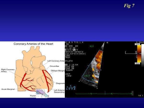
A dual-panel medical illustration and diagnostic image set demonstrating the transthoracic imaging of the right coronary artery (RCA). The left panel is an anatomical diagram of the 'Coronary Arteries of the Heart,' highlighting the major epicardial vessels including the Right Coronary Artery, Acute Marginal artery, Left Coronary Artery, Circumflex, Oblique Marginal, Diagonals, and Left Anterior Descending (LAD) artery. The right panel is a color Doppler transthoracic echocardiogram (TTE) in a specialized apical off-axis view. It displays real-time blood flow within the RCA, labeled as 'RCA' and 'CRUX' (indicating the crux of the heart), represented by a red and yellow color scale signifying flow velocity. A vertical color velocity bar and an electrocardiogram (ECG) trace are visible on the periphery. This comparison is used to guide non-invasive assessment of coronary flow reserve (CFR) by correlating anatomical landmarks with functional Doppler flow signals in a clinical cardiology setting.
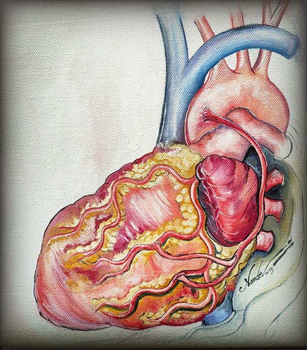
This medical illustration depicts the external anatomy of a human heart with a focus on surgical revascularization. The artwork shows the heart in an anterior view, highlighting the epicardial surface with prominent fatty tissue (yellow) and myocardium (red/pink). Key vascular structures include the superior vena cava (blue), the aortic arch with its branching vessels, and the pulmonary trunk. The illustration specifically demonstrates a 'baby Y' arterial anastomosis technique, where supplementary arterial conduits are grafted onto a parent bypass limb. Red-colored arteries, representing coronary bypass grafts, are shown in a branching pattern, with a proximal anastomosis connecting a secondary limb to the main conduit to supply multiple target vessels, such as the obtuse marginal or diagonal arteries. This anatomical diagram serves to educate on total arterial revascularization strategies in coronary artery bypass grafting (CABG) surgery, emphasizing the distribution and pattern of surgical anastomoses to restore myocardial blood flow.
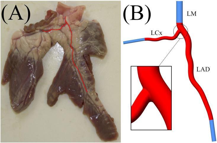
This educational graphic consists of two panels focusing on coronary artery anatomy and computational modeling. Panel (A) is a clinical photograph of a porcine ex vivo heart segment, illustrating the epicardial surface. The coronary artery geometry is highlighted with a red overlay, showing the vessel's origin, descent, and bifurcation into major branches across the cardiac tissue. Panel (B) presents a corresponding anatomical diagram and 3D reconstruction model of a human-scaled coronary artery bifurcation. It labels the Left Main (LM) coronary artery as the inlet, branching into the Left Anterior Descending (LAD) and the Left Circumflex (LCx) arteries. The diagram uses blue segments to represent computational domain extensions (inlets and outlets) and red to indicate the physiological vessel geometry. A magnified inset provides a detailed view of the bifurcation carina and the smooth transition between the parent vessel and daughter branches. This material is designed to demonstrate the translation of ex vivo anatomical findings into idealized geometries for hemodynamic and cardiovascular research.
mediastinum divisions diagram thorax
inguinal canal anatomy diagram layers
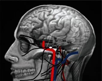
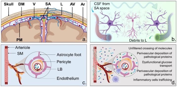
circle of Willis brain arteries diagram
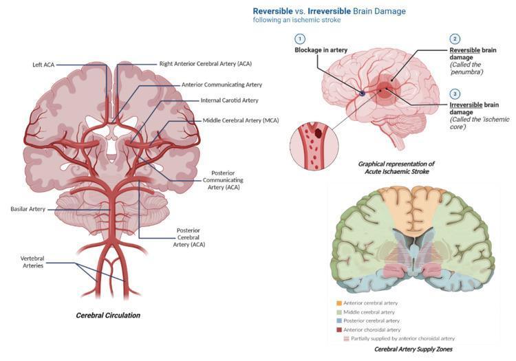
Educational medical infographic containing three panels illustrating cerebral neurovasculature and acute ischemic stroke pathophysiology. The first panel, 'Cerebral Circulation', features a detailed anatomical diagram of the Circle of Willis, labeling the vertebral arteries, basilar artery, and its bifurcation into the posterior cerebral arteries (PCA). It further illustrates the internal carotid arteries, middle cerebral arteries (MCA), and the anterior cerebral arteries (ACA) connected by the anterior communicating artery. The second panel, 'Graphical representation of Acute Ischaemic Stroke', depicts a sagittal view of the brain showing an arterial blockage. It differentiates between the 'ischemic core' (irreversible damage) and the surrounding 'penumbra' (reversible/salvageable tissue). The third panel, 'Cerebral Artery Supply Zones', is a coronal brain cross-section color-coded to show perfusion territories, specifically highlighting regions supplied by the ACA (orange), MCA (green), PCA (blue), and anterior choroidal artery (red). This resource is designed for students learning about stroke mechanisms, vascular anatomy, and clinical neuro-perfusion.
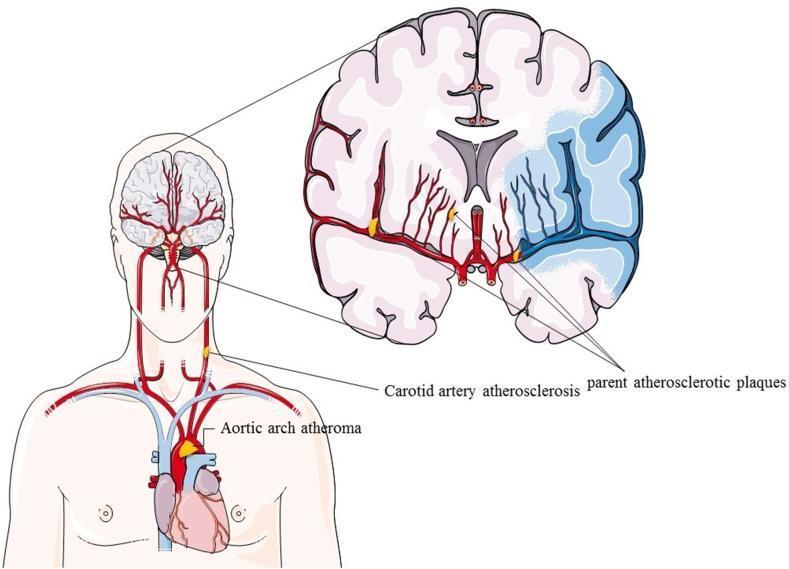
A medical illustration depicting the pathophysiological relationship between systemic atherosclerosis and ischemic stroke. The figure is divided into two primary views: a systemic vascular overview of the human torso and a detailed coronal section of the brain. The torso view displays the heart and great vessels, highlighting an 'Aortic arch atheroma' and 'Carotid artery atherosclerosis' with yellow plaques. The anatomical diagram shows the ascent of the carotid arteries toward the cerebral circulation. The enlarged coronal brain section illustrates a unilateral ischemic region, shaded in blue, representing a territory-specific stroke. 'Parent atherosclerotic plaques' are visualized at the origins of the cerebral arteries near the Circle of Willis. In the affected hemisphere, the distal vasculature appears dark and occluded compared to the healthy, red-colored branching vessels in the contralateral hemisphere. This diagram serves as an educational tool to demonstrate how proximal arterial plaques (aortic, carotid, and intracranial) contribute to distal cerebral infarction.
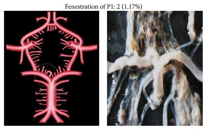
This composite image consists of an anatomical diagram and a corresponding gross clinical photograph illustrating a morphological variation of the Circle of Willis. The content focuses on the P1 segment of the posterior cerebral artery (PCA). The left panel is a schematic diagram of the cerebral arterial circle, highlighting a vascular fenestration within the right P1 segment, demarcated by a yellow dotted circle. The right panel is a macro-photograph of a cadaveric brain dissection showing the same anatomical region. In both images, the P1 segment exhibits a characteristic splitting into two distinct longitudinal channels that rejoin distally, creating a window-like defect rather than a single continuous lumen. This finding represents a P1 fenestration, an uncommon congenital vascular anomaly. Key structures visible include the basilar artery bifurcation, the superior cerebellar arteries, and the junction with the posterior communicating artery. This material is designed for neuroanatomy and neuroradiology education, specifically for understanding normal anatomical variants and potential sites for aneurysm formation.
pharyngeal branchial arches development diagram
nephron structure diagram kidney histology
A labeled anatomical diagram of the brachial plexus showing roots (C5-C8, T1), upper/middle/lower trunks, anterior and posterior divisions, lateral/posterior/medial cords, and the five terminal branches (musculocutaneous, axillary, radial, median, ulnar nerves), textbook style line drawing
A labeled anatomical diagram of the femoral triangle in the upper thigh showing its boundaries (inguinal ligament, sartorius, adductor longus) and contents from lateral to medial: femoral nerve, femoral artery, femoral vein, femoral canal, lymphatics, textbook style line drawing
A labeled cross-section anatomical diagram of the inguinal canal showing the deep ring, superficial ring, anterior wall (external oblique), posterior wall (transversalis fascia and conjoint tendon), roof, floor, and spermatic cord contents, textbook style line drawing
A labeled anatomical diagram of the hip joint showing the acetabulum, femoral head, ligamentum teres, iliofemoral ligament, pubofemoral ligament, ischiofemoral ligament, and blood supply from the medial circumflex femoral artery, textbook style line drawing
A labeled anatomical diagram of the six pharyngeal (branchial) arches in a human embryo showing the nerve, artery, cartilage, and muscle derivatives of each arch in a cross-sectional view, textbook style line drawing
A labeled histology diagram of compact bone microstructure showing the Haversian system with osteon, central Haversian canal, concentric lamellae, osteocytes in lacunae, canaliculi, and Volkmann canals, textbook style line drawing
2-Week Anatomy Revision Plan (MBBS 1st Year)
Ground Rules
- 2 sessions/day - morning (2 hrs) + evening (1 hr review/quiz)
- Each day = 1 main topic + evening recall/questions
- You tell me when you're on a topic and I'll quiz you or explain anything
WEEK 1 - Gross Anatomy (Regional)
| Day | Morning - Study | Evening - Review |
|---|---|---|
| Day 1 | Upper Limb - bones, joints (shoulder, elbow, wrist) | Quiz: brachial plexus roots & branches |
| Day 2 | Upper Limb - muscles, nerve supply, dermatomes | Quiz: muscle actions + clinical (wrist drop, claw hand, etc.) |
| Day 3 | Lower Limb - bones, joints (hip, knee, ankle) | Quiz: femoral triangle, popliteal fossa contents |
| Day 4 | Lower Limb - muscles, lumbosacral plexus, dermatomes | Quiz: common nerve injuries (foot drop, etc.) |
| Day 5 | Thorax - heart, great vessels, mediastinum | Quiz: heart borders, surface anatomy, coronary arteries |
| Day 6 | Thorax - lungs, pleura + Abdomen - peritoneum, GI tract | Quiz: lung segments, peritoneal vs retroperitoneal |
| Day 7 | Rest / Weak spots review | Light revision of Days 1-6 |
WEEK 2 - Head & Neck + Neuro + Histology/Embryology
| Day | Morning - Study | Evening - Review |
|---|---|---|
| Day 8 | Abdomen - liver, pancreas, spleen, kidneys | Quiz: porta hepatis, pancreatic ducts, suprarenal glands |
| Day 9 | Pelvis & Perineum - pelvic floor, bladder, uterus/prostate | Quiz: inguinal canal, femoral hernia vs inguinal |
| Day 10 | Head & Neck - skull, scalp, face, parotid, TMJ | Quiz: CN branches, dangerous area of face |
| Day 11 | Head & Neck - pharynx, larynx, thyroid, triangles of neck | Quiz: recurrent laryngeal nerve, carotid sheath |
| Day 12 | Neuroanatomy - spinal cord, brainstem, cerebellum | Quiz: tracts (DCML, spinothalamic), cerebellar signs |
| Day 13 | Neuroanatomy - cerebrum, cranial nerves, ventricles/CSF | Quiz: cranial nerve exam points, circle of Willis |
| Day 14 | Full mock revision - High-yield clinicals + past Qs | Cover all weak areas flagged during the week |
How I Can Help Each Day
- Study with you - paste a topic and I'll give you a tight, high-yield summary from anatomy textbooks
- Quiz you - I'll ask 5-10 MCQs or short-answer questions on the day's topic
- Explain on demand - stuck on a nerve plexus, a structure, a clinical correlation? Just ask
HighYield Anatomy QuickRef
PDF Document · PDF
| Section | Coverage |
|---|---|
| 1. Upper Limb | Brachial plexus table, nerve injury patterns (wrist drop, claw hand, ape hand), rotator cuff SITS |
| 2. Lower Limb | Lumbar/sacral plexus, femoral triangle NAVEL, hip joint blood supply, nerve injuries |
| 3. Thorax | Heart borders/apex, coronary arteries & MI territories, mediastinum divisions, lung lobes |
| 4. Abdomen | Retroperitoneal structures, porta hepatis, inguinal canal full table, hernia types |
| 5. Head & Neck | All 12 cranial nerves table, neck triangles, recurrent laryngeal nerve, accessory nerve |
| 6. Neuroanatomy | Spinal cord tracts (DCML vs spinothalamic), Brown-Sequard, Circle of Willis, ventricular system |
| 7. Embryology | Weekly development timeline, neural tube defects, common congenital anomalies |
| 8. Mnemonics | SITS, NAVEL, SCALP, diaphragm openings (I 8 10 eggs at 12), CN mnemonics + 12 quick-fire facts |
| 9. Pelvis | Pelvic floor muscles, ureter relations ('water under bridge'), stone lodgement sites |
| 10. Revision Tips | Active recall, spaced repetition, teaching back strategy |
HIGH-YIELD ANATOMY - MBBS Quick Reference
1. UPPER LIMB
Brachial Plexus
| Component | Roots | Key Nerves |
|---|---|---|
| Roots | C5-C8, T1 | Dorsal scapular (C5), Long thoracic (C5-7) |
| Trunks | Upper (C5-6), Middle (C7), Lower (C8-T1) | Suprascapular (upper trunk) |
| Cords | Lateral, Posterior, Medial | Named by relation to axillary artery |
| Terminal Branches | 5 nerves | Musculocutaneous, Axillary, Radial, Median, Ulnar |
Nerve Injury Patterns
| Nerve | Root | Cause | Deformity |
|---|---|---|---|
| Axillary | C5,6 | Surgical neck humerus fracture | Flattened deltoid; loss shoulder abduction >15° |
| Radial | C5-T1 | Midshaft humerus / Saturday night palsy | Wrist drop |
| Median | C6-T1 | Supracondylar fracture / carpal tunnel | Ape hand; Pope's blessing sign |
| Ulnar | C8-T1 | Medial epicondyle fracture | Claw hand (ring & little); Froment sign |
| Long thoracic | C5-7 | Mastectomy / axillary dissection | Winged scapula |
- Erb's palsy (C5-C6): "Waiter's tip" - arm adducted, medially rotated, forearm pronated
- Klumpke's palsy (C8-T1): Claw hand + Horner syndrome
Rotator Cuff - SITS
| Muscle | Nerve | Action |
|---|---|---|
| Supraspinatus | Suprascapular | Initiates abduction (0-15°) - most commonly torn |
| Infraspinatus | Suprascapular | Lateral rotation |
| Teres Minor | Axillary | Lateral rotation |
| Subscapularis | Subscapular | Medial rotation |
2. LOWER LIMB
Key Nerve Injuries
| Nerve | Root | Cause | Deficit |
|---|---|---|---|
| Femoral | L2-4 | Psoas haematoma | Weak knee extension |
| Obturator | L2-4 | Pelvic surgery | Weak hip adduction |
| Sciatic | L4-S3 | Posterior hip dislocation | Everything below knee |
| Common Fibular | L4-S2 | Fibular neck fracture | Foot drop; high-step gait |
| Superior Gluteal | L4-S1 | Hip arthroplasty | Trendelenburg gait |
- Femoral triangle contents (lateral→medial): NAVEL - Nerve, Artery, Vein, Empty space, Lymphatics
- Hip blood supply: Medial circumflex femoral artery → damaged in subcapital fracture → avascular necrosis
- Posterior hip dislocation: leg shortened, adducted, internally rotated; sciatic nerve at risk
3. THORAX
Heart Borders
| Border | Structure | Landmark |
|---|---|---|
| Right | Right atrium | Right sternal edge, 3rd-6th rib |
| Left | Left ventricle + auricle | Left sternal edge, 2nd rib to apex |
| Apex | Left ventricle | 5th ICS, midclavicular line |
| Base (posterior) | Left atrium | T5-T8 |
Coronary Arteries
| Artery | Supplies | Occlusion |
|---|---|---|
| LAD | Anterior LV, anterior 2/3 IVS | Anterior MI ("widow maker") |
| LCx | Lateral/posterior LV | Lateral/posterior MI |
| RCA | RV, SA node (60%), AV node (80%) | Inferior MI; heart block |
Mediastinum
| Division | Key Contents |
|---|---|
| Superior | Aortic arch, trachea, oesophagus, thymus, SVC, thoracic duct, vagus, left RLN |
| Middle | Heart, pericardium, ascending aorta, tracheal bifurcation, phrenic nerves |
| Posterior | Descending aorta, oesophagus, thoracic duct, azygos, sympathetic trunk |
- Right lung: 3 lobes, 10 segments | Left lung: 2 lobes, 8-10 segments
- Inhaled foreign body: most likely right lower lobe (wider, more vertical right main bronchus)
4. ABDOMEN
Retroperitoneal Structures - SAD PUCKER
Porta Hepatis
Inguinal Canal
| Feature | Detail |
|---|---|
| Deep ring | Lateral to inferior epigastric vessels |
| Superficial ring | Above pubic tubercle |
| Roof | Internal oblique + transversus abdominis arching fibres |
| Floor | Inguinal ligament + lacunar ligament (medially) |
| Posterior wall | Transversalis fascia + conjoint tendon (medially) |
| Male contents | Spermatic cord: vas deferens, testicular artery, pampiniform plexus |
| Female contents | Round ligament of uterus |
- Indirect hernia: lateral to inferior epigastric vessels; congenital
- Direct hernia: medial (through Hesselbach's triangle); acquired
- Femoral hernia: lateral to pubic tubercle, below inguinal ligament; more common in women; high strangulation risk
5. HEAD & NECK
12 Cranial Nerves
| CN | Name | Type | Key Function | Clinical |
|---|---|---|---|---|
| I | Olfactory | S | Smell | Anosmia → anterior fossa fracture |
| II | Optic | S | Vision | RAPD; visual field defects |
| III | Oculomotor | M+Para | Eye movement; pupil; lid | "Down & out"; ptosis; PComm aneurysm |
| IV | Trochlear | M | Superior oblique (intorsion) | Vertical diplopia; head tilt |
| V | Trigeminal | S+M | Face sensation; mastication | Corneal reflex afferent (V1); trigeminal neuralgia |
| VI | Abducens | M | Lateral rectus | Medial squint; false localising sign |
| VII | Facial | M+S+Para | Facial expression; taste ant. 2/3 | UMN (forehead spared) vs LMN (Bell's palsy) |
| VIII | Vestibulocochlear | S | Hearing + balance | SNHL; acoustic neuroma |
| IX | Glossopharyngeal | M+S+Para | Taste post. 1/3; parotid | Gag reflex afferent |
| X | Vagus | M+S+Para | Larynx; viscera | Hoarseness (RLN); uvula away from lesion |
| XI | Accessory | M | SCM + trapezius | Can't shrug or turn head |
| XII | Hypoglossal | M | Tongue movements | Tongue deviates TOWARD LMN lesion |
Neck Triangles
| Triangle | Key Contents |
|---|---|
| Anterior | Carotid arteries, IJV, vagus, thyroid, submandibular gland |
| Posterior | CN XI (accessory nerve), brachial plexus trunks, subclavian vessels |
- Recurrent laryngeal nerve: loops under aortic arch (left) / subclavian (right) → at risk in thyroid surgery → hoarseness
- Accessory nerve (XI) crosses posterior triangle → at risk in cervical lymph node biopsy → trapezius palsy
6. NEUROANATOMY
Spinal Cord Tracts
| Tract | Location | Carries | Decussation |
|---|---|---|---|
| Dorsal columns (DCML) | Posterior | Fine touch, vibration, proprioception | At medulla |
| Spinothalamic (ALS) | Anterolateral | Pain, temperature, crude touch | Within 1-2 segments of entry |
| Corticospinal (lateral) | Posterolateral | Voluntary motor | Pyramidal decussation (medulla) |
- Brown-Séquard (hemisection): ipsilateral motor + DCML loss; contralateral pain & temperature loss
Circle of Willis
- ACA (x2) joined by anterior communicating artery
- PCA (x2) joined to ICA by posterior communicating artery (PComm)
- PComm aneurysm → CN III palsy (fixed dilated pupil, "down and out")
- MCA stroke (most common): contralateral face & arm > leg; aphasia if dominant hemisphere
Ventricular System
- CSF produced by choroid plexus (~500 mL/day); absorbed by arachnoid granulations
7. HIGH-YIELD EMBRYOLOGY
| Week | Key Events |
|---|---|
| Week 1 | Fertilisation → blastocyst → implantation (day 6-7) |
| Week 2 | Bilaminar disc; primitive streak |
| Week 3 | Gastrulation → trilaminar disc; notochord |
| Week 3-4 | Neurulation; neural tube closes by day 28; somites |
| Week 4-8 | Organogenesis - most sensitive to teratogens |
| Defect | Basis | Feature |
|---|---|---|
| Spina bifida | Posterior neural tube closure failure | Folate deficiency; meningocele/myelomeningocele |
| Anencephaly | Cranial neural tube closure failure | Elevated AFP; incompatible with life |
| Cleft lip | Failure maxillary + medial nasal process fusion | Unilateral or bilateral |
| Meckel's diverticulum | Persistent vitello-intestinal duct | 2 inches, 2 feet from ileocaecal valve, 2% population |
| PDA | Ductus arteriosus fails to close | Continuous murmur; treat with indomethacin |
| VSD | IVS formation failure | Most common congenital heart defect |
8. MNEMONICS
| Topic | Mnemonic |
|---|---|
| Brachial plexus | Real Teens Drink Cold Beer (Roots, Trunks, Divisions, Cords, Branches) |
| Proximal carpals | She Likes To Party (Scaphoid, Lunate, Triquetrum, Pisiform) |
| Distal carpals | Then The Carpal Hurts (Trapezium, Trapezoid, Capitate, Hamate) |
| Femoral triangle | NAVEL (Nerve, Artery, Vein, Empty, Lymphatics) |
| Scalp layers | SCALP (Skin, Connective tissue, Aponeurosis, Loose areolar, Pericranium) |
| Diaphragm openings | I 8 (ate) 10 eggs AT 12 → T8=IVC, T10=Oesophagus+Vagus, T12=Aorta+Thoracic duct+Azygos |
| Rotator cuff | SITS (Supraspinatus, Infraspinatus, Teres minor, Subscapularis) |
| CN types | Some Say Marry Money But My Brother Says Big Brains Matter More |
9. QUICK-FIRE FACTS
- Longest nerve: Sciatic | Smallest bone: Stapes | Only bone not articulating: Hyoid
- Right kidney lower than left (displaced by liver)
- Most common Berry aneurysm site: Anterior communicating artery (~35%)
- Foramen ovale closes at birth (LA pressure > RA)
- Portal-systemic anastomoses: oesophageal, para-umbilical, rectal, retroperitoneal
- McBurney's point: 1/3 from ASIS to umbilicus (appendicitis tenderness)
- Ureter in females passes under uterine artery ("water under the bridge") - at risk in hysterectomy
- Ureter narrowings: PUJ → pelvic brim → VUJ (narrowest; stones most lodge here)
Anatomy FrequentQA
PDF Document · PDF
| Chapter | Questions Covered |
|---|---|
| 1. Upper Limb | Brachial plexus, Erb's palsy, Radial nerve injuries, Carpal tunnel syndrome, Anatomical snuffbox, Rotator cuff |
| 2. Lower Limb | Femoral triangle, Hip joint, Popliteal fossa, Foot drop, Trendelenburg sign |
| 3. Thorax | Coronary arteries, Mediastinum, Heart surface markings, Pericardium |
| 4. Abdomen | Inguinal canal + hernias, Portal vein + anastomoses, Liver, Diaphragm, Kidneys |
| 5. Head & Neck | Facial nerve (CN VII), Thyroid gland, Parotid gland, Carotid sheath |
| 6. Neuroanatomy | Meninges + haematomas, Ventricular system + CSF, Circle of Willis, Spinal cord tracts |
| 7. Embryology | Pharyngeal arches, Neural tube defects, Heart development + CHDs |
| 8. Histology | Bone structure + cells, Kidney nephron, Liver lobule (acinus zones) |
| 9. Pelvis | Male urethra, Pelvic floor muscles |
| Bonus Viva Table | 50 one-liner Q&As for rapid revision |
- A structured answer with bullet points
- A clinical correlation (★) - exactly what examiners look for in vivas
Error calling model 'gemini-3-pro-image' (RESOURCE_EXHAUSTED): 429 RESOURCE_EXHAUSTED. {'error': {'code': 429, 'message': 'Resource has been exhausted (e.g. check quota).', 'status': 'RESOURCE_EXHAUSTED'}}
We encountered an error while processing your request. Please try again or reach out to us for help.
Error calling model 'gemini-3-pro-image' (RESOURCE_EXHAUSTED): 429 RESOURCE_EXHAUSTED. {'error': {'code': 429, 'message': 'Resource has been exhausted (e.g. check quota).', 'status': 'RESOURCE_EXHAUSTED'}}
We encountered an error while processing your request. Please try again or reach out to us for help.
are you done yet?
A labeled anatomical diagram of the brachial plexus showing roots (C5-C8, T1), upper/middle/lower trunks, anterior and posterior divisions, lateral/posterior/medial cords, and the five terminal branches (musculocutaneous, axillary, radial, median, ulnar nerves), textbook style line drawing
A labeled anatomical diagram of the femoral triangle in the upper thigh showing its boundaries (inguinal ligament, sartorius, adductor longus) and contents from lateral to medial: femoral nerve, femoral artery, femoral vein, femoral canal, lymphatics, textbook style line drawing
A labeled cross-section anatomical diagram of the inguinal canal showing the deep ring, superficial ring, anterior wall (external oblique), posterior wall (transversalis fascia and conjoint tendon), roof, floor, and spermatic cord contents, textbook style line drawing
A labeled anatomical diagram of the hip joint showing the acetabulum, femoral head, ligamentum teres, iliofemoral ligament, pubofemoral ligament, ischiofemoral ligament, and blood supply from the medial circumflex femoral artery, textbook style line drawing
A labeled anatomical diagram of the six pharyngeal (branchial) arches in a human embryo showing the nerve, artery, cartilage, and muscle derivatives of each arch in a cross-sectional view, textbook style line drawing
2-Week Anatomy Revision Plan (MBBS 1st Year)
Ground Rules
- 2 sessions/day - morning (2 hrs) + evening (1 hr review/quiz)
- Each day = 1 main topic + evening recall/questions
- You tell me when you're on a topic and I'll quiz you or explain anything
WEEK 1 - Gross Anatomy (Regional)
| Day | Morning - Study | Evening - Review |
|---|---|---|
| Day 1 | Upper Limb - bones, joints (shoulder, elbow, wrist) | Quiz: brachial plexus roots & branches |
| Day 2 | Upper Limb - muscles, nerve supply, dermatomes | Quiz: muscle actions + clinical (wrist drop, claw hand, etc.) |
| Day 3 | Lower Limb - bones, joints (hip, knee, ankle) | Quiz: femoral triangle, popliteal fossa contents |
| Day 4 | Lower Limb - muscles, lumbosacral plexus, dermatomes | Quiz: common nerve injuries (foot drop, etc.) |
| Day 5 | Thorax - heart, great vessels, mediastinum | Quiz: heart borders, surface anatomy, coronary arteries |
| Day 6 | Thorax - lungs, pleura + Abdomen - peritoneum, GI tract | Quiz: lung segments, peritoneal vs retroperitoneal |
| Day 7 | Rest / Weak spots review | Light revision of Days 1-6 |
WEEK 2 - Head & Neck + Neuro + Histology/Embryology
| Day | Morning - Study | Evening - Review |
|---|---|---|
| Day 8 | Abdomen - liver, pancreas, spleen, kidneys | Quiz: porta hepatis, pancreatic ducts, suprarenal glands |
| Day 9 | Pelvis & Perineum - pelvic floor, bladder, uterus/prostate | Quiz: inguinal canal, femoral hernia vs inguinal |
| Day 10 | Head & Neck - skull, scalp, face, parotid, TMJ | Quiz: CN branches, dangerous area of face |
| Day 11 | Head & Neck - pharynx, larynx, thyroid, triangles of neck | Quiz: recurrent laryngeal nerve, carotid sheath |
| Day 12 | Neuroanatomy - spinal cord, brainstem, cerebellum | Quiz: tracts (DCML, spinothalamic), cerebellar signs |
| Day 13 | Neuroanatomy - cerebrum, cranial nerves, ventricles/CSF | Quiz: cranial nerve exam points, circle of Willis |
| Day 14 | Full mock revision - High-yield clinicals + past Qs | Cover all weak areas flagged during the week |
How I Can Help Each Day
- Study with you - paste a topic and I'll give you a tight, high-yield summary from anatomy textbooks
- Quiz you - I'll ask 5-10 MCQs or short-answer questions on the day's topic
- Explain on demand - stuck on a nerve plexus, a structure, a clinical correlation? Just ask
HighYield Anatomy QuickRef
PDF Document · PDF
| Section | Coverage |
|---|---|
| 1. Upper Limb | Brachial plexus table, nerve injury patterns (wrist drop, claw hand, ape hand), rotator cuff SITS |
| 2. Lower Limb | Lumbar/sacral plexus, femoral triangle NAVEL, hip joint blood supply, nerve injuries |
| 3. Thorax | Heart borders/apex, coronary arteries & MI territories, mediastinum divisions, lung lobes |
| 4. Abdomen | Retroperitoneal structures, porta hepatis, inguinal canal full table, hernia types |
| 5. Head & Neck | All 12 cranial nerves table, neck triangles, recurrent laryngeal nerve, accessory nerve |
| 6. Neuroanatomy | Spinal cord tracts (DCML vs spinothalamic), Brown-Sequard, Circle of Willis, ventricular system |
| 7. Embryology | Weekly development timeline, neural tube defects, common congenital anomalies |
| 8. Mnemonics | SITS, NAVEL, SCALP, diaphragm openings (I 8 10 eggs at 12), CN mnemonics + 12 quick-fire facts |
| 9. Pelvis | Pelvic floor muscles, ureter relations ('water under bridge'), stone lodgement sites |
| 10. Revision Tips | Active recall, spaced repetition, teaching back strategy |
HIGH-YIELD ANATOMY - MBBS Quick Reference
1. UPPER LIMB
Brachial Plexus
| Component | Roots | Key Nerves |
|---|---|---|
| Roots | C5-C8, T1 | Dorsal scapular (C5), Long thoracic (C5-7) |
| Trunks | Upper (C5-6), Middle (C7), Lower (C8-T1) | Suprascapular (upper trunk) |
| Cords | Lateral, Posterior, Medial | Named by relation to axillary artery |
| Terminal Branches | 5 nerves | Musculocutaneous, Axillary, Radial, Median, Ulnar |
Nerve Injury Patterns
| Nerve | Root | Cause | Deformity |
|---|---|---|---|
| Axillary | C5,6 | Surgical neck humerus fracture | Flattened deltoid; loss shoulder abduction >15° |
| Radial | C5-T1 | Midshaft humerus / Saturday night palsy | Wrist drop |
| Median | C6-T1 | Supracondylar fracture / carpal tunnel | Ape hand; Pope's blessing sign |
| Ulnar | C8-T1 | Medial epicondyle fracture | Claw hand (ring & little); Froment sign |
| Long thoracic | C5-7 | Mastectomy / axillary dissection | Winged scapula |
- Erb's palsy (C5-C6): "Waiter's tip" - arm adducted, medially rotated, forearm pronated
- Klumpke's palsy (C8-T1): Claw hand + Horner syndrome
Rotator Cuff - SITS
| Muscle | Nerve | Action |
|---|---|---|
| Supraspinatus | Suprascapular | Initiates abduction (0-15°) - most commonly torn |
| Infraspinatus | Suprascapular | Lateral rotation |
| Teres Minor | Axillary | Lateral rotation |
| Subscapularis | Subscapular | Medial rotation |
2. LOWER LIMB
Key Nerve Injuries
| Nerve | Root | Cause | Deficit |
|---|---|---|---|
| Femoral | L2-4 | Psoas haematoma | Weak knee extension |
| Obturator | L2-4 | Pelvic surgery | Weak hip adduction |
| Sciatic | L4-S3 | Posterior hip dislocation | Everything below knee |
| Common Fibular | L4-S2 | Fibular neck fracture | Foot drop; high-step gait |
| Superior Gluteal | L4-S1 | Hip arthroplasty | Trendelenburg gait |
- Femoral triangle contents (lateral→medial): NAVEL - Nerve, Artery, Vein, Empty space, Lymphatics
- Hip blood supply: Medial circumflex femoral artery → damaged in subcapital fracture → avascular necrosis
- Posterior hip dislocation: leg shortened, adducted, internally rotated; sciatic nerve at risk
3. THORAX
Heart Borders
| Border | Structure | Landmark |
|---|---|---|
| Right | Right atrium | Right sternal edge, 3rd-6th rib |
| Left | Left ventricle + auricle | Left sternal edge, 2nd rib to apex |
| Apex | Left ventricle | 5th ICS, midclavicular line |
| Base (posterior) | Left atrium | T5-T8 |
Coronary Arteries
| Artery | Supplies | Occlusion |
|---|---|---|
| LAD | Anterior LV, anterior 2/3 IVS | Anterior MI ("widow maker") |
| LCx | Lateral/posterior LV | Lateral/posterior MI |
| RCA | RV, SA node (60%), AV node (80%) | Inferior MI; heart block |
Mediastinum
| Division | Key Contents |
|---|---|
| Superior | Aortic arch, trachea, oesophagus, thymus, SVC, thoracic duct, vagus, left RLN |
| Middle | Heart, pericardium, ascending aorta, tracheal bifurcation, phrenic nerves |
| Posterior | Descending aorta, oesophagus, thoracic duct, azygos, sympathetic trunk |
- Right lung: 3 lobes, 10 segments | Left lung: 2 lobes, 8-10 segments
- Inhaled foreign body: most likely right lower lobe (wider, more vertical right main bronchus)
4. ABDOMEN
Retroperitoneal Structures - SAD PUCKER
Porta Hepatis
Inguinal Canal
| Feature | Detail |
|---|---|
| Deep ring | Lateral to inferior epigastric vessels |
| Superficial ring | Above pubic tubercle |
| Roof | Internal oblique + transversus abdominis arching fibres |
| Floor | Inguinal ligament + lacunar ligament (medially) |
| Posterior wall | Transversalis fascia + conjoint tendon (medially) |
| Male contents | Spermatic cord: vas deferens, testicular artery, pampiniform plexus |
| Female contents | Round ligament of uterus |
- Indirect hernia: lateral to inferior epigastric vessels; congenital
- Direct hernia: medial (through Hesselbach's triangle); acquired
- Femoral hernia: lateral to pubic tubercle, below inguinal ligament; more common in women; high strangulation risk
5. HEAD & NECK
12 Cranial Nerves
| CN | Name | Type | Key Function | Clinical |
|---|---|---|---|---|
| I | Olfactory | S | Smell | Anosmia → anterior fossa fracture |
| II | Optic | S | Vision | RAPD; visual field defects |
| III | Oculomotor | M+Para | Eye movement; pupil; lid | "Down & out"; ptosis; PComm aneurysm |
| IV | Trochlear | M | Superior oblique (intorsion) | Vertical diplopia; head tilt |
| V | Trigeminal | S+M | Face sensation; mastication | Corneal reflex afferent (V1); trigeminal neuralgia |
| VI | Abducens | M | Lateral rectus | Medial squint; false localising sign |
| VII | Facial | M+S+Para | Facial expression; taste ant. 2/3 | UMN (forehead spared) vs LMN (Bell's palsy) |
| VIII | Vestibulocochlear | S | Hearing + balance | SNHL; acoustic neuroma |
| IX | Glossopharyngeal | M+S+Para | Taste post. 1/3; parotid | Gag reflex afferent |
| X | Vagus | M+S+Para | Larynx; viscera | Hoarseness (RLN); uvula away from lesion |
| XI | Accessory | M | SCM + trapezius | Can't shrug or turn head |
| XII | Hypoglossal | M | Tongue movements | Tongue deviates TOWARD LMN lesion |
Neck Triangles
| Triangle | Key Contents |
|---|---|
| Anterior | Carotid arteries, IJV, vagus, thyroid, submandibular gland |
| Posterior | CN XI (accessory nerve), brachial plexus trunks, subclavian vessels |
- Recurrent laryngeal nerve: loops under aortic arch (left) / subclavian (right) → at risk in thyroid surgery → hoarseness
- Accessory nerve (XI) crosses posterior triangle → at risk in cervical lymph node biopsy → trapezius palsy
6. NEUROANATOMY
Spinal Cord Tracts
| Tract | Location | Carries | Decussation |
|---|---|---|---|
| Dorsal columns (DCML) | Posterior | Fine touch, vibration, proprioception | At medulla |
| Spinothalamic (ALS) | Anterolateral | Pain, temperature, crude touch | Within 1-2 segments of entry |
| Corticospinal (lateral) | Posterolateral | Voluntary motor | Pyramidal decussation (medulla) |
- Brown-Séquard (hemisection): ipsilateral motor + DCML loss; contralateral pain & temperature loss
Circle of Willis
- ACA (x2) joined by anterior communicating artery
- PCA (x2) joined to ICA by posterior communicating artery (PComm)
- PComm aneurysm → CN III palsy (fixed dilated pupil, "down and out")
- MCA stroke (most common): contralateral face & arm > leg; aphasia if dominant hemisphere
Ventricular System
- CSF produced by choroid plexus (~500 mL/day); absorbed by arachnoid granulations
7. HIGH-YIELD EMBRYOLOGY
| Week | Key Events |
|---|---|
| Week 1 | Fertilisation → blastocyst → implantation (day 6-7) |
| Week 2 | Bilaminar disc; primitive streak |
| Week 3 | Gastrulation → trilaminar disc; notochord |
| Week 3-4 | Neurulation; neural tube closes by day 28; somites |
| Week 4-8 | Organogenesis - most sensitive to teratogens |
| Defect | Basis | Feature |
|---|---|---|
| Spina bifida | Posterior neural tube closure failure | Folate deficiency; meningocele/myelomeningocele |
| Anencephaly | Cranial neural tube closure failure | Elevated AFP; incompatible with life |
| Cleft lip | Failure maxillary + medial nasal process fusion | Unilateral or bilateral |
| Meckel's diverticulum | Persistent vitello-intestinal duct | 2 inches, 2 feet from ileocaecal valve, 2% population |
| PDA | Ductus arteriosus fails to close | Continuous murmur; treat with indomethacin |
| VSD | IVS formation failure | Most common congenital heart defect |
8. MNEMONICS
| Topic | Mnemonic |
|---|---|
| Brachial plexus | Real Teens Drink Cold Beer (Roots, Trunks, Divisions, Cords, Branches) |
| Proximal carpals | She Likes To Party (Scaphoid, Lunate, Triquetrum, Pisiform) |
| Distal carpals | Then The Carpal Hurts (Trapezium, Trapezoid, Capitate, Hamate) |
| Femoral triangle | NAVEL (Nerve, Artery, Vein, Empty, Lymphatics) |
| Scalp layers | SCALP (Skin, Connective tissue, Aponeurosis, Loose areolar, Pericranium) |
| Diaphragm openings | I 8 (ate) 10 eggs AT 12 → T8=IVC, T10=Oesophagus+Vagus, T12=Aorta+Thoracic duct+Azygos |
| Rotator cuff | SITS (Supraspinatus, Infraspinatus, Teres minor, Subscapularis) |
| CN types | Some Say Marry Money But My Brother Says Big Brains Matter More |
9. QUICK-FIRE FACTS
- Longest nerve: Sciatic | Smallest bone: Stapes | Only bone not articulating: Hyoid
- Right kidney lower than left (displaced by liver)
- Most common Berry aneurysm site: Anterior communicating artery (~35%)
- Foramen ovale closes at birth (LA pressure > RA)
- Portal-systemic anastomoses: oesophageal, para-umbilical, rectal, retroperitoneal
- McBurney's point: 1/3 from ASIS to umbilicus (appendicitis tenderness)
- Ureter in females passes under uterine artery ("water under the bridge") - at risk in hysterectomy
- Ureter narrowings: PUJ → pelvic brim → VUJ (narrowest; stones most lodge here)
Anatomy FrequentQA
PDF Document · PDF
| Chapter | Questions Covered |
|---|---|
| 1. Upper Limb | Brachial plexus, Erb's palsy, Radial nerve injuries, Carpal tunnel syndrome, Anatomical snuffbox, Rotator cuff |
| 2. Lower Limb | Femoral triangle, Hip joint, Popliteal fossa, Foot drop, Trendelenburg sign |
| 3. Thorax | Coronary arteries, Mediastinum, Heart surface markings, Pericardium |
| 4. Abdomen | Inguinal canal + hernias, Portal vein + anastomoses, Liver, Diaphragm, Kidneys |
| 5. Head & Neck | Facial nerve (CN VII), Thyroid gland, Parotid gland, Carotid sheath |
| 6. Neuroanatomy | Meninges + haematomas, Ventricular system + CSF, Circle of Willis, Spinal cord tracts |
| 7. Embryology | Pharyngeal arches, Neural tube defects, Heart development + CHDs |
| 8. Histology | Bone structure + cells, Kidney nephron, Liver lobule (acinus zones) |
| 9. Pelvis | Male urethra, Pelvic floor muscles |
| Bonus Viva Table | 50 one-liner Q&As for rapid revision |
- A structured answer with bullet points
- A clinical correlation (★) - exactly what examiners look for in vivas
Error calling model 'gemini-3-pro-image' (RESOURCE_EXHAUSTED): 429 RESOURCE_EXHAUSTED. {'error': {'code': 429, 'message': 'Resource has been exhausted (e.g. check quota).', 'status': 'RESOURCE_EXHAUSTED'}}
We encountered an error while processing your request. Please try again or reach out to us for help.
Error calling model 'gemini-3-pro-image' (RESOURCE_EXHAUSTED): 429 RESOURCE_EXHAUSTED. {'error': {'code': 429, 'message': 'Resource has been exhausted (e.g. check quota).', 'status': 'RESOURCE_EXHAUSTED'}}
We encountered an error while processing your request. Please try again or reach out to us for help.
Error calling model 'gemini-3-pro-image' (RESOURCE_EXHAUSTED): 429 RESOURCE_EXHAUSTED. {'error': {'code': 429, 'message': 'Resource has been exhausted (e.g. check quota).', 'status': 'RESOURCE_EXHAUSTED'}}
We encountered an error while processing your request. Please try again or reach out to us for help.
okay do it quick
A labeled anatomical diagram of the femoral triangle in the upper thigh showing its boundaries (inguinal ligament, sartorius, adductor longus) and contents from lateral to medial: femoral nerve, femoral artery, femoral vein, femoral canal, lymphatics, textbook style line drawing
A labeled cross-section anatomical diagram of the inguinal canal showing the deep ring, superficial ring, anterior wall, posterior wall, roof, floor, and spermatic cord contents, textbook style line drawing
A labeled anatomical diagram of the hip joint showing the acetabulum, femoral head, ligamentum teres, iliofemoral ligament, pubofemoral ligament, ischiofemoral ligament, and blood supply from the medial circumflex femoral artery, textbook style line drawing
mkdir -p /tmp/workspace/anatomy-pdf/images && cd /tmp/workspace/anatomy-pdf/images && \ curl -sL -o bone_histology.png "https://cdn.orris.care/image-gen/8bab1dec99314111b14ddc2cd940ed0a.png" && \ curl -sL -o brachial_plexus.png "https://cdn.orris.care/image-gen/aab6094d51704311bfe389fc5d1e880d.png" && \ curl -sL -o pharyngeal_arches.png "https://cdn.orris.care/image-gen/aa3a787060134b3c8c473bf57465fcb8.png" && \ curl -sL -o femoral_triangle.png "https://cdn.orris.care/image-gen/83f3266041914470a55c7bea3c6ef34a.png" && \ curl -sL -o inguinal_canal.png "https://cdn.orris.care/image-gen/a7c14b9ab67b4efd92768ec2a17e5ce2.png" && \ curl -sL -o hip_joint.png "https://cdn.orris.care/image-gen/9c7e139c9492456e92b2de41b6067ad7.png" && \ curl -sL -o mediastinum.png "https://cdn.orris.care/cdss_images/BAILY_1760530350667_8f4bf062-973f-43cb-b161-2b8e0caa657a.png" && \ curl -sL -o nephron.png "https://cdn.orris.care/cdss_images/HARRISON_1763034261030_590f2792-b44d-41fd-9951-0836bbcd314e.png" && \ curl -sL -o thyroid.png "https://cdn.orris.care/cdss_images/BAILY_1760543794286_106065d9-9507-4b55-a59f-9ee865e54b8a.png" && \ curl -sL -o liver_lobule.png "https://cdn.orris.care/cdss_images/pmc_clinical_VQA_4e01c93071b8a72b1a1bf83cd7adaefb89bbb37709ba0907bd9c72ac148da9fe.jpg" && \ curl -sL -o circle_willis.png "https://cdn.orris.care/cdss_images/pmc_clinical_VQA_5b0c63c589b83cd4a8a834d9cd053d6d6d23a903d94592bb5c4bf6e30448f4c5.jpg" && \ curl -sL -o cranial_nerves.png "https://cdn.orris.care/cdss_images/pmc_clinical_VQA_322aaf0cb9e96402745c40a490659e7167d992b814b84c84bb9b7fc1e43534dc.jpg" && \ curl -sL -o meninges.png "https://cdn.orris.care/cdss_images/pmc_clinical_VQA_3d2c0701338a0c6b6bbac1e9932f23b366e4e39c4f2d78ec10d36dd1c3053e84.jpg" && \ curl -sL -o coronary.png "https://cdn.orris.care/cdss_images/pmc_clinical_VQA_1e8b18b061c6ff13c1fb6658b2dc1464fac253c2535d3fac67ba1c4d30420c13.jpg" && \ ls -la
~/anatomy-pdf/gen_qa_illustrated.py
import copy
from reportlab.lib.pagesizes import A4
from reportlab.lib import colors
from reportlab.lib.styles import ParagraphStyle
from reportlab.lib.units import mm, cm
from reportlab.platypus import (
    Paragraph, Spacer, Table, TableStyle, PageBreak, KeepTogether, Image
)
from reportlab.lib.enums import TA_CENTER, TA_JUSTIFY
from reportlab.platypus.doctemplate import BaseDocTemplate, PageTemplate
from reportlab.platypus.frames import Frame
from reportlab.lib.utils import ImageReader
import os

IMG = "/tmp/workspace/anatomy-pdf/images"
OUTPUT = "/tmp/workspace/anatomy-pdf/Anatomy_FrequentQA_Illustrated.pdf"

NAVY=colors.HexColor("#1a2c5b"); TEAL=colors.HexColor("#0d7a8a"); GREEN=colors.HexColor("#1a6b3a")
AMBER=colors.HexColor("#e07b1a"); RED=colors.HexColor("#8b0000"); LGREY=colors.HexColor("#f0f4f8")
LGREEN=colors.HexColor("#e8f5ec"); WHITE=colors.white; DKGREY=colors.HexColor("#2b2b2b")

class QADoc(BaseDocTemplate):
    def __init__(self, filename, **kwargs):
        super().__init__(filename, **kwargs)
        frame = Frame(1.5*cm, 2*cm, 18*cm, 25.5*cm, id='normal')
        self.addPageTemplates([PageTemplate(id='main', frames=[frame], onPage=self._draw_page)])
    def _draw_page(self, c, doc):
        c.saveState()
        c.setFillColor(NAVY); c.rect(0, A4[1]-1.5*cm, A4[0], 1.5*cm, fill=1, stroke=0)
        c.setFillColor(WHITE); c.setFont("Helvetica-Bold", 10)
        c.drawString(1.5*cm, A4[1]-1.0*cm, "ANATOMY | Frequently Repeated Q&A - Illustrated Edition")
        c.setFont("Helvetica", 8); c.drawRightString(A4[0]-1.5*cm, A4[1]-1.0*cm, f"Page {doc.page}")
        c.setFillColor(NAVY); c.rect(0, 0, A4[0], 1.2*cm, fill=1, stroke=0)
        c.setFillColor(WHITE); c.setFont("Helvetica-Oblique", 7)
        c.drawCentredString(A4[0]/2, 0.45*cm, "MBBS First Year | All Chapters | High-Yield Exam Q&A with Diagrams")
        c.restoreState()

TITLE_S=ParagraphStyle("ts",fontName="Helvetica-Bold",fontSize=24,textColor=WHITE,alignment=TA_CENTER,spaceAfter=4)
SUB_S=ParagraphStyle("ss",fontName="Helvetica-Oblique",fontSize=11,textColor=colors.HexColor("#cce0ff"),alignment=TA_CENTER)
H1_S=ParagraphStyle("h1",fontName="Helvetica-Bold",fontSize=12,textColor=WHITE,backColor=NAVY,spaceAfter=6,spaceBefore=12,borderPadding=(5,8,5,8),leading=18)
BOLD_CELL=ParagraphStyle("bc",fontName="Helvetica-Bold",fontSize=8,textColor=WHITE)
REG_CELL=ParagraphStyle("rc",fontName="Helvetica",fontSize=8,textColor=DKGREY,leading=11)
A_S=ParagraphStyle("as",fontName="Helvetica",fontSize=9,textColor=DKGREY,leading=13,spaceAfter=2,leftIndent=4)
BULLET_S=ParagraphStyle("bs",fontName="Helvetica",fontSize=9,textColor=DKGREY,leading=13,leftIndent=14,spaceAfter=1)
CLIN_S=ParagraphStyle("cs",fontName="Helvetica-Oblique",fontSize=8.5,textColor=RED,leftIndent=4,leading=12,spaceAfter=3)
CAP_S=ParagraphStyle("cap",fontName="Helvetica-Oblique",fontSize=7.5,textColor=TEAL,alignment=TA_CENTER,spaceAfter=6,spaceBefore=2)

def h1(t): return Paragraph(f"  {t}", H1_S)
def sp(n=3): return Spacer(1, n*mm)

def fig(path, caption, max_w=13*cm, max_h=8*cm):
    """Return an image flowable scaled to fit, centered, with caption."""
    if not os.path.exists(path):
        return Paragraph(f"[Diagram unavailable: {caption}]", CAP_S)
    try:
        ir = ImageReader(path)
        iw, ih = ir.getSize()
        scale = min(max_w/iw, max_h/ih)
        w, h = iw*scale, ih*scale
        img = Image(path, width=w, height=h)
        img.hAlign = 'CENTER'
        tbl = Table([[img]], colWidths=[18*cm])
        tbl.setStyle(TableStyle([("ALIGN",(0,0),(-1,-1),"CENTER")]))
        return KeepTogether([sp(2), tbl, Paragraph(caption, CAP_S)])
    except Exception as e:
        return Paragraph(f"[Diagram load error: {caption}]", CAP_S)

def qa_block(n, question, answer_lines, clinical=None, figure=None):
    items = [sp(2)]
    q_data = [[Paragraph(f"Q{n}", BOLD_CELL), Paragraph(question, ParagraphStyle("qi", fontName="Helvetica-Bold", fontSize=9.5, textColor=NAVY, leading=14))]]
    q_tbl = Table(q_data, colWidths=[1.2*cm, 16.8*cm])
    q_tbl.setStyle(TableStyle([("BACKGROUND",(0,0),(0,0),TEAL),("BACKGROUND",(1,0),(1,0),colors.HexColor("#dceef5")),
        ("VALIGN",(0,0),(-1,-1),"MIDDLE"),("TOPPADDING",(0,0),(-1,-1),5),("BOTTOMPADDING",(0,0),(-1,-1),5),("LEFTPADDING",(0,0),(-1,-1),6)]))
    items.append(q_tbl)
    ans_content = []
    for line in answer_lines:
        ans_content.append(Paragraph(f"  {line}" if line.startswith("•") else line, BULLET_S if line.startswith("•") else A_S))
    if clinical:
        ans_content.append(Paragraph(f"★ Clinical: {clinical}", CLIN_S))
    a_tbl = Table([[ans_content]], colWidths=[18*cm])
    a_tbl.setStyle(TableStyle([("BACKGROUND",(0,0),(-1,-1),LGREEN),("TOPPADDING",(0,0),(-1,-1),6),("BOTTOMPADDING",(0,0),(-1,-1),6),
        ("LEFTPADDING",(0,0),(-1,-1),8),("RIGHTPADDING",(0,0),(-1,-1),8),("LINEABOVE",(0,0),(-1,0),1.5,GREEN),
        ("LINEBELOW",(0,-1),(-1,-1),0.5,colors.HexColor("#aad4b5"))]))
    items.append(a_tbl)
    if figure:
        items.append(figure)
    return items

story = []
cd=[[Paragraph("ANATOMY", TITLE_S)],[Paragraph("Frequently Repeated Q&A - Illustrated Edition", TITLE_S)],[Paragraph("MBBS First Year | All Chapters | With Diagrams", SUB_S)]]
ct=Table(cd, colWidths=[18*cm]); ct.setStyle(TableStyle([("BACKGROUND",(0,0),(-1,-1),NAVY),("TOPPADDING",(0,0),(-1,-1),12),("BOTTOMPADDING",(0,0),(-1,-1),8),("LINEBELOW",(0,-1),(-1,-1),4,AMBER)]))
story += [sp(10), ct, sp(6)]
story.append(Paragraph("This illustrated booklet contains the most frequently asked anatomy questions across all MBBS first-year chapters, now with labeled diagrams for the highest-yield structures. Each question has a structured answer, a clinical correlation, and a supporting figure where relevant.", ParagraphStyle("intro",fontName="Helvetica",fontSize=9,textColor=DKGREY,leading=14,alignment=TA_JUSTIFY)))
story.append(PageBreak())

# ---------- CH1 UPPER LIMB ----------
story.append(h1("CHAPTER 1: UPPER LIMB"))
for item in qa_block(1,"Describe the brachial plexus - roots, trunks, divisions, cords, and terminal branches.",
    ["Formed by ventral rami of C5, C6, C7, C8, T1.","• Roots → Trunks: Upper (C5-C6), Middle (C7), Lower (C8-T1)",
     "• Each trunk → anterior + posterior division (6 total)","• Cords: Lateral (C5-7), Posterior (C5-T1), Medial (C8-T1)",
     "• 5 Terminal branches: Musculocutaneous, Axillary, Radial, Median, Ulnar","Mnemonic: Real Teens Drink Cold Beer"],
    "Erb's palsy = C5-C6 (waiter's tip). Klumpke's = C8-T1 (claw hand + Horner's)",
    fig(f"{IMG}/brachial_plexus.png","Fig 1.1 Brachial plexus - roots, trunks, cords, and terminal branches")):
    story.append(item)

for item in qa_block(2,"What is Erb's palsy? Causes and clinical features?",
    ["Injury to upper trunk of brachial plexus (C5-C6).","Causes: Excessive lateral neck flexion (birth injury, RTA)",
     "Clinical - Waiter's tip position:","• Arm: adducted + medially rotated","• Forearm: pronated, wrist flexed",
     "• Loss of shoulder abduction (axillary N), elbow flexion (musculocutaneous N)"],
    "Moro reflex absent on affected side in neonate"):
    story.append(item)

for item in qa_block(3,"Describe the radial nerve - course and effects of injury at different levels.",
    ["Radial nerve (C5-T1): largest branch of posterior cord.","Course: Axilla → spiral groove of humerus → lateral to elbow → superficial + deep (PIN) branches",
     "Injury at axilla: wrist drop + loss of elbow extension","Injury at spiral groove (most common): WRIST DROP; triceps spared",
     "Injury at lateral epicondyle: finger drop only; no wrist drop","Saturday night palsy = compression in spiral groove"],
    "Test: extend wrist against resistance. Sensory: first dorsal web space"):
    story.append(item)

for item in qa_block(4,"What is carpal tunnel syndrome?",
    ["Carpal tunnel: roof = flexor retinaculum; floor/walls = carpal bones.","Contents: 4x FDS + 4x FDP + FPL + median nerve",
     "Features: pain/tingling lateral 3.5 fingers, thenar wasting, Tinel's + Phalen's positive",
     "Causes: idiopathic, pregnancy, hypothyroidism, RA, diabetes","Treatment: splinting, steroid injection, surgical release"],
    "Most common entrapment neuropathy"):
    story.append(item)

for item in qa_block(5,"Describe the anatomical snuffbox.",
    ["Medial boundary: EPL. Lateral boundary: APL + EPB.","Floor: radial styloid, scaphoid, trapezium",
     "Contents: radial artery, cephalic vein, superficial radial nerve","Clinical: tenderness = scaphoid fracture until proven otherwise"],
    "Blood supply enters distally → proximal pole AVN risk"):
    story.append(item)

for item in qa_block(6,"Describe the rotator cuff.",
    ["SITS = Supraspinatus, Infraspinatus, Teres minor, Subscapularis","• Supraspinatus: abduction 0-15°; most commonly torn",
     "• Infraspinatus + Teres minor: lateral rotation","• Subscapularis: medial rotation"],
    "Painful arc 60-120° = supraspinatus tendinopathy"):
    story.append(item)
story.append(PageBreak())

# ---------- CH2 LOWER LIMB ----------
story.append(h1("CHAPTER 2: LOWER LIMB"))
for item in qa_block(7,"Describe the femoral triangle - boundaries and contents.",
    ["Boundaries: Superior = inguinal ligament; Lateral = sartorius; Medial = adductor longus","Roof: Fascia lata; Floor: Iliopsoas + pectineus",
     "Contents (lateral → medial) - NAVEL:","• Nerve (femoral)","• Artery (femoral)","• Vein (femoral)","• Empty space (femoral canal)","• Lymphatics"],
    "Femoral pulse: mid-inguinal point (ASIS to pubic symphysis)",
    fig(f"{IMG}/femoral_triangle.png","Fig 2.1 Femoral triangle - boundaries and NAVEL contents")):
    story.append(item)

for item in qa_block(8,"Describe the hip joint.",
    ["Type: Synovial ball-and-socket.","Ligaments: Iliofemoral (strongest), Pubofemoral, Ischiofemoral, Ligamentum teres",
     "Blood supply: Medial circumflex femoral artery (main)","Nerve supply: Femoral, obturator, sciatic, superior gluteal"],
    "Subcapital fracture → AVN of femoral head. Posterior dislocation → shortened, adducted, internally rotated; sciatic nerve at risk",
    fig(f"{IMG}/hip_joint.png","Fig 2.2 Hip joint - ligaments and blood supply")):
    story.append(item)

for item in qa_block(9,"Describe the popliteal fossa.",
    ["Boundaries: biceps femoris, semimembranosus/tendinosus, gastrocnemius heads","Contents (superficial→deep): Tibial nerve, common fibular nerve, popliteal vein, popliteal artery"],
    "Popliteal aneurysm: most common peripheral artery aneurysm"):
    story.append(item)

for item in qa_block(10,"What is foot drop?",
    ["Common fibular (peroneal) nerve injury (L4-S2)","Vulnerable at fibular neck","Loss of dorsiflexion + eversion; steppage gait"],
    "Causes: fibular neck fracture, prolonged squatting, tight cast"):
    story.append(item)

for item in qa_block(11,"Explain Trendelenburg's sign.",
    ["Standing on one leg - pelvis drops on unsupported side = positive sign","Cause: failure of gluteus medius/minimus (superior gluteal nerve, L4-S1) on standing leg"],
    "Seen in hip arthroplasty (posterior approach), DDH, painful hip"):
    story.append(item)
story.append(PageBreak())

# ---------- CH3 THORAX ----------
story.append(h1("CHAPTER 3: THORAX"))
for item in qa_block(12,"Describe the coronary arteries.",
    ["LCA: left aortic sinus → LAD (anterior LV, IVS) + LCx (lateral/posterior LV)","RCA: right aortic sinus → RV, SA node (60%), AV node (80%)",
     "Right dominant (85%): RCA gives posterior descending artery"],
    "LAD occlusion = anterior MI ('widow maker'). RCA occlusion = inferior MI + heart block",
    fig(f"{IMG}/coronary.png","Fig 3.1 Coronary arteries of the heart")):
    story.append(item)

for item in qa_block(13,"Describe the mediastinum.",
    ["Superior mediastinum: thymus, aortic arch, trachea, oesophagus, thoracic duct","Inferior - Anterior: thymus remnant. Middle: heart + pericardium. Posterior: descending aorta, oesophagus, sympathetic chain"],
    "Posterior mediastinal mass: neurogenic tumour (commonest)",
    fig(f"{IMG}/mediastinum.png","Fig 3.2 Mediastinal compartments and typical tumours by region")):
    story.append(item)

for item in qa_block(14,"Surface markings of the heart?",
    ["Right border (RA): right sternal edge 3rd-6th costal cartilage","Left border (LV): left 2nd costal cartilage to apex",
     "Apex (LV): 5th ICS midclavicular line"],
    "Displaced apex = cardiomegaly"):
    story.append(item)

for item in qa_block(15,"Describe the pericardium.",
    ["Fibrous pericardium (outer) + serous pericardium (parietal + visceral/epicardium)","Sinuses: transverse (surgical clamp site), oblique",
     "Nerve: phrenic (C3-5) → referred shoulder pain"],
    "Cardiac tamponade: Beck's triad - muffled sounds, raised JVP, hypotension"):
    story.append(item)
story.append(PageBreak())

# ---------- CH4 ABDOMEN ----------
story.append(h1("CHAPTER 4: ABDOMEN"))
for item in qa_block(16,"Describe the inguinal canal.",
    ["Openings: deep ring (lateral to inf. epigastric vessels), superficial ring (above pubic tubercle)","Walls: anterior = ext. oblique; posterior = transversalis fascia + conjoint tendon",
     "Male contents: spermatic cord (vas deferens, testicular artery, pampiniform plexus)"],
    "Indirect hernia = lateral to inf. epigastrics (congenital). Direct = medial, through Hesselbach's triangle (acquired)",
    fig(f"{IMG}/inguinal_canal.png","Fig 4.1 Inguinal canal - walls and spermatic cord contents")):
    story.append(item)

for item in qa_block(17,"Describe the portal vein and portal-systemic anastomoses.",
    ["Formed by SMV + splenic vein behind pancreas neck","Anastomoses: oesophageal (varices), umbilical (caput medusae), rectal (haemorrhoids), retroperitoneal"],
    "Oesophageal varices: most dangerous - risk of fatal haemorrhage"):
    story.append(item)

for item in qa_block(18,"Describe the liver and porta hepatis.",
    ["Lobes: right, left, caudate, quadrate","Porta hepatis (right→left, anterior→posterior): Bile duct, Hepatic artery, Portal vein",
     "Blood supply: portal vein (75-80%) + hepatic artery (20-25%)"],
    "Pringle's manoeuvre: compress hepatoduodenal ligament to control haemorrhage",
    fig(f"{IMG}/liver_lobule.png","Fig 4.2 Hepatic lobule - central vein and portal triads")):
    story.append(item)

for item in qa_block(19,"Describe the diaphragm openings.",
    ["T8: IVC","T10: Oesophagus + vagus nerves","T12: Aorta + thoracic duct + azygos vein","Mnemonic: I 8 Ten Eggs At 12","Motor nerve: Phrenic (C3,4,5)"],
    "Hiatus hernia: sliding (95%) vs rolling/paraoesophageal"):
    story.append(item)

for item in qa_block(20,"Describe kidney position and hilum contents.",
    ["Retroperitoneal, T12-L3; right kidney lower than left","Hilum (ant→post): Vein, Artery, Pelvis (VAP)"],
    "Horseshoe kidney: fused lower poles, isthmus anterior to aorta at L3-4"):
    story.append(item)
story.append(PageBreak())

# ---------- CH5 HEAD & NECK ----------
story.append(h1("CHAPTER 5: HEAD & NECK"))
for item in qa_block(21,"Describe the facial nerve (CN VII).",
    ["Course: pons → IAM → facial canal → geniculate ganglion → stylomastoid foramen → parotid","Branches: greater petrosal, nerve to stapedius, chorda tympani",
     "Terminal branches: Temporal, Zygomatic, Marginal mandibular, Buccal, Cervical (TZMBC)"],
    "UMN lesion: contralateral lower face only. LMN (Bell's palsy): all face same side",
    fig(f"{IMG}/cranial_nerves.png","Fig 5.1 Cranial nerves at the base of the skull")):
    story.append(item)

for item in qa_block(22,"Describe the thyroid gland - blood supply and surgical risk.",
    ["Superior thyroid artery (ECA) - near external laryngeal nerve","Inferior thyroid artery (thyrocervical trunk) - near recurrent laryngeal nerve (RLN)"],
    "RLN damage: hoarseness (unilateral); stridor (bilateral)",
    fig(f"{IMG}/thyroid.png","Fig 5.2 Thyroid gland - posterior view with vessels, nerves, parathyroids")):
    story.append(item)

for item in qa_block(23,"Describe the parotid gland.",
    ["Duct (Stensen's): crosses masseter, pierces buccinator, opens opposite upper 2nd molar","Structures through gland (superficial→deep): Facial nerve, retromandibular vein, external carotid artery"],
    "Frey's syndrome: gustatory sweating post-parotidectomy"):
    story.append(item)

for item in qa_block(24,"Describe the carotid sheath.",
    ["Contents: Common carotid artery (medial), IJV (lateral), Vagus nerve (posterior between)","Carotid body = chemoreceptor; carotid sinus = baroreceptor"],
    "Central line: needle lateral to carotid pulse"):
    story.append(item)
story.append(PageBreak())

# ---------- CH6 NEUROANATOMY ----------
story.append(h1("CHAPTER 6: NEUROANATOMY"))
for item in qa_block(25,"Describe the meninges.",
    ["Dura mater (outer, tough) → Arachnoid mater (avascular) → Pia mater (vascular, closely applied)","Subdural space: bridging veins. Subarachnoid space: CSF + vessels"],
    "Extradural haematoma: arterial, lens-shaped. Subdural: venous, crescent-shaped",
    fig(f"{IMG}/meninges.png","Fig 6.1 Meningeal layers and CSF drainage pathway")):
    story.append(item)

for item in qa_block(26,"Describe CSF circulation.",
    ["Lateral ventricles → foramen of Monro → 3rd ventricle → aqueduct of Sylvius → 4th ventricle → foramina of Luschka/Magendie → subarachnoid space → arachnoid granulations → dural sinuses"],
    "Obstructive vs communicating hydrocephalus",
    fig(f"{IMG}/nephron.png","Fig 6.2 (reference) Nephron structure for comparison of tubular systems - see Ch.8 for renal histology")):
    story.append(item)

for item in qa_block(27,"Describe the circle of Willis.",
    ["Formed by ICAs + basilar artery","ACA x2 joined by AComm. PCA x2 joined to ICA by PComm x2"],
    "AComm aneurysm = most common berry aneurysm (~35%). PComm aneurysm → CN III palsy",
    fig(f"{IMG}/circle_willis.png","Fig 6.3 Circle of Willis and cerebral artery supply zones")):
    story.append(item)

for item in qa_block(28,"Describe the spinal cord tracts.",
    ["DCML: fine touch, vibration, proprioception - crosses at medulla","Spinothalamic: pain, temperature - crosses within 1-2 segments",
     "Corticospinal: motor - crosses at pyramidal decussation"],
    "Brown-Séquard: ipsilateral motor+DCML loss, contralateral pain/temp loss"):
    story.append(item)
story.append(PageBreak())

# ---------- CH7 EMBRYOLOGY ----------
story.append(h1("CHAPTER 7: EMBRYOLOGY"))
for item in qa_block(29,"Describe the pharyngeal arches.",
    ["Arch 1 (CN V3): mandible, muscles of mastication","Arch 2 (CN VII): stapes, styloid, facial muscles",
     "Arch 3 (CN IX): stylopharyngeus","Arch 4/6 (CN X): laryngeal cartilages + muscles"],
    "Branchial cyst = 2nd arch remnant → lateral neck cyst",
    fig(f"{IMG}/pharyngeal_arches.png","Fig 7.1 Pharyngeal arches - nerve, artery, cartilage and muscle derivatives")):
    story.append(item)

for item in qa_block(30,"What are neural tube defects?",
    ["Spina bifida occulta → meningocele → myelomeningocele (increasing severity)","Anencephaly: failure of cranial neuropore closure"],
    "Prevention: folic acid 400 mcg/day preconception. Screening: elevated maternal serum AFP"):
    story.append(item)

for item in qa_block(31,"Describe common congenital heart defects.",
    ["VSD: most common CHD","ASD: fixed split S2","PDA: continuous machinery murmur, Rx indomethacin",
     "TOF: VSD + overriding aorta + pulmonary stenosis + RVH - boot-shaped heart"],
    "Eisenmenger syndrome: shunt reversal → cyanosis"):
    story.append(item)
story.append(PageBreak())

# ---------- CH8 HISTOLOGY ----------
story.append(h1("CHAPTER 8: HISTOLOGY"))
for item in qa_block(32,"Describe the histology of bone.",
    ["Compact bone: Haversian system (osteon) - concentric lamellae around central canal","Cells: osteoprogenitor, osteoblasts (form), osteocytes (maintain), osteoclasts (resorb)"],
    "Paget's disease: mosaic bone pattern, raised ALP",
    fig(f"{IMG}/bone_histology.png","Fig 8.1 Compact bone - Haversian system (osteon)")):
    story.append(item)

for item in qa_block(33,"Describe the nephron histology.",
    ["Renal corpuscle (glomerulus + Bowman's capsule) → PCT (brush border) → Loop of Henle → DCT → Collecting duct","PCT reabsorbs 65-70% of filtrate; all glucose/amino acids"],
    "JGA: JG cells (renin) + macula densa (NaCl sensor)",
    fig(f"{IMG}/nephron.png","Fig 8.2 Nephron structure - cortex to medulla")):
    story.append(item)

for item in qa_block(34,"Describe the liver lobule histology.",
    ["Classic lobule: central vein at centre, portal triads at 6 corners","Acinus of Rappaport: Zone 1 (periportal, resistant) → Zone 3 (centrilobular, vulnerable)"],
    "Zone 3 necrosis: right heart failure, paracetamol overdose, alcohol",
    fig(f"{IMG}/liver_lobule.png","Fig 8.3 Liver lobule - hexagonal architecture with central vein and portal triad")):
    story.append(item)
story.append(PageBreak())

# ---------- CH9 PELVIS ----------
story.append(h1("CHAPTER 9: PELVIS & PERINEUM"))
for item in qa_block(35,"Describe the male urethra.",
    ["Parts: Pre-prostatic, Prostatic (3-4cm), Membranous (shortest, external sphincter), Spongy (longest)","Narrowings: internal meatus, membranous urethra, external meatus (narrowest)"],
    "Straddle injury → bulbous urethra rupture → extravasation limited by Colles' fascia"):
    story.append(item)

for item in qa_block(36,"Describe the pelvic floor.",
    ["Levator ani: pubococcygeus (puborectalis sling), iliococcygeus","Nerve supply: S3-S4 + inferior rectal nerve (pudendal)"],
    "Childbirth injury → stress urinary incontinence, prolapse"):
    story.append(item)
story.append(PageBreak())

# ---------- VIVA TABLE ----------
story.append(h1("BONUS: HIGH-YIELD VIVA ONE-LINERS"))
story.append(sp(2))
viva_rows = [
    ["Most common fracture in adults","Distal radius (Colles' fracture)"],
    ["Most common carpal bone fractured","Scaphoid"],
    ["Nerve injured in surgical neck humerus fracture","Axillary nerve"],
    ["Nerve at risk in midshaft humerus fracture","Radial nerve → wrist drop"],
    ["Artery at risk in supracondylar humerus fracture","Brachial artery"],
    ["Most common rotator cuff muscle torn","Supraspinatus"],
    ["Blood supply to femoral head (main)","Medial circumflex femoral artery"],
    ["Nerve at risk in posterior hip dislocation","Sciatic nerve"],
    ["Nerve at risk at fibular neck","Common fibular nerve → foot drop"],
    ["Vertebral level of aortic bifurcation","L4"],
    ["Vertebral level of tracheal bifurcation","T4-T5 (sternal angle)"],
    ["Vertebral level of carotid bifurcation","C3-C4"],
    ["Most common coronary artery causing MI","LAD ('widow maker')"],
    ["Most common site of berry aneurysm","Anterior communicating artery"],
    ["Nerve at risk in thyroid surgery","Recurrent laryngeal nerve"],
    ["Nerve at risk in parotidectomy","Facial nerve (CN VII)"],
    ["Only CN to exit dorsal brainstem","Trochlear (CN IV)"],
    ["Spinal cord ends at (adult)","L1-L2"],
    ["Safe level for lumbar puncture","L3-L4 or L4-L5"],
    ["Most common CHD","VSD"],
    ["Most dangerous portal-systemic anastomosis","Oesophageal varices"],
    ["Deficiency causing neural tube defects","Folic acid"],
    ["Most abundant bone cell","Osteocyte"],
    ["Cell responsible for bone resorption","Osteoclast"],
    ["Narrowest part of male urethra","External urethral meatus"],
]
viva_tbl = Table([[Paragraph("<b>Question</b>", BOLD_CELL), Paragraph("<b>Answer</b>", BOLD_CELL)]] +
    [[Paragraph(r[0], REG_CELL), Paragraph(r[1], REG_CELL)] for r in viva_rows], colWidths=[8*cm,10*cm], repeatRows=1)
viva_tbl.setStyle(TableStyle([("BACKGROUND",(0,0),(-1,0),TEAL),("ROWBACKGROUNDS",(0,1),(-1,-1),[LGREY,WHITE]),
    ("GRID",(0,0),(-1,-1),0.3,colors.HexColor("#bbccd8")),("VALIGN",(0,0),(-1,-1),"TOP"),
    ("TOPPADDING",(0,0),(-1,-1),4),("BOTTOMPADDING",(0,0),(-1,-1),4),("LEFTPADDING",(0,0),(-1,-1),5)]))
story.append(viva_tbl)
story.append(PageBreak())

story.append(h1("HOW TO USE THIS BOOKLET"))
story.append(sp(2))
tips = [("Cover and recall","Hide the answer, try to state it, then check."),
    ("Use the diagrams","Redraw each figure from memory - labeling structures cements retention."),
    ("Clinical anchoring","Every Q&A ends with a clinical note - link anatomy to practice."),
    ("Spaced repetition","Review each chapter Day 1 → Day 3 → Day 7 → Day 14.")]
for title, tip in tips:
    story.append(Paragraph(f"<b>{title}:</b>  {tip}", ParagraphStyle("tp",fontName="Helvetica",fontSize=9.5,textColor=DKGREY,leading=14,spaceAfter=7,leftIndent=4)))
story.append(sp(4))
sign_tbl = Table([[Paragraph("Generated by Orris AI | MBBS Anatomy Revision - Illustrated Edition | August 2026\nDiagrams are educational aids; cross-reference with Gray's Anatomy, BD Chaurasia, and Snell.", ParagraphStyle("sg",fontName="Helvetica-Oblique",fontSize=8,textColor=TEAL,alignment=TA_CENTER,leading=13))]], colWidths=[18*cm])
sign_tbl.setStyle(TableStyle([("BACKGROUND",(0,0),(-1,-1),LGREY),("TOPPADDING",(0,0),(-1,-1),10),("BOTTOMPADDING",(0,0),(-1,-1),10),("LINEABOVE",(0,0),(-1,0),2,AMBER)]))
story.append(sign_tbl)

doc = QADoc(OUTPUT, pagesize=A4, topMargin=1.8*cm, bottomMargin=1.5*cm, leftMargin=1.5*cm, rightMargin=1.5*cm, title="Anatomy Frequently Repeated Q&A - Illustrated", author="Orris AI")
doc.build(story)
print(f"PDF created: {OUTPUT}")
cd /tmp/workspace/anatomy-pdf && python3 gen_qa_illustrated.py 2>&1
ls -lh /tmp/workspace/anatomy-pdf/Anatomy_FrequentQA_Illustrated.pdf && python3 -c " import pypdf r = pypdf.PdfReader('/tmp/workspace/anatomy-pdf/Anatomy_FrequentQA_Illustrated.pdf') print('Pages:', len(r.pages)) "
Anatomy FrequentQA Illustrated
PDF Document · PDF
| Diagram | Chapter |
|---|---|
| Brachial plexus (roots→branches) | Upper Limb |
| Femoral triangle (NAVEL contents) | Lower Limb |
| Hip joint (ligaments + blood supply) | Lower Limb |
| Coronary arteries of the heart | Thorax |
| Mediastinal compartments + tumours | Thorax |
| Inguinal canal (walls + spermatic cord) | Abdomen |
| Liver lobule (central vein/portal triad) | Abdomen (used twice - also in Histology) |
| Cranial nerves at skull base | Head & Neck |
| Thyroid gland (posterior view, vessels/nerves) | Head & Neck |
| Meningeal layers + CSF drainage | Neuroanatomy |
| Circle of Willis + artery territories | Neuroanatomy |
| Pharyngeal arches (nerve/artery/muscle) | Embryology |
| Compact bone Haversian system | Histology |
| Nephron structure (cortex→medulla) | Histology |