
ADHD
What aspect of ADHD would you like to explore?
"attention deficit hyperactivity disorder"[MeSH Terms] AND management
ADHD brain neurobiology dopamine prefrontal cortex diagram
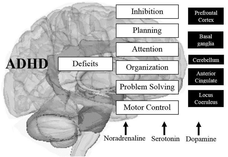
This medical infographic illustrates the neurobiological basis of Attention-Deficit/Hyperactivity Disorder (ADHD) by mapping cognitive deficits to specific neuroanatomical structures and neurotransmitter systems. The central visual is a translucent sagittal-view diagram of the human brain. Labeled 'ADHD' and 'Deficits,' the diagram connects executive function impairments—including inhibition, planning, attention, organization, problem-solving, and motor control—to a highlight of the prefrontal-striatal-cerebellar circuitry. Key anatomical regions identified as involved in the pathophysiology include the Prefrontal Cortex, Basal Ganglia, Cerebellum, Anterior Cingulate, and Locus Coeruleus. At the base of the diagram, the catecholamine and monoamine pathways are represented through labels for Noradrenaline, Serotonin, and Dopamine, indicating their role in modulating these cognitive processes. This educational visualization demonstrates the multi-regional and neurochemical complexity of ADHD, emphasizing how structural and biochemical alterations manifest as clinical deficits in executive functioning. It is designed for clinical neurobiology education and diagnostic conceptualization.
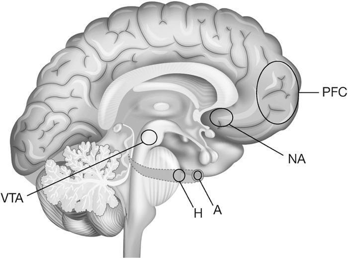
This grayscale anatomical diagram illustrates the mesolimbic dopamine system, often referred to as the reward or addiction circuit, within a human brain. The illustration utilizes a midsagittal view to map key functional regions. Specific structures are highlighted with circles and alphanumeric labels: the Prefrontal Cortex (PFC) at the anterior frontal lobe, the Nucleus Accumbens (NA) in the ventral striatum, the Amygdala (A) and Hippocampus (H) within the medial temporal region, and the Ventral Tegmental Area (VTA) in the midbrain. The diagram emphasizes the spatial relationships and connectivity between these cortical and subcortical structures, which collectively form the final common pathway for the neurobiology of addiction. The cerebellum is visible posteriorly with its characteristic arbor vitae pattern, and the brainstem and corpus callosum provide central anatomical landmarks. This visual is designed for medical education regarding the psychiatric and neurological mechanisms of substance use disorders and reward processing.
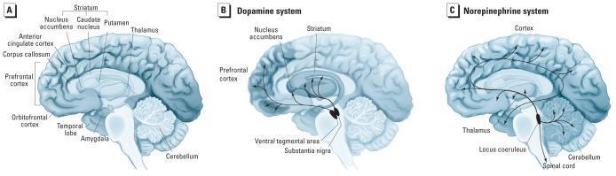
A three-panel anatomical diagram illustrating the neuroanatomy and neurotransmitter pathways implicated in Attention Deficit Hyperactivity Disorder (ADHD) using medial views of a hemisected human brain. Panel A labels key anatomical structures: the prefrontal cortex, orbitofrontal cortex, anterior cingulate cortex, corpus callosum, striatum (including nucleus accumbens, caudate nucleus, and putamen), thalamus, temporal lobe, amygdala, and cerebellum. Panel B details the dopamine system, mapping projections from the ventral tegmental area (VTA) to the prefrontal cortex and nucleus accumbens, and from the substantia nigra to the striatum. Panel C illustrates the norepinephrine system, showing projections originating in the locus coeruleus of the brainstem and extending to the cortex, thalamus, cerebellum, and spinal cord. This set of illustrations serves as a pathophysiological model for understanding how altered volumes and functional connectivity in these catecholaminergic circuits contribute to ADHD symptoms.
ADHD: Full Clinical Summary
1. Definition & Overview
2. Epidemiology
| Population | Prevalence |
|---|---|
| Children & adolescents (worldwide) | 6-9% |
| US children aged 2-17 (NSCH data) | 9.4% |
| Adults (persistence from childhood) | ~50% |
| Sex ratio | Boys diagnosed more frequently and earlier than girls |
- The Harriet Lane Handbook, 23rd ed.; Kaplan & Sadock
3. DSM-5 Diagnostic Criteria
- Symptoms: Inattention and/or hyperactivity-impulsivity that are more frequent and severe than expected for developmental age
- Duration: Symptoms persist for ≥6 months
- Onset: Symptoms present before age 12
- Pervasiveness: Evidence in two or more settings (e.g., home and school)
- Impairment: Clear interference with functioning or development
Subtypes (DSM-5)
- Combined presentation - both inattention and hyperactivity-impulsivity
- Predominantly inattentive presentation
- Predominantly hyperactive-impulsive presentation
Note: ICD-11 supports first-time diagnosis in adults, provided symptoms were present in childhood. - Maudsley Prescribing Guidelines, 15th ed.
4. Pathophysiology & Neurobiology
- Prefrontal cortex (PFC): Governs executive functions (inhibition, planning, attention, organization, problem-solving, motor control)
- Dopamine system: Projections from the ventral tegmental area (VTA) to the PFC and nucleus accumbens are hypoactive
- Norepinephrine system: Locus coeruleus projections to the cortex and thalamus are dysregulated
- Basal ganglia / striatum: Involved in motor control and reward processing; structurally and functionally altered in ADHD
- Anterior cingulate cortex: Impaired error-monitoring and sustained attention
5. Clinical Features
Inattention symptoms (≥6 for children; ≥5 for adults ≥17 years)
- Fails to give close attention to details / makes careless mistakes
- Difficulty sustaining attention in tasks or play
- Does not seem to listen when spoken to directly
- Does not follow through on instructions; fails to finish tasks
- Difficulty organizing tasks and activities
- Avoids tasks requiring sustained mental effort
- Often loses things necessary for tasks
- Easily distracted by extraneous stimuli
- Often forgetful in daily activities
Hyperactivity-Impulsivity symptoms (≥6 for children; ≥5 for adults ≥17 years)
- Fidgets, taps hands/feet, squirms
- Leaves seat when expected to remain seated
- Runs or climbs excessively (in adults: subjective restlessness)
- Unable to play/engage in activities quietly
- "On the go" as if driven by a motor
- Talks excessively
- Blurts out answers before questions completed
- Difficulty awaiting turn
- Interrupts or intrudes on others
6. Evaluation & Diagnosis
- Rating scales: Vanderbilt Diagnostic Rating Scales (parent and teacher versions) are recommended for children with academic/behavioral concerns. Also used to monitor treatment response. - Harriet Lane Handbook
- Adult diagnostic interview: DIVA-5 (Diagnostic Interview for ADHD in Adults, based on DSM-5 criteria) is the recommended validated tool for adults. - Maudsley
- Lab/imaging: Not routinely required if medical history is unremarkable. Neuropsychological testing is not required for diagnosis but is useful if academic/developmental concerns are also present.
- Before starting stimulants: Exclude cardiac symptoms, Wolff-Parkinson-White syndrome, family history of sudden death, hypertrophic cardiomyopathy, and long QT syndrome. Cardiology referral if cardiac history exists. Routine ECG is not required without personal/family cardiac history.
Differential Diagnosis
| Condition | Key Distinguishing Feature |
|---|---|
| Conduct disorder | Deliberate rule violations, aggression |
| Oppositional defiant disorder | Defiance toward authority, anger |
| Anxiety disorder | Worry-driven inattention, not impulsivity |
| Depression | Low mood, anhedonia, cognitive slowing |
| Learning disorder | Domain-specific deficit without core ADHD symptoms |
| Trauma/ACEs | ADHD-like symptoms with clear trauma history |
7. Management
Step 1: Non-Pharmacological (always adjunct; first-line in preschoolers)
- Behavioral therapy: Best combined with pharmacotherapy in school-age children
- Preschoolers (age 4-5): Behavioral therapy alone should be tried first before medications
- Cognitive Behavioral Therapy (CBT): Especially useful in adults; can be used when there is partial medication response or intolerance
- Environmental modifications: Structured routines, classroom accommodations, parent training
Step 2: Pharmacotherapy
First-Line - Stimulants (65-75% response rate)
- Mechanism: Blocks DA and NE reuptake transporters
- Forms: Immediate release (IR) - BID/TID; Extended release (ER) preferred for adherence and coverage
- Adequate trial dose: >0.8 mg/kg/day
- Key formulations: Concerta (IR 22%/ER 78%), Aptensio XR, Cotempla XR-ODT, Azstarys (prodrug)
- Mechanism: Block reuptake AND promote DA/NE release (more potent)
- Adequate trial dose: ≥0.5 mg/kg/day for mixed amphetamine salts
- Key formulations: Adderall XR, Vyvanse (lisdexamfetamine - prodrug, lower abuse potential)
Second-Line - Non-Stimulants
| Drug | Class | Notes |
|---|---|---|
| Atomoxetine | Selective NE reuptake inhibitor (SNRI) | Non-controlled; good for abuse concern; monitor for liver dysfunction and suicidal thinking |
| Guanfacine ER | Alpha-2A adrenergic agonist | RCT evidence as monotherapy; 2023 meta-analysis showed ~60% response (vs 30% placebo); well tolerated in adults |
| Clonidine ER | Alpha-2 adrenergic agonist | Supported by multiple RCTs in children/adolescents |
| Viloxazine | NE reuptake inhibitor | Effective per limited data; less evidence than atomoxetine |
| Bupropion | DA/NE reuptake inhibitor | Off-label; limited comparative evidence |
Atomoxetine is preferred when stimulants are not tolerated, are contraindicated, or two separate 6-week trials fail. - Maudsley Prescribing Guidelines
Common Stimulant Side Effects
- Appetite suppression (most common)
- Abdominal pain / nausea
- Headache
- Sleep disturbance (especially insomnia)
- Palpitations / elevated HR and BP
- Growth deceleration (with long-term use in children)
8. Monitoring
- Weight, blood pressure, heart rate: All ADHD medications
- Liver function + suicidal ideation: Atomoxetine specifically
- Annual medication review: Required - reassess whether medication should be continued
- Cardiovascular surveillance: A 2024 population study found a dose-related increase in cardiovascular disease risk with stimulants (earlier 2022 meta-analysis found no adverse CV effect) - monitor cardiovascular signs and symptoms throughout treatment. - Maudsley, 15th ed.
- "Drug holidays": Consider ad hoc periods of stopping or dose reduction to minimize adverse outcomes and re-evaluate need
9. Special Populations
Children (age 4-5 years)
- Behavioral therapy is first-line; medications only if behavioral therapy fails or is unavailable
- Stimulant titration is weight-based
Adolescents
- Most affected children continue to meet diagnostic criteria through adolescence
- Extended-release formulations preferred to reduce abuse/diversion
Adults
- ADHD is under-recognized in adults; rates rising globally
- DIVA-5 recommended for assessment
- Modified-release (MR) preparations or non-stimulants preferred to reduce diversion risk
- Comorbid bipolar disorder or psychosis: ADHD medications may worsen these; exercise caution
- Evidence for long-term treatment benefit in adults is currently rated "low" or "very low" in quality - short-term trials show symptom improvement but limited quality-of-life data beyond 52 weeks
Pregnancy
- Separate ADHD-in-pregnancy guidelines exist (Maudsley block 5, line 2310); stimulants carry fetal risk and should be carefully reviewed
10. Comorbidities
- Oppositional Defiant Disorder (ODD)
- Conduct Disorder (CD)
- Anxiety disorders
- Depression / mood disorders
- Learning disabilities
- Tic disorders / Tourette syndrome
- Substance use disorders (untreated ADHD is a risk factor)
Key Sources
- Kaplan & Sadock's Comprehensive Textbook of Psychiatry - pharmacotherapy & neurobiology
- The Harriet Lane Handbook, 23rd ed. (Johns Hopkins) - pediatric diagnosis & management
- Maudsley Prescribing Guidelines in Psychiatry, 15th ed. - adult treatment, monitoring, NICE guidance