
Varicose vveins
varicose veins lower limb clinical appearance
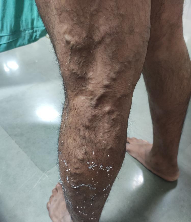
This clinical photograph displays a 50-year-old male's left lower limb with extensive varicose veins. The affected veins are highly tortuous, dilated, and exhibit a prominent, rope-like bulging appearance characteristic of Chronic Venous Insufficiency (CVI). The distribution is primarily located along the anterior and medial aspects of the distal thigh and proximal lower leg. Multiple interconnected, distended venous segments are visible beneath the skin's surface. Additionally, the lower portion of the leg shows secondary skin changes including visible dryness, scaling, and white flakiness, which can be associated with venous stasis dermatitis or the residue of topical treatments. This image serves as a textbook example of CEAP classification grade C2 (varicose veins) and illustrates clinical manifestations often assessed using the Venous Clinical Severity Score (VCSS).
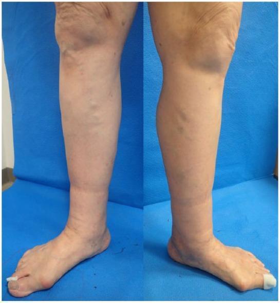
Clinical photograph of the lower extremities of a 79-year-old female presenting with bilateral lower leg edema. The image displays a side-by-side view of the left and right legs from the knee to the foot. Key findings include diffuse non-pitting edema primarily localized below the knees, most prominent in the calf and ankle regions, resulting in an increased limb circumference and a taut, shiny skin appearance. Prominent varicose veins are visible along the medial aspect of both legs, with greater severity noted on the left. The clinical presentation is consistent with CEAP classification grade C3 (venous edema) and secondary lymphedema, indicated by the loss of normal anatomical contours at the ankles. These visual features are characteristic of chronic venous insufficiency and comorbid lymphatic drainage impairment (phlebolymphedema). This material is intended for instruction on vascular assessment, clinical grading of chronic venous disease, and the differential diagnosis of lower extremity swelling.
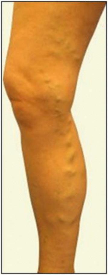
This clinical photograph displays a lateral view of a right lower limb exhibiting varicose syndrome. The image demonstrates significant venous insufficiency characterized by prominent, dilated, and tortuous superficial veins. These enlarged vessels are particularly visible along the posterior and medial aspects of the calf and around the knee joint. The veins appear as irregular, bulging, rope-like elevations beneath the skin surface. The skin over the affected areas shows mild distention due to the underlying venous pressure, but there are no overt signs of chronic venous stasis such as hyperpigmentation, stasis dermatitis, or ulceration in this specific view. The anatomical distribution is consistent with involvement of the saphenous system. This visual represents a classic presentation of severe varicose veins, which clinically correlates with symptoms like heaviness, pain, or edema, and often serves as a preoperative baseline for surgical interventions such as saphenectomy or vein stripping.
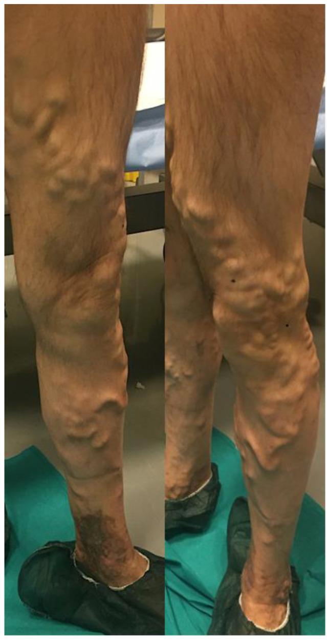
A preoperative clinical photograph showing the posterior and medial views of a lower left limb with severe chronic venous insufficiency. The image demonstrates extensive varicose veins involving the great saphenous vein distribution and its collateral branches. The veins appear markedly enlarged, tortuous, and protuberant, extending from the distal thigh through the popliteal region and down to the mid-calf. In the lower calf and ankle region, there is significant hyperpigmentation and skin discoloration consistent with venous stasis dermatitis or lipodermatosclerosis. The morphology of the veins is characteristic of advanced primary or secondary varicosities, indicating venous hypertension. This visual serves as an educational example of advanced peripheral vascular disease, highlighting the physical examination findings of varicosity, tortuosity, and secondary cutaneous changes associated with chronic venous stasis.
"varicose veins"[MeSH Terms] AND treatment
Varicose Veins
Definition
Epidemiology
- Adult prevalence of visible varicose veins is 30-50% in the general population
- Up to 20% of males and one-third of females develop lower extremity varicose veins
- Prevalence rises steeply with age: from ~11.5% in the 18-24 age group to ~55.7% in the 55-64 age group (Edinburgh Vein Study)
- Higher prevalence in women, likely due to pregnancy-related inferior vena cava compression
- Bailey and Love's Surgery, 28th Ed.
| Factor | Detail |
|---|---|
| Female sex | Hormonal and gravid uterus effects |
| Pregnancy | IVC compression raises venous pressure |
| Obesity (raised BMI) | Increased intra-abdominal pressure |
| Family history | Faulty venous wall development |
| Age | Increasing prevalence with each decade |
| Prolonged standing | Occupational risk |
| Inactivity | Reduced calf muscle pump |
Pathophysiology
- Primary varicose veins: intrinsic abnormalities of the venous wall
- Secondary varicose veins: associated with deep and/or superficial venous insufficiency (e.g., post-DVT)
- Schwartz's Principles of Surgery, 11th Ed.
Clinical Presentation
Symptoms
- Aching, heaviness, throbbing, burning, or bursting over the affected area
- Pruritus over varicose clusters
- Ankle swelling (especially by end of day)
- Symptoms worsen with prolonged standing/sitting and improve with leg elevation or compression hosiery
- Bailey and Love's Surgery
Signs
- Tortuous, dilated subcutaneous veins (rope-like bulging)
- CEAP Classification is used clinically:
| Class | Finding |
|---|---|
| C0 | No visible venous disease |
| C1 | Telangiectasias / reticular veins |
| C2 | Varicose veins |
| C3 | Oedema |
| C4 | Skin changes (pigmentation, eczema, lipodermatosclerosis) |
| C5 | Healed venous ulcer |
| C6 | Active venous ulcer |
Complications
- Stasis dermatitis ("brawny induration") - from haemolysis of extravasated red cells
- Lipodermatosclerosis - chronic inflammation and fibrosis of subcutaneous fat
- Hyperpigmentation - haemosiderin deposition
- Venous ulceration - venous disease accounts for ~85% of chronic lower limb ulcers
- Superficial vein thrombosis (thrombophlebitis) - painful, cord-like induration
- Bleeding - from attenuated vein clusters (even spontaneous rupture)
Note: Embolism from superficial varicose veins is very rare, unlike DVT. - Robbins
- Oesophageal varices - portal hypertension (liver cirrhosis)
- Haemorrhoids - anorectal venous plexus
- Caput medusae - periumbilical veins (portal hypertension)
Investigations
- Duplex Ultrasound - gold standard; maps reflux, identifies GSV/SSV incompetence, perforator incompetence; essential before treatment planning
- Trendelenburg test (clinical) - tourniquet test to assess saphenofemoral junction incompetence
- Perthe's test - assesses deep vein patency before intervention
Management
1. Conservative
- Compression hosiery: 20-30, 30-40, or 40-50 mmHg graduated stockings; knee-high to waist-high depending on distribution
- Leg elevation, weight loss, exercise
- Provides symptom relief; recommended as first-line in many guidelines
2. Sclerotherapy
- Destroys venous endothelium via chemical injection
- Agents: hypertonic saline, sodium tetradecyl sulfate (STS), polidocanol
- Suitable for telangiectasias (low concentrations) and small-to-medium varicosities (higher concentrations)
- Foam sclerotherapy: increased contact area; effective for larger veins - RCT evidence shows significant symptom relief vs placebo
- Post-injection: elastic bandage x 3-5 days, then stockings x 2+ weeks
- Complications: hyperpigmentation, allergic reaction, thrombophlebitis, DVT, skin necrosis
3. Endovenous Thermal Ablation (preferred modern approach)
- Endovenous Laser Ablation (EVLA) and Radiofrequency Ablation (RFA)
- Under ultrasound guidance, a catheter is introduced into the GSV/SSV; tumescent anaesthesia is injected around the vein; thermal energy ablates the vein as the catheter is withdrawn
- Equally effective but faster recovery than surgical stripping
- Complications: DVT, ecchymosis, saphenous nerve injury
A 2024 meta-analysis (Jiang et al., 2024) comparing RFA vs EVLA found both equally effective for lower extremity varicose veins. A systematic review on recurrent varicose veins at the saphenofemoral junction (Bontinis et al., 2024) also supports endovenous techniques for recurrence.
4. Non-thermal, Non-sclerosant Ablation
- Cyanoacrylate glue (VenaSeal): proprietary adhesive injected to seal the vein; no tumescent anaesthesia required; promising early results
- Mechanochemical ablation (MOCA): combines mechanical endoluminal injury with sclerosant
5. Surgical Treatment
- GSV ligation and stripping: still preferred for very large GSVs (>2 cm diameter)
- Incisions at groin and below knee; vein removed via stripper
- Lower recurrence rate vs ligation alone
- Complications: ecchymosis, haematoma, lymphocele, DVT, infection, saphenous nerve injury
- Stab avulsion (phlebectomy): 2-mm incisions directly over branch varicosities; vein avulsed with no ligation; bleeding controlled by elevation and tumescent anaesthesia
Clinical Images
Key Takeaways
- Varicose veins affect 30-50% of adults; women > men
- Caused by venous valve incompetence leading to reflux and hypertension in superficial veins
- GSV system involved in 60% of cases
- Duplex ultrasound is essential for diagnosis and pre-treatment planning
- Modern treatment favours endovenous ablation (EVLA/RFA) over open surgery - similar efficacy, quicker recovery
- Venous disease causes ~85% of all chronic leg ulcers
Bisgard regime