
Role of embolization orthopaedics recent advances paper4 theory knruhs 10 marks
embolization orthopedic surgery advances
role of embolization in orthopaedics recent advances KNRUHS paper 4 theory 10 marks
therapeutic embolization bone tumors musculoskeletal
embolization in orthopaedics indications preoperative bone tumors aneurysmal bone cyst pelvic fracture recent advances
PMID: 39727678
geniculate artery embolization knee osteoarthritis
Role of Embolization in Orthopaedics - Recent Advances
Introduction
Mechanism
- Permanent - using coils, polyvinyl alcohol (PVA) particles, Onyx (ethylene-vinyl alcohol copolymer), or glue (n-BCA)
- Temporary - using Gelfoam (gelatin sponge), which recanalize over weeks
Indications in Orthopaedics
1. Benign Bone Tumors
- Serial arterial embolization is the preferred treatment for sacral GCT where en-bloc excision risks neurological injury
- Embolization can be primary (standalone), preoperative to reduce vascularity, or repeated for local control
- Lackman et al. showed successful control of sacral GCTs by serial embolization (JBJS 2002)
- Also used for GCT in the spine (vertebral body) and inaccessible pelvis
- Campbell's (2026): "Selective arterial embolization is recommended in the treatment of giant cell tumor, aneurysmal bone cyst of spine, and aggressive hemangiomas."
- Embolization reduces vascularity and promotes healing, especially in surgically challenging locations (spine, pelvis, sacrum)
- Can be used as primary treatment or preoperative adjunct to reduce intraoperative bleeding
- Repeat sessions (3-4) may be required for complete regression
- Recent evidence (2023, ScienceDirect) confirms selective embolization as effective primary or adjuvant treatment for ABC
- Embolization reduces pain, prevents pathological fracture, and can be combined with vertebroplasty or surgery
- Must carefully identify the artery of Adamkiewicz (usually at T10-T12 on the left side) - inadvertent embolization causes anterior spinal artery syndrome and paraplegia
2. Malignant Bone Tumors and Metastases
- Reduces intraoperative blood loss in hypervascular tumors before surgical resection
- Most useful for: renal cell carcinoma (RCC) metastases, thyroid metastases, hepatocellular carcinoma metastases (all highly vascular)
- RCC bone metastases in the femur, acetabulum, and spine are the classic indication
- Timing: surgery is optimally performed within 24-72 hours after embolization before collateral re-vascularization occurs
- Acuna et al. (2024, J Surg Oncol): retrospective study confirmed timing between embolization and surgery impacts blood loss in hypervascular bone metastases
- Used for painful bone metastases that are not resectable
- Provides pain relief by tumor devascularization and reduction in size
- Protocol-level RCT ongoing (Zenda et al., BMC Cancer 2023)
- Osteosarcoma and chondrosarcoma: embolization used adjunctively, not as standalone treatment
- Reduces surgical difficulty, especially in pelvic tumors
3. Pelvic Fractures with Hemorrhage
- Life-saving indication - pelvic ring disruptions (type C/Young-Burgess pattern) can cause fatal hemorrhage from the internal iliac artery branches (superior gluteal, obturator, pudendal arteries)
- Protocol: ATLS resuscitation -> pelvic binder/external fixator to reduce pelvic volume -> if hemodynamically unstable despite resuscitation: angiography + embolization (or pelvic packing)
- Time-sensitive: delay >60 minutes from presentation to embolization increases mortality
- Packing vs. embolization debate: Suzuki et al. - these are "complementary, not competitive" approaches (Injury 2009), cited in Rockwood & Green (2025)
- CT-angiography is now routinely used to identify "blush" (active extravasation) before proceeding to embolization
4. Spine Tumors (Vertebral)
- Benign sacral/vertebral tumors (GCT, ABC, hemangioma) treated by serial embolization to avoid major surgery
- Malignant vertebral metastases: preoperative embolization before decompression and stabilization surgery
- Fischer's Mastery of Surgery: "Aneurysmal bone cysts may be treated with arterial embolization, avoiding the morbidity of surgical excision. Benign sacral tumors involving S3 and above may be treated with serial embolization."
5. Post-Traumatic Hemorrhage (Extremity)
- Pseudoaneurysms following orthopaedic trauma or surgery can be treated by coil or glue embolization
- Avoids repeat open surgery in already compromised tissues
Recent Advances (2020-2026)
a) Genicular Artery Embolization (GAE) for Knee Osteoarthritis
- The most exciting recent advance in musculoskeletal embolization
- The synovium of OA knee is hypervascular; embolizing the genicular arteries reduces synovial hyperemia and inflammation, decreasing pain
- Embolic agents: imipenem/cilastatin microspheres (Embosphere), calibrated PVA particles
- Meta-analysis by Epelboym et al. (Cardiovasc Intervent Radiol 2023, PMID 36991094): systematic review confirmed significant pain reduction in knee OA, with durable effects up to 12 months
- Papalexis et al. (Curr Oncol 2024, PMID 39727678): "Its use has expanded globally in treating chronic pain syndromes and osteoarthritis... it offers symptom relief, reduces tumor size, and improves quality of life"
- Also being trialed for shoulder (adhesive capsulitis / frozen shoulder) and hip OA
b) Shoulder Embolization for Adhesive Capsulitis
- Systematic review (Orthopedics Journal 2023): arterial embolization targeting the shoulder synovial vessels shows promising early results for pain and range of motion in frozen shoulder
c) Improved Embolic Agents
- Drug-eluting beads (DEB): loaded with doxorubicin or irinotecan - deliver chemotherapy locally while causing mechanical ischemia (TACE - transarterial chemoembolization concept applied to bone mets)
- Liquid embolics (Onyx/EVOH): better control, can fill complex vascular spaces, used for AVM and GCT
- Calibrated microspheres: allow precise vessel sizing, reduce non-target embolization risk
d) Cone-Beam CT + 3D Roadmap Navigation
- Intraoperative cone-beam CT with 3D roadmap allows real-time, high-resolution guidance during embolization
- Reduces contrast use, improves precision, and reduces complications
- Enables superselective embolization avoiding collateral damage
e) Robotic and AI-Assisted Angiography
- AI-based algorithms for pre-procedure planning, real-time target identification, and radiation dose optimization
- Emerging, still experimental in 2025-26
f) Osteoid Osteoma
- Though primarily treated by radiofrequency ablation (RFA), embolization is a backup in select cases
Embolic Agents: Classification
| Category | Examples | Duration |
|---|---|---|
| Mechanical | Coils, detachable balloons | Permanent |
| Particulate | PVA particles, microspheres, Gelfoam | Permanent / Temp |
| Liquid | Onyx, n-BCA glue, ethanol | Permanent |
| Drug-eluting | DEB-TACE beads | Permanent + Chemo |
Complications
- Post-embolization syndrome - fever, pain, elevated WBC (expected, managed with NSAIDs)
- Non-target embolization - inadvertent occlusion of normal vessels
- Anterior spinal artery syndrome - devastating complication from spinal embolization if artery of Adamkiewicz is involved - paraplegia
- Infection/abscess of embolized tissue
- Contrast nephropathy - especially in elderly or pre-existing renal disease
- Recanalization and recurrence - particularly with Gelfoam; requires repeat sessions
- Skin necrosis - from non-target peripheral embolization
Key Points for Exam
- Embolization = therapeutic vascular occlusion via catheter; not to be confused with pathological embolism
- Classic orthopaedic indications: sacral GCT, ABC of spine, pelvic fracture hemorrhage, hypervascular bone metastases (especially RCC)
- Most exciting recent advance: genicular artery embolization for knee OA pain (strong systematic review evidence, 2023)
- Artery of Adamkiewicz (T10-T12, left side) must be identified before spinal embolization
- Surgery after preoperative embolization should occur within 24-72 hours
- Packing and embolization are complementary, not competing, for pelvic fracture hemorrhage
- Campbell's Operative Orthopaedics, 15th Ed 2026
- Rockwood & Green's Fractures in Adults, 10th Ed 2025
- Papalexis N et al. "How Arterial Embolization Is Transforming Treatment of Oncologic and Degenerative Musculoskeletal Disease." Curr Oncol 2024 [PMID 39727678]
- Epelboym Y et al. "Genicular Artery Embolization for Osteoarthritis Related Knee Pain: Systematic Review and Meta-analysis." Cardiovasc Intervent Radiol 2023 [PMID 36991094]
- Acuna AJ et al. "Effect of timing between preoperative embolization and surgery in hypervascular bone metastases." J Surg Oncol 2024 [PMID 37781952]
Reamer irrigation aspirator 10 marks
reamer irrigator aspirator RIA bone graft
reamer irrigator aspirator RIA system orthopaedics 10 marks KNRUHS theory exam
PMID: 36114869
PMID: 34851462
https://pmc.ncbi.nlm.nih.gov/articles/PMC3167404
Reamer-Irrigator-Aspirator (RIA)
Definition and Concept
Device Components
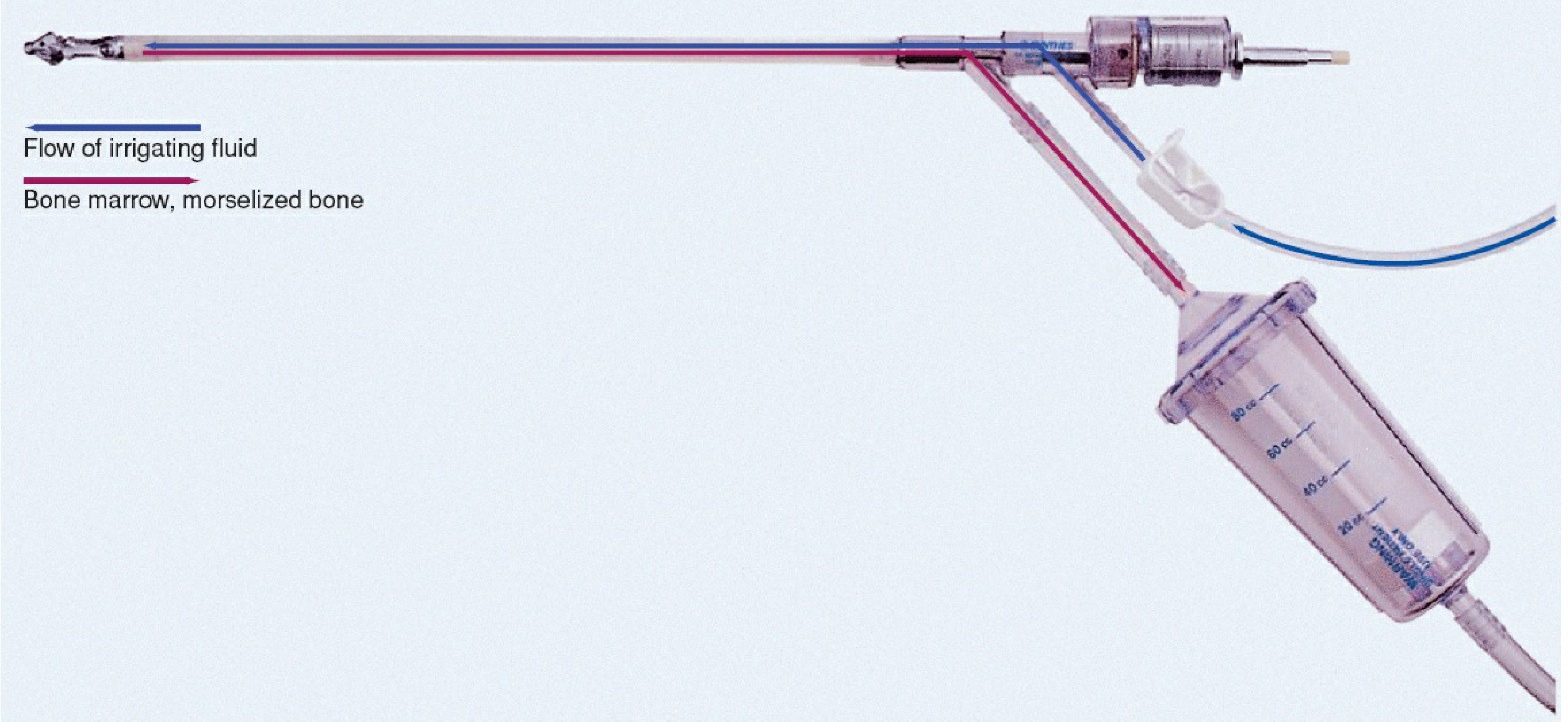
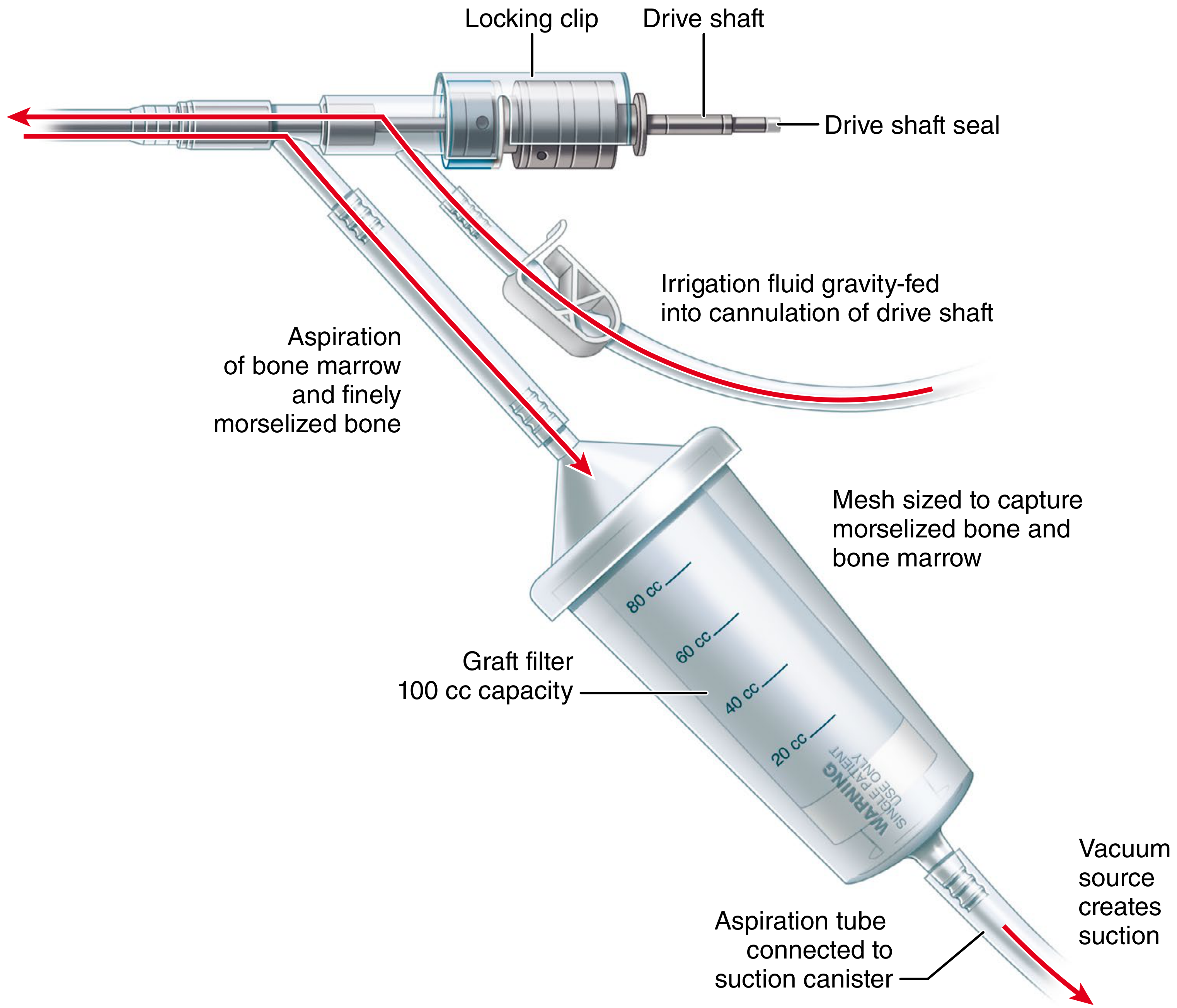
- Powered reamer head - cutter heads ranging 10-18 mm in 0.5 mm increments (RIA-2)
- Drive shaft with locking clip and drive shaft seal
- Inner irrigation tube - delivers saline to the reamer tip (gravity-fed)
- Outer aspiration tube - evacuates bone marrow, morselized bone, fat, and debris under vacuum
- Graft filter canister - 100 cc capacity, mesh-lined to capture bone and marrow while allowing fluid to pass through
- Vacuum source - creates suction via aspiration tube connected to suction canister
Mechanism of Action
- Fat embolism - elevated intramedullary pressure forces fat globules and debris into emissary veins and the venous system
- Thermal necrosis - heat generated by reaming (without cooling) causes cortical bone necrosis
- Reams the canal with a rotating fluted cutter
- Irrigates with saline (reducing heat and intramedullary pressure)
- Aspirates the mixture of saline, fat, bone marrow, and morselized bone - reducing canal pressure, preventing embolism, and collecting usable graft
Indications
1. Autogenous Bone Graft Harvesting (Primary Use)
- Fracture nonunion - segmental defects averaging 5-8 cm
- Critical-size bone defects after debridement of osteomyelitis
- Spinal fusion (revision cases with pseudarthrosis)
- Ankle and hindfoot arthrodesis
- Tibial plateau and distal femur reconstructions
- Mesenchymal stem cells (MSCs) with pluripotent osteogenic potential
- Osteoinductive growth factors (BMP-2, BMP-7, TGF-beta, IGF-1, VEGF) - in concentrations superior to or comparable to ICBG
- Viable osteoblast precursors
- "Reamings contain pluripotent stem cells with the possibility of dedifferentiation into osteoblasts." - Rockwood & Green (2025)
2. Prevention of Fat Embolism and Thermal Necrosis
- Used instead of conventional reaming before intramedullary nailing in polytrauma patients, chest trauma, and those at risk of fat embolism syndrome
- Continuous irrigation lowers intramedullary temperature and pressure during reaming
3. Treatment of Intramedullary Osteomyelitis
- In chronic osteomyelitis of the femur or tibia, RIA is used to debride the infected medullary canal
- Followed by antibiotic cement rod (antibiotic-loaded PMMA intramedullary rod) placement
- Campbell's (2026) references: "Treatment of intramedullary osteomyelitis of the femur and tibia using the Reamer-Irrigator-Aspirator system and antibiotic cement rods"
- Allows thorough mechanical debridement without open cortical windowing
4. Canal Preparation for Nailing
- Canal enlargement before insertion of a large-diameter nail or prosthesis
- Reduces the "push" effect that forces fat emboli during conventional reaming
Surgical Technique (Campbell's Technique 58.3)
- Confirm reaming diameter by measuring the diaphyseal isthmus on radiograph
- Select reamer head no larger than 1.5 mm more than the measured isthmus diameter (prevents cortical perforation)
- Blood should be available - anticipate potential significant blood loss
- Position as for standard IM nailing (supine or lateral for femur, supine for tibia)
- Gain access to canal as for IM nailing
- Insert guidewire (reaming wire) to the physeal scar; confirm on AP and lateral fluoroscopy
- Assemble the RIA: attach drive shaft, locking clip, drive shaft seal, drive unit
- Connect irrigation tube (smaller port) and aspiration tube with graft filter (larger port)
- Start irrigation and aspiration before insertion to confirm functioning
- Insert RIA over guidewire; confirm position with image intensification
- Ream 20-30 mm, then retract 50-80 mm to allow irrigation fluid to fill the space
- Repeat slow advancement until resistance felt; never ream without irrigation
- Reamer can be reversed if reaming becomes difficult
- After removal, stop irrigation and suction; compress graft with plunger; record volume
- Fluoroscopic check of donor bone for perforation or iatrogenic fracture before closing
- Cadaver studies show RIA does not significantly reduce femoral mechanical properties
- Weight bearing to tolerance is generally permitted in the donor limb
RIA vs. Iliac Crest Bone Graft (ICBG) - Comparison
| Parameter | RIA | ICBG |
|---|---|---|
| Volume of graft | 38-48 cc (larger) | 20-30 cc |
| Donor site pain | Less (P < 0.004) | More (chronic in 10-30%) |
| Infection rate | Lower | Higher |
| Adverse events | Lower overall | Up to 30% reported |
| Bone union rate | Comparable | Slightly higher in some studies |
| Additional incision | No | Yes |
| Graft quality (growth factors) | Equal or superior | Gold standard |
| Harvest site morbidity | Intramedullary | Iliac crest (nerve, hernia, fracture) |
Complications
- Overall complication rate: 1.7% (95% CI 0.4-3.6%)
- Most common: cortical perforation (34/1834 cases)
| Complication | Prevention |
|---|---|
| Cortical perforation - most common | Use reamer head ≤1.5 mm over isthmus; avoid thin-cortex bones; preoperative radiograph evaluation |
| Donor bone fracture (intra/postoperative) | Protected weight bearing; avoid osteoporotic bone; correct reamer sizing |
| Significant blood loss (mean Hb drop 3.74 g/dL; transfusion in 9.72%) | Turn off aspirator when not reaming; check Hb post-op; have blood available |
| Fat embolism (rare - system designed to prevent it) | Ensure irrigation running before reaming; single-pass technique |
| Femoral neck fracture (rare) | Correct entry point; avoid proximal perforation |
| Hypertrophic scar | Standard wound care |
| RIA head breakage (RIA-2, rare) | Novel retrieval: laparoscopic suction device (JOCR 2025) |
Recent Advances (RIA-2 System)
- Exchangeable cutter heads (10-18 mm, in 0.5 mm increments) - precise sizing for any canal
- Improved irrigation flow control for better intramedullary pressure management
- Reduced heat generation
- Better ergonomic drive unit
- Intended for adults and adolescents
- RIA aspirate combined with low-intensity pulsed ultrasound (LIPUS) shown to enhance osteogenic differentiation of RIA-derived cells in vitro (Sawauchi et al., 2022, PMID 34785092)
- RIA graft + BMP-2 scaffold combinations for large segmental defects
- RIA as a source of MSCs for tissue engineering - pluripotent cells can be expanded and differentiated for orthobiologic strategies
- Spinal pseudarthrosis revision surgery (Elhessy et al., Cureus 2022)
- Finite element analysis (2026) confirms minimal femoral strength reduction after RIA harvest
Clinical Outcomes
- Nonunion: 70% union at 6 months, 90% at 12 months in segmental defects averaging 5.8 cm (Stafford & Norris)
- Large defects (avg. 6.6 cm): 17/21 healed at 11 months (McCall et al.)
- Osteomyelitis: Favorable results when combined with antibiotic cement rods
- Cost: Less expensive than demineralized bone matrix (DBM) allograft and BMP per 10 cc
Key Points for Exam
- RIA = reams + irrigates + aspirates simultaneously in a single pass
- Original purpose: prevent fat embolism and thermal necrosis during IM nailing
- Now primarily used for: autogenous bone graft harvesting from the femur/tibia
- Yield: 38-48 cc per femoral harvest; superior growth factor content vs. ICBG
- Reamer head size: no more than 1.5 mm over the measured isthmus diameter
- Most common complication: cortical perforation (1.7% overall complication rate - Laubach meta-analysis 2023)
- Significant blood loss (mean Hb drop 3.74 g/dL) must be anticipated
- Advantages over ICBG: less donor site pain, fewer infections, no additional incision
- RIA-2 = current generation; exchangeable cutter heads 10-18 mm
- Contraindications: thin cortices (osteoporosis), children (open physes), bony deformity of canal, very narrow isthmus
- Campbell's Operative Orthopaedics, 15th Ed 2026 (Technique 58.3, Table 58.5)
- Rockwood & Green's Fractures in Adults, 10th Ed 2025
- Laubach M et al. "Complications associated using the RIA system: systematic review and meta-analysis." Arch Orthop Trauma Surg 2023 [PMID 36114869]
- Oliva F et al. "RIA versus traditional ICBG harvesting: systematic review and meta-analysis." J Orthop Traumatol 2021 [PMID 34851462]
- AO Foundation RIA-2 System Technical Documentation, 2020
Meniscal transplantation , meniscal repair techniques, recent advances
meniscal transplantation repair recent advances
meniscus allograft transplantation outcomes
meniscus repair all-inside inside-out outside-in techniques outcomes
PMID: 41549414
PMID: 38880182
PMID: 38742839
Meniscal Repair Techniques, Meniscal Transplantation & Recent Advances
PART 1: MENISCAL REPAIR
Anatomy and Vascular Zones (Basis for Repairability)
| Zone | Description | Vascularity | Healing Potential |
|---|---|---|---|
| Red-Red zone | Outer 1/3 (peripheral) | Well vascularized | Excellent |
| Red-White zone | Middle 1/3 | Moderately vascular | Good |
| White-White zone | Inner 1/3 (avascular) | Avascular | Poor - repair not indicated |
Indications for Meniscal Repair
- Tear length 1-4 cm
- Vertical (longitudinal) tear configuration
- Red-red or red-white zone tear
- Patient age < 40 years
- Meniscal root tear (now considered a special indication)
- Concomitant ACL reconstruction extends indications significantly
- Augmentation techniques (fibrin clot, PRP) may extend indications further - Miller's Review of Orthopaedics, 9th Ed
- White-white zone (avascular) tears
- Horizontal cleavage / degenerative tears
- Very small tears (< 1 cm)
- Radial tears (relative; midsubstance radial tears now being repaired)
- Advanced knee OA
Four Techniques of Meniscal Repair
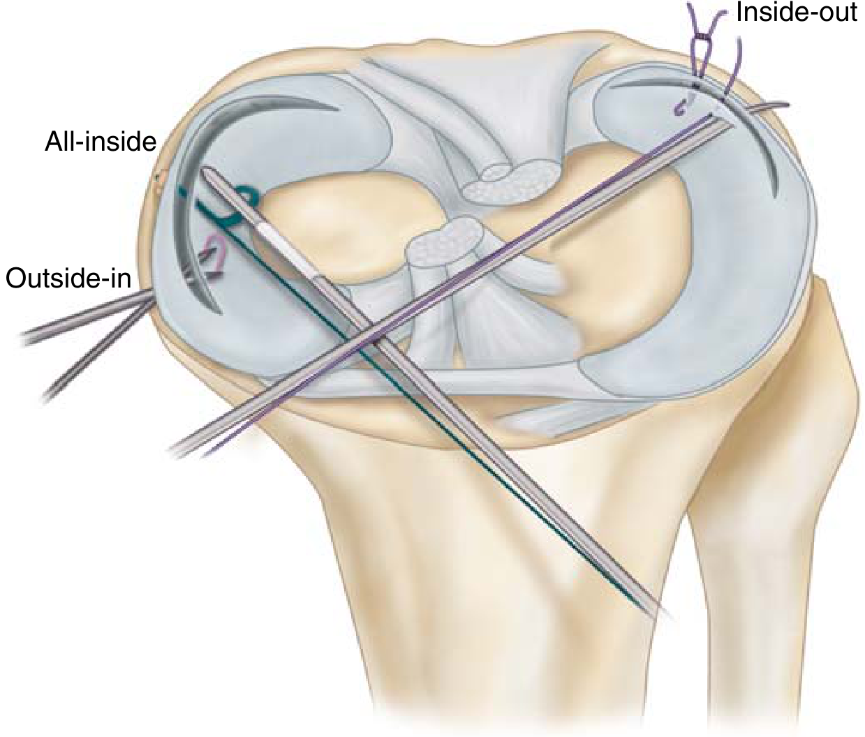
1. Open Repair (Historical)
- Arthrotomy-based; the original technique
- Now rarely used except in combined open procedures (e.g., combined with HTO)
- Allows direct visualization and suture placement
- Sutures: 0-PDS or 2-0 non-absorbable vertical mattress sutures placed through posteromedial or posterolateral capsule
2. Inside-Out Technique (Gold Standard)
- Gold standard for meniscal repair, particularly for posterior horn and body tears
- Long flexible cannulated needles passed from inside the joint, through the tear, and retrieved through a small posteromedial or posterolateral incision
- Suture configuration: vertical mattress (preferred) > horizontal mattress
- Sutures tied over the capsule extraarticularly
- Arthroscopic setup; posteromedial or posterolateral mini-open incision made
- Retractor (popliteal retractor) placed to protect neurovascular structures
- Zone-specific cannula introduced (posterior horn cannula)
- Long flexible needles (single or double barrel) passed from inside-out through tear
- Sutures retrieved and tied over capsule
- Vertical mattress sutures placed 3-4 mm apart
- Medial repairs: protect saphenous nerve (anterior to semitendinosus/gracilis, posterior to inferior sartorius border)
- Lateral repairs: protect common peroneal nerve (posterior to biceps femoris)
3. Outside-In Technique
- Especially useful for anterior horn and body tears; anterior root repairs
- Spinal needles passed from outside through skin, capsule, into joint to exit through the tear
- A suture or wire loop is passed through the needle, retrieved arthroscopically, brought out through an anterior portal, and tied
- Avoids risk to posterior neurovascular structures
- 2026 Review (Pace & Garra, Curr Rev Musculoskelet Med): outside-in remains preferred for anterior horn pathology including meniscal root repairs
4. All-Inside Technique (Most Popular Currently)
- Fully arthroscopic; no accessory incisions needed
- Uses proprietary implant-based devices (FasT-Fix, MaxFire, CrossFix, etc.)
- A flexible shaft deploys a pre-loaded suture anchor/cinch/locking mechanism across both leaves of the tear
- Latest generation devices allow tensioning of the construct (adjustable loop - similar to cortical button concept)
- Best for posterior horn tears, radial tears, and root repairs
- Advantage: no mini-incision, faster, less risk to neurovascular structures
- Disadvantage: device cost; learning curve; risk of chondral injury from implant
Repair Outcomes
- Overall success: 80-90% in published series
- 90% success when repair performed with concurrent ACL reconstruction
- 60% success in repair with intact ACL
- 30% success in repair with ACL-deficient knee
- Results best in: acute peripheral tears, young patients, concurrent ACL reconstruction
Augmentation Techniques
- Fibrin clot - autologous blood clot prepared from the patient's own venous blood; placed at repair site to deliver growth factors and scaffold
- PRP (Platelet-Rich Plasma) clot - concentrated platelets with growth factors (TGF-beta, PDGF, IGF-1); injected at repair site
- Vascular access channels (trephination) - small channels drilled from vascular peripheral zone into avascular tear to allow ingrowth of vessels
- Synovial rasping - mechanical abrasion of the synovial tissue adjacent to the tear to stimulate bleeding and growth factor release
- Bone marrow aspirate concentrate (BMAC) - recent advance; MSCs delivered to repair site
Postoperative Rehabilitation
- Avoid knee flexion beyond 90 degrees for 6 weeks
- Weight bearing: controversial; typically partial to full weight bearing in extension with brace
- Return to sport: 4-6 months
- Posterior horn repairs: more cautious ROM restriction
PART 2: MENISCAL ALLOGRAFT TRANSPLANTATION (MAT)
Concept and Background
Indications and Contraindications
- Age < 40 years (up to 50 if highly active with minimal OA)
- Skeletally mature
- Meniscal deficiency (absent or nonfunctioning) with pain in affected compartment
- Failure of conservative management
- Cause of meniscal damage must be mechanical, not degenerative or synovial
- Normal mechanical alignment
- Stable knee (or stabilizable)
- Outerbridge grade I or II articular cartilage changes only
- Knee instability (relative - can address concurrent ACL)
- Varus/valgus malalignment >2-4° (relative - can correct with osteotomy)
- Advanced osteoarthritis (Outerbridge III-IV)
- Inflammatory or synovial disease (will destroy the graft)
- Obesity
- Systemic infection
Graft Procurement and Processing
- Fresh (least common now): viable cells preserved but disease transmission risk, logistics difficulty
- Fresh-frozen (most common): cells lost but structural collagen maintained; low immunogenicity
- Cryopreserved: 10-40% viable cells preserved; however necessity of preserving donor cells questioned (allograft repopulated by host cells)
- Freeze-dried + gamma irradiation: risk of graft shrinkage; irradiation >2.5 Mrad compromises collagen mechanical properties; secondary sterilization with gamma irradiation not currently recommended
- Cell viability - cryopreservation preserves 10-40% cells; necessity questioned
- Immunogenicity - deep freezing reduces it; cryopreservation maintains HLA antigens (more immunogenic)
- Immunologic host response - occurs but clinical significance unknown
- Disease transmission - freeze-drying + irradiation eliminate viral risk but risk graft shrinkage
Sizing
- Radiographic sizing: tibial plateau AP and ML dimensions on weight-bearing radiographs
- Tibial plateau width and length measured; graft matched within 5 mm
- MRI sizing also used (anterior-posterior and medial-lateral tibial plateau)
- Undersizing leads to instability; oversizing leads to extrusion
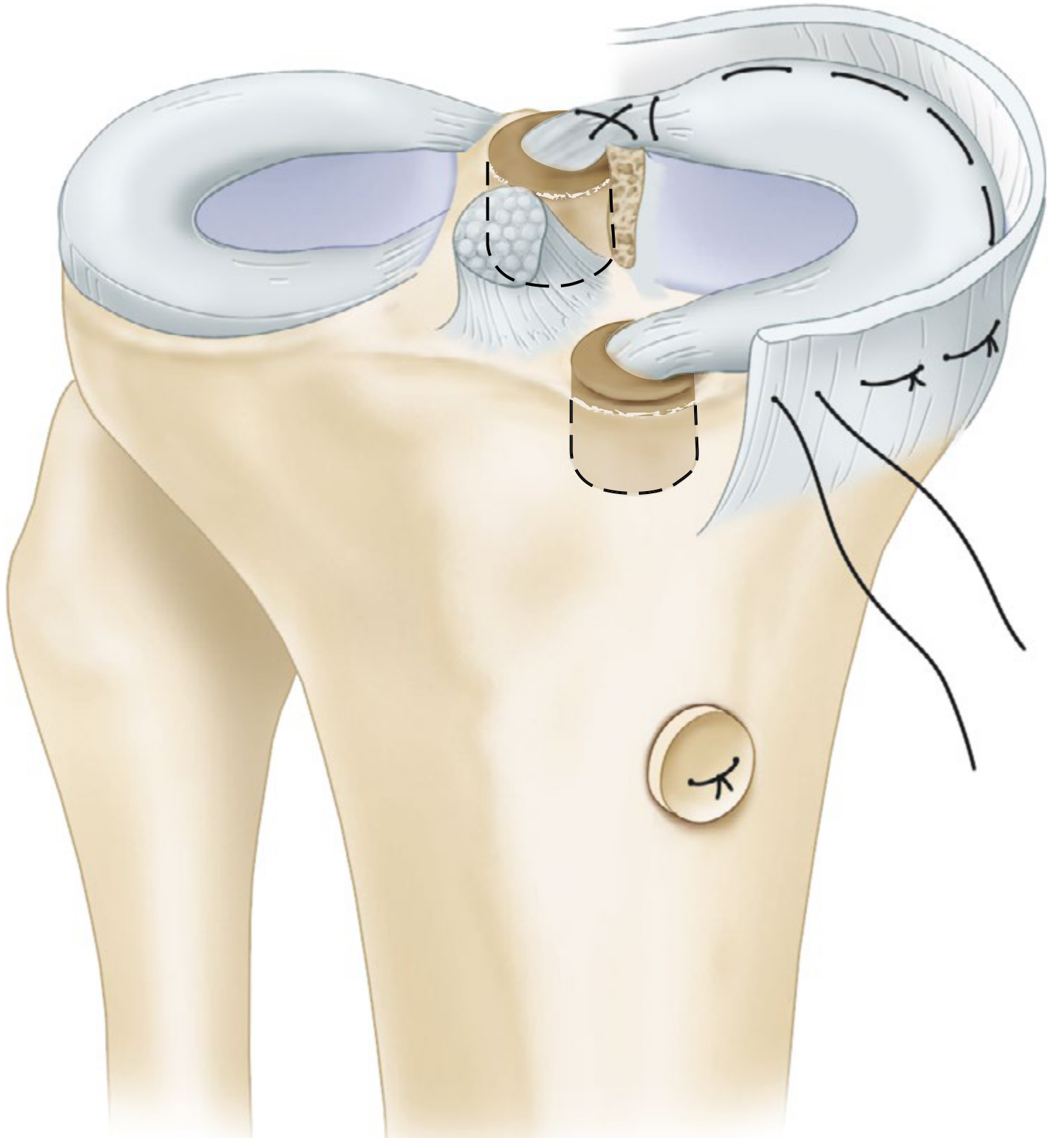
Fixation Techniques
| Technique | Description | Best for |
|---|---|---|
| Bone plug | Separate anterior and posterior bone plugs secured in individual bone tunnels | Lateral MAT (well-separated horn insertions) |
| Bone bridge / bone trough | Bone bridge connecting anterior and posterior horns slid into a trough cut in the tibial plateau | Medial MAT; ensures precise anatomic spacing |
| Soft tissue fixation | Suture-only fixation of horns without bone; used when bone preservation needed | Revision cases |
Outcomes of MAT
- 13 studies, 619 patients, 632 MATs, mean follow-up 11.1-20 years
- Significant improvements in Lysholm, Tegner, VAS, KOOS
- 10-year MAT survival: 45-100% (most studies ≥73%)
- 15-year survival: 19-87% (highly variable)
- Patient Acceptable Symptom State (PASS): 70-71% for Lysholm, 58-82% for KOOS
PART 3: RECENT ADVANCES
1. Meniscal Scaffold Implantation (MSI)
- CMI (Collagen Meniscus Implant / Menaflex) - collagen-based scaffold from bovine Achilles tendon; for partial medial meniscus deficiency
- Actifit (polyurethane scaffold) - synthetic, porous scaffold; for lateral or medial partial defects; allows tissue ingrowth
- Both MAT and MSI showed significant improvement in clinical scores
- MSI had higher 10-year survival and lower complication rate than MAT
- MAT showed greater pain reduction (VAS improvement)
- MSI: higher IKDC, KOOS improvements
2. Meniscal Root Repair
- Posterior meniscal root tears (equivalent biomechanically to total meniscectomy) are now repaired rather than ignored
- Transtibial pullout repair technique - sutures through root passed through tibial tunnel and tied at cortex
- All-inside root repair with knotless anchors gaining popularity
- Evidence shows root repair prevents rapid OA progression vs meniscectomy
3. Radial Tear Repair
- Previously considered unrepairable; now repaired with specialized techniques
- "Cinch stitch" / horizontal mattress technique
- All-inside devices with ramp configurations
- Evidence supports repair over meniscectomy for radial tears in younger patients
4. Ramp Lesion Repair
- Posterior horn medial meniscus capsular attachment tears (ramp lesions) - common with ACL tears (missed in 9-17% of ACL cases)
- Repaired with all-inside technique using retrograde curved cannulas through intercondylar notch
5. Biologic Augmentation
- PRP injection at repair site - now routinely used; delivers PDGF, TGF-beta, VEGF
- BMAC (Bone Marrow Aspirate Concentrate) - MSCs applied to repair site
- Growth factor-impregnated scaffolds - collagen scaffolds loaded with BMP-7, TGF-beta
- In vitro evidence: low-intensity pulsed ultrasound (LIPUS) enhances meniscal cell osteogenic potential
6. 3D-Printed Personalized Meniscal Scaffolds
- Computed 3D models from patient MRI used to design patient-specific polyurethane or hydrogel scaffolds
- Seeded with autologous chondrocytes or MSCs before implantation
- Experimental phase; promising early results in animal and first-in-human studies
7. Gene Therapy for Meniscal Healing
- Viral vector-mediated delivery of HGF (hepatocyte growth factor), TGF-beta1, or BMP-7 to avascular white-white zone tears
- Aim: convert avascular zone into healing environment
- Preclinical studies only; not yet clinical
8. Adjustable Loop All-Inside Devices
- Latest generation all-inside devices (FiberStitch, MaxFire Meniscal Repair System) with adjustable cinching loops similar to suture button technology
- Allow intraoperative tensioning after placement
- Campbell's (2026): "The latest generation of all-inside devices allows tensioning of the construct"
9. Synthetic Meniscus (NUsurface, Neovasc)
- NUsurface (Active Implants): polycarbonate-urethane total meniscus replacement; does not require fixation (free-floating)
- Approved in Europe; FDA investigational device status in USA
- For patients too old for MAT and not yet needing TKA - the "bridge" population
10. MAT with Concurrent Procedures
- MAT + HTO (High Tibial Osteotomy): for varus knee with medial meniscal deficiency; osteotomy offloads the reconstructed compartment
- MAT + cartilage restoration (OATS, ACI): combined procedures for the young arthritic knee
- MAT + ACL reconstruction: well-established; systematic review (2023) confirms good outcomes
- Note: combined with OCA or ACL reconstruction shows poorer results in some studies (Campbell's)
Summary Table: Repair Techniques
| Technique | Access | Best Zone | Best Tear Location | Gold Standard? |
|---|---|---|---|---|
| Open | Arthrotomy | Any | Any | Historical |
| Inside-Out | Mini-open posteromedial/lateral | Red-red, Red-white | Posterior horn, body | YES |
| Outside-In | Mini-open anterior | Red-red | Anterior horn, anterior root | For anterior tears |
| All-Inside | Fully arthroscopic | Red-red, Red-white | Posterior horn, body | Most popular now |
Key Points for Exam
- Gold standard repair: Inside-out with vertical mattress sutures
- Vertical > horizontal mattress sutures in biomechanical testing (Desai 2025)
- Repair zone: Red-red and red-white; never white-white
- Best repair outcomes: Young patient + peripheral tear + concurrent ACL reconstruction = 90% success
- MAT candidate: Age <40, mechanical deficiency, Outerbridge ≤II, aligned, stable knee
- MAT fixation: bone bridge (medial) vs bone plugs (lateral) vs soft tissue
- MAT 10-year survival: 45-100%; most studies report ≥73% (Kodra 2026)
- MSI has higher 10-year survival and fewer complications than MAT (Dong 2024)
- Meniscal extrusion present in most MAT patients; reduces chondroprotection at >10 years
- Augmentation: fibrin clot, PRP, trephination, synovial rasping extend repair indications
- Campbell's Operative Orthopaedics, 15th Ed 2026 (Chapter 50, Box 50.1)
- Miller's Review of Orthopaedics, 9th Ed (Chapter 4, Fig 4.12)
- Desai SS et al. "Neither AI, IO, nor OI Repair demonstrates superior biomechanical properties." Arthroscopy 2025 [PMID 38880182]
- Kodra JD et al. "Long-term Outcomes and Survivorship of MAT: minimum 10-year follow-up." Am J Sports Med 2026 [PMID 41549414]
- Dong J et al. "MAT vs MSI outcome comparison: systematic review." Int J Surg 2024 [PMID 38742839]
- Leite CBG et al. "Medial MAT: >5-year systematic review." Arthroscopy 2023 [PMID 36543661]
- Pace JL & Garra S. "Outside-In Meniscal Repair." Curr Rev Musculoskelet Med 2026 [PMID 41709039]
Autologous chondrocyte implantation
autologous chondrocyte implantation ACI outcomes
autologous chondrocyte implantation versus microfracture MACI
PMID: 39497425
PMID: 37957845
PMID: 39859006
MACI matrix autologous chondrocyte implantation long term
PMID: 38312085
PMID: 35716187
Autologous Chondrocyte Implantation (ACI)
Introduction and Historical Background
Biology of Articular Cartilage and the Need for ACI
- Avascular, aneural, alymphatic - extremely limited intrinsic healing
- Composed of chondrocytes in a type II collagen / proteoglycan matrix
- Full-thickness defects (reaching subchondral bone) do not heal spontaneously with hyaline cartilage
Indications
- Age: Typically 15-50 years (skeletally mature, not yet a candidate for arthroplasty)
- Defect size: Single defects >2 cm² (small defects managed with microfracture/OATS)
- For larger defects: up to 10 cm² (MACI)
- Focal, symptomatic full-thickness chondral defects (Outerbridge grade III-IV)
- Femoral condyle (medial or lateral) - most common; also trochlea, patella, tibial plateau
- Failed prior cartilage repair (salvage)
- Osteochondritis dissecans (OCD)
- Meniscal deficiency, malalignment, and ligamentous instability must be corrected before or at the time of ACI
Contraindications
- Advanced osteoarthritis (diffuse, not focal)
- Inflammatory joint disease (RA, crystalline arthropathy) - will destroy implanted cells
- Uncorrected malalignment
- Uncorrected ligamentous instability
- Significant subchondral bone loss without bone grafting
- Severe obesity
- Skeletal immaturity (relative)
- Inability to comply with postoperative rehabilitation
Three Generations of ACI
Generation 1: Classic ACI (Periosteal Patch)
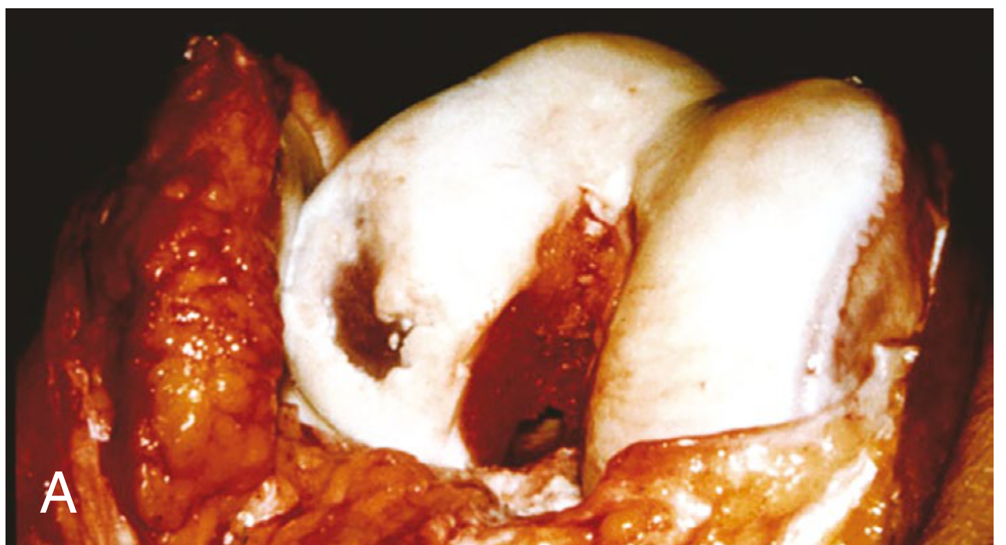
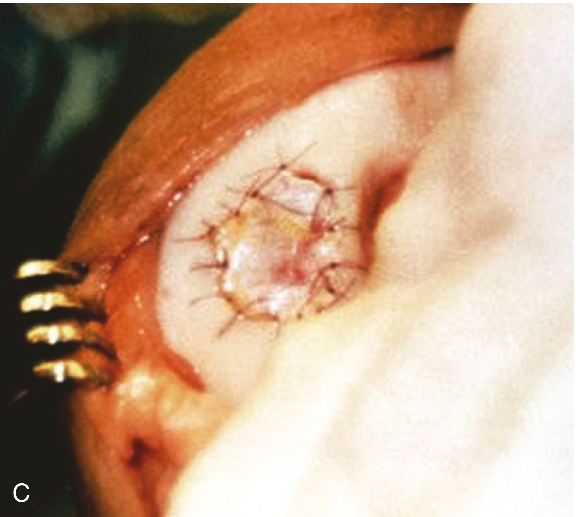
- Arthroscopy to confirm defect suitability
- Harvest 200-300 mg cartilage biopsy from a non-weight-bearing area (intercondylar notch, superomedial femoral condyle)
- Specimen sent to laboratory for chondrocyte isolation and expansion
- Enzymatic digestion of cartilage (collagenase) releases chondrocytes
- Monolayer culture expansion for 3-6 weeks until 12-48 million cells obtained (6-8 population doublings)
- Cell viability confirmed before implantation
- Mini-arthrotomy (medial or lateral parapatellar)
- Defect debridement to stable cartilage margins; vertical walls created; subchondral bone intact
- Periosteal graft harvested from medial tibial metaphysis
- Periosteal patch sutured (watertight) over the defect with 2-0 Vicryl sutures, cambium layer facing down
- Small gap left; cell suspension injected under the patch via cannula
- Gap sealed with fibrin glue
- Periosteal patch hypertrophy (up to 25-36% of cases) requiring arthroscopic shaving
- Periosteal graft delamination and calcification
- Cell suspension leakage
- Requires two procedures + arthrotomy
- Technically demanding, longer surgery
Generation 2: ACI with Collagen Membrane (ACI-C)
- Porcine type I/III collagen membrane (Bio-Gide, Chondro-Gide) replaces periosteal patch
- Reduced hypertrophy rate significantly
- Otherwise same technique as Gen 1
- Cells injected under membrane; fibrin glue seal
- ChondroCelect (TiGenix, Belgium): first EU-approved ACI cell product with characterized biomarkers of stable cartilage-forming potential (EMA registered under Advanced Therapy Medicinal Product regulation)
- RCT: ChondroCelect produced structurally superior repair tissue at 12 months and superior clinical outcome at 3 years vs microfracture; non-inferior at 5 years overall, but superior in patients with lesions <3 years old - Firestein & Kelley's Rheumatology (2022)
Generation 3: MACI - Matrix-Induced ACI (Current Standard)
- Mini-arthrotomy or arthroscopic (newer approach)
- Defect prepared: debridement to stable walls, subchondral bone preserved
- Defect measured, membrane trimmed to exact defect size/shape
- MACI membrane (cells seeded on one side) placed cell-side down into defect
- Fixed with fibrin glue only - no sutures needed
- Membrane conforms to irregular defect shapes
- Campbell's (2026): "Although prior generations required a periosteal graft sutured over the defect, the current iteration requires fixation with fibrin glue only."
- No periosteal harvest (no donor site morbidity)
- No graft hypertrophy
- Improved cell distribution (3D scaffold)
- Fibrin glue only - simpler, faster
- Can be applied arthroscopically
- Reduced cell leakage
- 3D environment prevents chondrocyte dedifferentiation
Cartilage Treatment Algorithm by Defect Size
| Defect Size | Preferred Treatment |
|---|---|
| <1 cm² | Marrow stimulation (microfracture, drilling) |
| 1-2 cm² | Microfracture or OATS (mosaicplasty) |
| 2-3.5 cm² | OATS, OCA, MACI |
| >3.5 cm² (up to 10 cm²) | MACI (ACI), fresh osteochondral allograft |
| Multiple/large complex | MACI preferred |
Postoperative Rehabilitation
| Phase | Duration | Activity |
|---|---|---|
| Phase 1 (maturation) | 0-6 weeks | Continuous passive motion (CPM); non-weight-bearing or toe-touch |
| Phase 2 | 6-12 weeks | Gradual progressive weight bearing; closed chain exercises |
| Phase 3 | 3-6 months | Strengthening; low-impact activities |
| Phase 4 | 6-12 months | Sport-specific training |
| Return to sport | ~12-18 months | After clinical and MRI confirmation of graft maturation |
- Optimal outcome reached at 18-24 months
- Lack of low-load activities after surgery (in both ACI and microfracture groups) adversely affects functional outcomes - Campbell's (2026)
Outcomes
Long-Term Results
- 188 chondral defects, mean age 37, mean BMI 26.2
- Significant, durable improvements in all PRO measures at 10-17 years
- MRI: satisfactory defect fill and intact graft in majority
- All-cause reoperation rate: 9.0%
- Progression to TKA: 7.4% at 10-17 years
- "These data support the use of MACI as a long-term treatment of focal cartilage defects."
- 235 patients; defects 2.2-15.1 cm² (ACI) and 2.0-7.6 cm² (MACI)
- Stable improvements up to 11 years (ACI) and 15 years (MACI)
- Failure rate ~10% at 11 years
- Both procedures delayed progression to arthroplasty
- 47 studies, 1,993 patients, mean follow-up 57 months
- All four procedures showed significant improvements in Lysholm, IKDC, Tegner, and VAS scores
- No significant differences between ACI, MACI, OAT, and OCA
- OAT surpassed PASS threshold for IKDC; all surpassed Lysholm and Tegner PASS
- Conclusion: Technique selection should be individualized based on patient and defect characteristics
- 965 patients; OR = 8.75 (95% CI 7.1-10.7) for significant improvement in osteochondral defects
Positive Predictors of Good Outcome
- Young patient age
- Femoral condyle location (vs patella/trochlea)
- Early intervention (<3 years after becoming symptomatic)
- Good quality harvested chondrocytes
- Well-trained surgeon
- Strict rehabilitation adherence
- No radiographic signs of OA
Complications
| Complication | Generation | Incidence |
|---|---|---|
| Graft/periosteal hypertrophy | Gen 1 (periosteal) | 25-36%; reduced to <5% with membrane |
| Graft delamination | Gen 1 | Uncommon |
| Cell leakage | Gen 1 > Gen 3 | Rare with MACI/fibrin glue |
| Infection | All | <1% |
| Calcification of periosteal patch | Gen 1 | Rare |
| Failure / non-integration | All | ~10% at 10 years |
| Progression to TKA | All | ~7% at 10-17 years |
| Reoperation (any cause) | MACI | 9% |
| Adhesions / stiffness | All | Uncommon |
ACI vs Microfracture - Key Comparison
| Parameter | Microfracture | ACI/MACI |
|---|---|---|
| Tissue type produced | Fibrocartilage (Type I collagen) | Hyaline-like (Type II collagen) |
| Defect size | <2 cm² | >2 cm² |
| Stages | Single stage | Two stages |
| Durability | Declines after 18-24 months | Durable up to 15+ years |
| Reoperation | Lower | ~9% (MACI) |
| Cost | Lower | Higher |
| Rehabilitation | Shorter | Longer (18 months to full RTS) |
| First RCT | ACI not superior to microfracture at 2 years (NEJM 2005) | ChondroCelect superior at 3 years, non-inferior at 5 years |
Recent Advances
1. Arthroscopic MACI
- MACI now deliverable fully arthroscopically (no arthrotomy)
- Reduced soft tissue trauma, faster recovery, better cosmesis
- Statistically better outcomes vs microfracture at 2 years in arthroscopic delivery study - Firestein & Kelley (2022)
2. High-Density ACI (HD-ACI)
- Guillén-García et al. (Bioengineering 2023, PMID 37760185): increased cell density implantation improves matrix production and mechanical properties
- May reduce need for extended culture periods
3. Nasal Chondrocytes
- Chondrocytes from nasal septum have known stable chondrogenic capacity (do not dedifferentiate with age/mechanical unloading as readily as articular chondrocytes)
- Safe and effective in 10-patient pilot study for knee defects - Firestein & Kelley
- Also trialled in two patients with advanced OA (KL grade 3-4) with promising early results
- Large-scale controlled trials needed
4. MSC-Based Approaches (Cell-Free or Alternative Cell Sources)
- MSCs from bone marrow, adipose tissue, synovial membrane can differentiate into chondrocytes
- Bone marrow MSCs = at least as effective as chondrocytes for symptomatic improvement
- Adipose-derived stem cells (ADSCs): abundant source, minimal donor site morbidity
- Synovial MSCs: highest chondrogenic potential among MSC sources
- Challenge: MSCs produce transient fibrocartilage rather than stable hyaline cartilage without scaffold guidance - Rheumatology 2-Vol Set (2022)
5. Hydrogel-Enhanced ACI
- Hydrogels (alginate, fibrin, hyaluronic acid, PEG-based) as 3D injectable carriers
- Provide structural support and prevent chondrocyte dedifferentiation
- Can be injected minimally invasively - no arthrotomy needed
- Ahmadpoor et al. (Bioengineering 2024, PMID 39593824): updated preclinical review of hydrogel-enhanced ACI showing improved cell viability and matrix production
6. 3D Bioprinting / Bioengineered Constructs
- Patient-specific 3D-printed collagen/bioink scaffolds seeded with autologous chondrocytes
- Precise anatomical match to defect geometry
- Combined with growth factors (TGF-beta3, BMP-6, IGF-1)
7. Gene Therapy Augmentation
- CRISPR/Cas9 gene editing used to modify harvested chondrocytes to upregulate type II collagen synthesis, overexpress SOX9 (master chondrogenic transcription factor), or knock out catabolic factors (IL-1, MMP-13)
- Focsa et al. (Medicina 2024, PMID 39859006): CRISPR combined with ACI shows targeted cellular modification potential
- Still experimental/preclinical phase
8. Biophysical Stimulation
- LIPUS (Low-Intensity Pulsed Ultrasound): enhances chondrogenic differentiation and ECM production
- Electromagnetic fields (EMFs): promote chondrogenesis; combined with ACI in preclinical models
- Both being trialled as intraoperative or postoperative adjuncts to accelerate graft maturation
9. Non-Autologous / Off-The-Shelf Alternatives
- CARTIMAX / Cartiform: allogeneic chondrocytes/acellular scaffolds (FDA approved)
- Agili-C (aragonite + hyaluronan scaffold, Cartiheal): FDA approved 2022; shown to outperform microfracture/debridement in mild-moderate OA in RCT; good MRI filling - Campbell's (2026)
- Juvenile particulate cartilage (IMPACT/DeNovo NT): juvenile allogeneic chondrocytes with higher metabolic activity than adult cells; single-stage procedure
- Cryopreserved viable osteochondral allografts: small cohort studies show good results, low failure rate
Key Points for Exam
- ACI = two-stage procedure: biopsy → lab expansion → reimplantation
- Pioneer: Brittberg et al., 1994; first commercial product: Carticel, 1997
- Defect size: suitable for >2 cm² single focal full-thickness defects
- Three generations: Gen 1 (periosteal patch) → Gen 2 (collagen membrane) → Gen 3/MACI (cell-seeded matrix, fibrin glue only)
- MACI = current gold standard; no periosteal harvest, no sutures, fibrin glue fixation
- Regenerates hyaline-like (type II collagen) tissue vs fibrocartilage from microfracture
- MACI at 10+ years: 9% reoperation, 7.4% TKA progression, durable PRO improvements (Wang 2024)
- All four procedures (ACI, MACI, OAT, OCA) show comparable outcomes (Nassar 2025)
- Optimal outcome at 18-24 months; return to sport ~12-18 months
- Must correct: malalignment, ligament instability, meniscal deficiency before/during ACI
- Positive predictors: young age, femoral condyle, early intervention (<3 years), good chondrocyte quality
- Most exciting recent advances: arthroscopic MACI, nasal chondrocytes, hydrogel carriers, 3D bioprinting, CRISPR-modified chondrocytes, Agili-C (FDA 2022)
- Campbell's Operative Orthopaedics, 15th Ed 2026 (Chapter 50, MACI section)
- Firestein & Kelley's Textbook of Rheumatology (2022)
- Rheumatology 2-Vol Set, Elsevier (2022)
- Wang AS et al. "Minimum 10-Year Outcomes of MACI." Am J Sports Med 2024 [PMID 38312085]
- Nassar JE et al. "ACI, MACI, OAT and OCA improve knee function: meta-analysis." KSSTA 2025 [PMID 39497425]
- Colombini A et al. "ACI provides good long-term results in knee OA." KSSTA 2023 [PMID 35716187]
- Almohaileb FI & Rasheed Z. "Clinical Applicability of ACI: meta-analysis." Curr Rheumatol Rev 2024 [PMID 37957845]
- Focsa MA et al. "Emerging Strategies in Cartilage Repair." Medicina 2024 [PMID 39859006]