
Type 2 hypersensitive reaction
Reading File
Now let me search the medical library for Type 2 hypersensitivity reactions in parallel.
Finding Sources
Finding Sources
Finding Sources
Excellent results. Now let me read the key sections from Robbins, Roitt's, and Goldman-Cecil in parallel.
Reading File
Reading File
Reading File
I have rich content. Let me also read the Robbins continuation (Table 5.4, Fig 5.12 details) and fetch the Goodpasture image.
Reading File
Loading Image
Let me also fetch the Robbins mechanism diagram (Fig 5.12).
Loading Image
I now have comprehensive, well-illustrated content from multiple authoritative sources. Here is a full answer:
Type II Hypersensitivity (Antibody-Mediated Cytotoxic Hypersensitivity)
Definition
Type II hypersensitivity reactions are caused by antibodies (IgG or IgM) directed against target antigens on the surface of cells or other tissue components. The antigens may be:
- Normal molecules intrinsic to cell membranes or the extracellular matrix (as in autoimmune diseases)
- Adsorbed exogenous antigens - e.g., a drug metabolite bound to a cell surface (as in drug reactions)
It is also called antibody-mediated cytotoxic hypersensitivity in the Gell-Coombs classification.
- Robbins & Kumar Basic Pathology, p. 169
Three Core Mechanisms of Tissue Injury
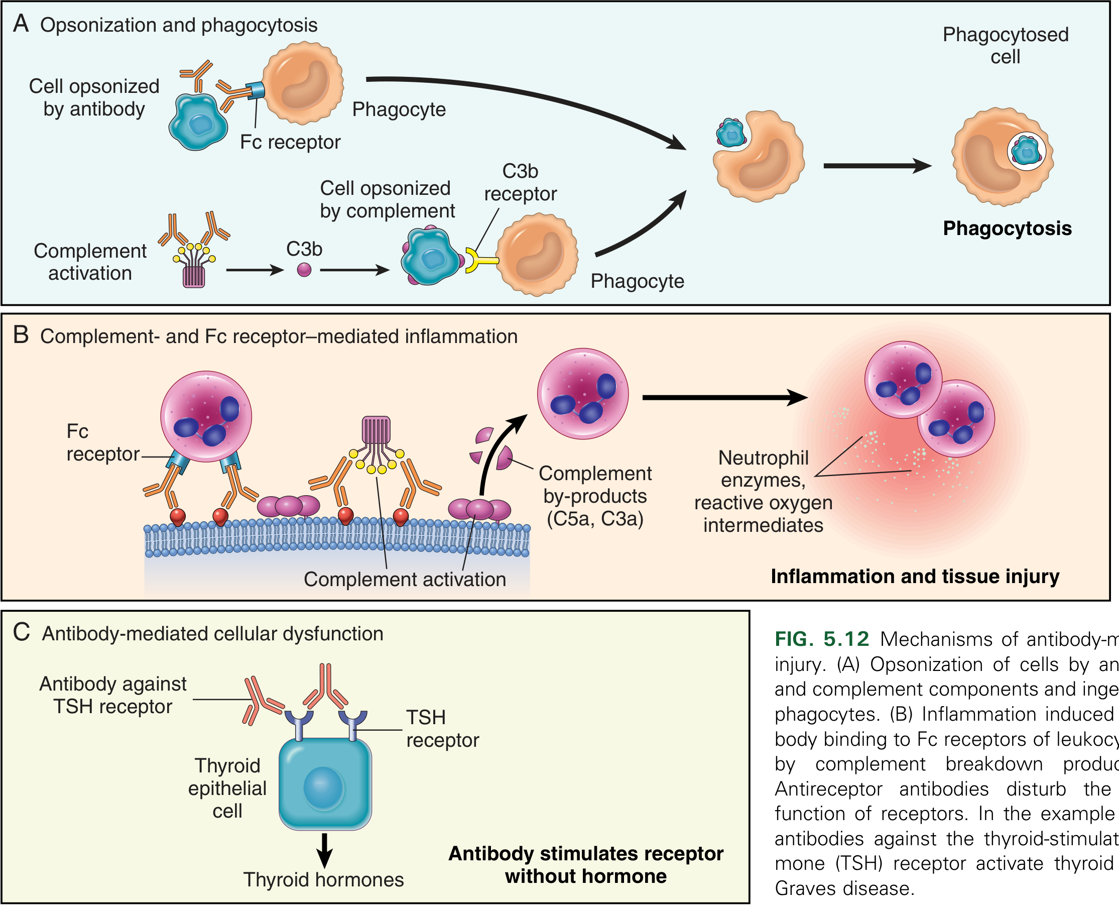
Fig 5.12 - Mechanisms of antibody-mediated (Type II) injury: (A) Opsonization and phagocytosis, (B) Complement- and Fc receptor-mediated inflammation, (C) Antibody-mediated cellular dysfunction. (Robbins & Kumar Basic Pathology)
1. Opsonization and Phagocytosis
When circulating cells (red cells, platelets) are coated (opsonized) with autoantibodies, with or without complement proteins, they become targets for phagocytosis by neutrophils and macrophages. Phagocytes express:
- Fc receptors (for IgG antibody tails)
- C3b receptors (for complement breakdown products)
Opsonized blood cells are usually eliminated by macrophages in the spleen - this is why splenectomy is clinically beneficial in some antibody-mediated diseases.
2. Complement- and Fc Receptor-Mediated Inflammation
Antibodies bound to tissue antigens activate the complement cascade. This generates:
- C3b - promotes opsonization
- C5a, C3a - anaphylatoxins that recruit and activate neutrophils and macrophages
- MAC (C5b-C9) - membrane attack complex causing direct lysis
Leukocytes recruited via Fc receptors and complement products release proteases, reactive oxygen intermediates, and cytokines causing local tissue damage. C1 through C9 activation can cause intravascular lysis of red cells.
3. Antibody-Mediated Cellular Dysfunction (without direct cell killing)
Antibodies directed at cell surface receptors can:
-
Block the receptor (e.g., anti-acetylcholine receptor antibodies in myasthenia gravis block neuromuscular transmission)
-
Stimulate the receptor inappropriately (e.g., anti-TSH receptor antibodies in Graves' disease cause hyperthyroidism)
-
Strip receptors from the cell surface
-
Robbins & Kumar Basic Pathology, p. 169-170
Immunoglobulin Classes Involved
- IgG (most common) - activates complement via classical pathway, binds Fc receptors on phagocytes, mediates ADCC
- IgM - very efficient at activating complement; involved in ABO transfusion reactions and cold hemagglutinin disease
Classic Clinical Examples
| Disease | Target Antigen | Mechanism | Manifestation |
|---|---|---|---|
| Autoimmune hemolytic anemia | Red cell membrane proteins | Opsonization, phagocytosis | Hemolysis, anemia |
| Autoimmune thrombocytopenic purpura (ITP) | Platelet glycoproteins (GpIIb:IIIa) | Opsonization, phagocytosis | Bleeding |
| Goodpasture syndrome | Type IV collagen (GBM, alveolar BM) | Complement + Fc receptor-mediated inflammation | Nephritis, lung hemorrhage |
| Myasthenia gravis | Acetylcholine receptor | Receptor blockade, stripping, complement | Muscle weakness, paralysis |
| Graves' disease | TSH receptor | Receptor stimulation | Hyperthyroidism |
| Pemphigus vulgaris | Desmoglein 3 (intercellular junctions) | Protease activation, disruption of cell adhesion | Skin bullae |
| Acute rheumatic fever | Streptococcal antigen cross-reacting with myocardium | Inflammation, macrophage activation | Myocarditis, arthritis |
| ANCA vasculitis | Neutrophil granule proteins | Neutrophil degranulation | Vasculitis |
- Robbins & Kumar Basic Pathology, Table 5.4, p. 169
Goodpasture Syndrome - Immunofluorescence Pattern
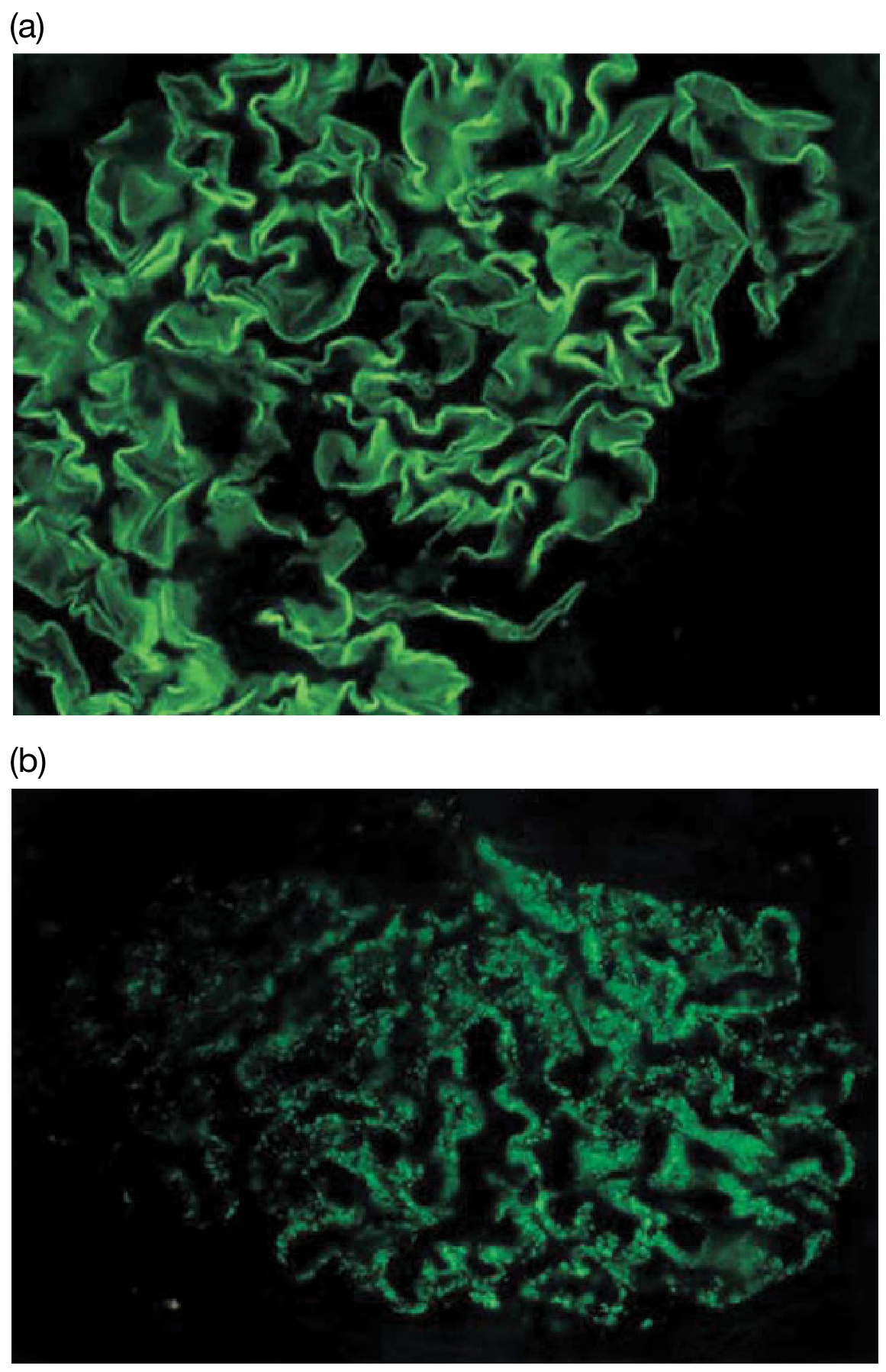
Figure 14.19 (Roitt's Essential Immunology): (a) Goodpasture syndrome (Type II) - LINEAR IgG deposition along the glomerular basement membrane. (b) SLE (Type III) - GRANULAR/lumpy-bumpy immune complex deposition. This distinction is diagnostically important.
Drug-Induced Type II Reactions
Drugs can act as haptens - they bind covalently to the surface of circulating blood cells (red cells, platelets, neutrophils), creating new epitopes. The immune system recognizes these conjugates as foreign and generates IgG/IgM antibodies. Upon drug withdrawal, the reaction resolves.
Mechanism steps:
- Drug binds to red cell surface protein
- Penicillin-specific IgG binds to the drug-protein conjugate
- Complement activated (C1 through C9), C3b deposited on the red cell
- Red cells susceptible to phagocytosis in spleen/liver, or intravascular lysis by MAC
Examples:
-
Penicillin, quinidine, methyldopa - hemolytic anemia and/or thrombocytopenia
-
Amidopyrine, quinidine - agranulocytosis
-
Chlorpromazine, phenacetin - hemolytic anemia
-
Heparin-induced thrombocytopenia (HIT) - involves heparin + platelet factor 4 complex, anti-PF4-heparin IgG, and FcγRIIA-mediated platelet activation; affects 1-3% of heparin-exposed patients
-
Goldman-Cecil Medicine, p. 1552-1554; Roitt's Essential Immunology, p. 441
Comparison with Other Types (Gell-Coombs)
| Feature | Type I | Type II | Type III | Type IV |
|---|---|---|---|---|
| Mediator | IgE | IgG, IgM | IgG, IgM (complexes) | T cells |
| Antigen location | Soluble | Cell surface / ECM | Soluble (circulating) | Cell-associated / soluble |
| Complement | No | Yes | Yes | No |
| Onset | Minutes | Minutes-hours | Hours-days | 48-72 hours |
| Example | Anaphylaxis | Goodpasture, MG, Graves' | SLE, serum sickness | Contact dermatitis, TB |
Key Diagnostic Points
- Linear immunofluorescence on basement membrane = Type II (e.g., Goodpasture) vs. granular = Type III (e.g., SLE)
- Coombs' (DAT) test detects antibodies or complement on red cell surfaces - used for autoimmune hemolytic anemia and hemolytic transfusion reactions
- Splenectomy helps in ITP and autoimmune hemolytic anemia because the spleen is the major site where opsonized cells are cleared
This is a shared conversation. Sign in to Orris to start your own chat.