
urticaria
urticaria hives wheals skin
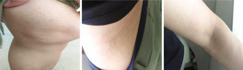
A series of three clinical photographs demonstrating the dermatological resolution of urticaria (hives) across three anatomical regions: the knee joint (A), the lateral waist (B), and the elbow (C). The images show characteristic wheals—edematous, circumscribed, and slightly elevated skin lesions. Panel A displays the knee area with subtle, flesh-colored to slightly erythematous elevations and an uneven skin texture consistent with resolving wheals. Panel B shows the lateral waist with faint, linear, and confluent dermal edema. Panel C depicts the elbow where the lesions have largely flattened, leaving only minimal, faint markings on the skin surface. The progression illustrates the transient nature of urticarial lesions as they gradually diminish in size and elevation. This visual serves as an educational reference for identifying skin morphology in the recovery phase of an acute urticarial reaction, emphasizing the characteristic lack of secondary skin changes (such as scaling or scarring) during the resolution of wheals.
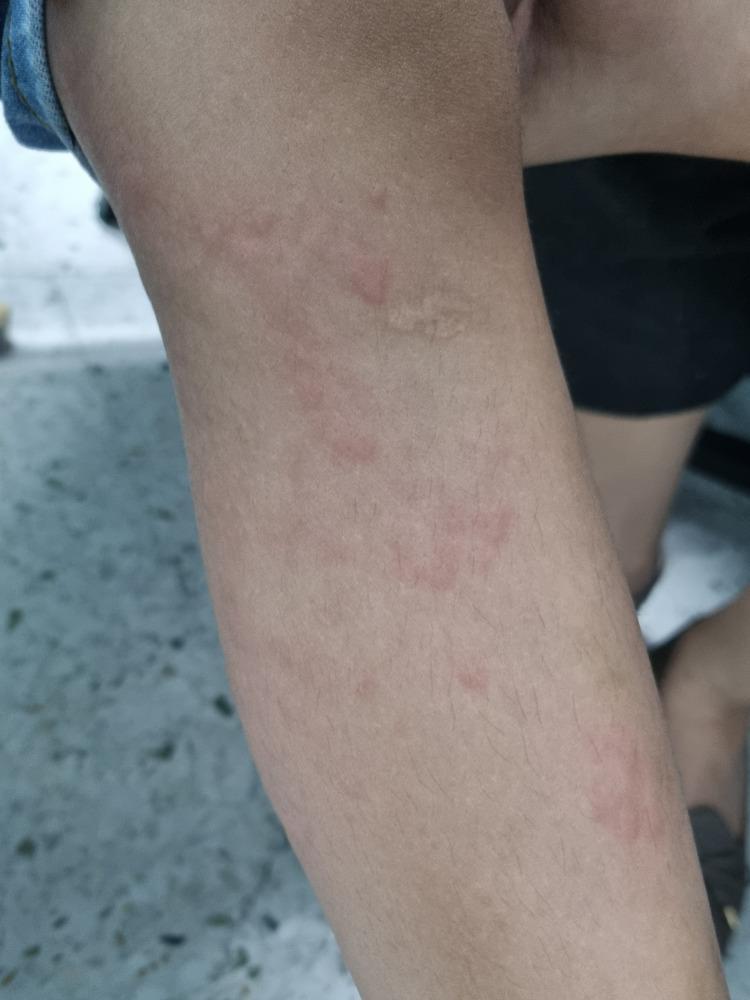
A clinical photograph showing a close-up of a child's lower extremity demonstrating urticaria. The image displays multiple raised, edematous skin lesions known as wheals or hives. These lesions are characterized by pale-to-skin-colored central swellings of varying sizes and irregular shapes, surrounded by a distinct zone of erythema (redness). The distribution is scattered across the leg, illustrating the typical appearance of a hypersensitivity reaction or allergic response. This image serves as an educational reference for identifying cutaneous manifestations of urticaria, focusing on the morphology of superficial dermal edema and associated inflammatory flare.
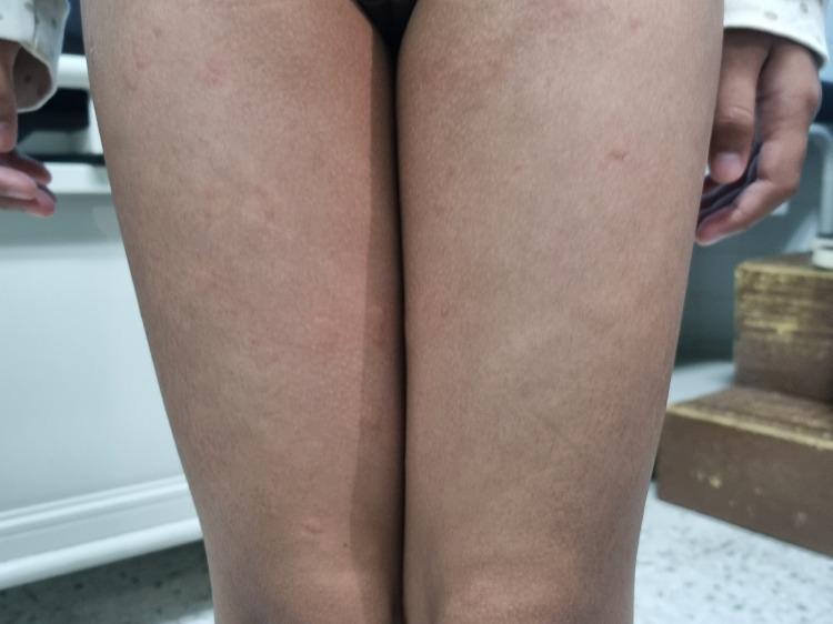
Clinical photograph of a pediatric patient's bilateral lower limbs demonstrating classic urticaria (hives). The image shows multiple discrete and confluent wheals distributed across the thighs. Morphologically, the lesions are characterized by raised, edematous, superficial swellings of varying sizes. The central portions of the wheals appear pale or skin-colored, while the peripheries exhibit varying degrees of erythema. These findings represent a type I hypersensitivity reaction or mast cell degranulation, leading to dermal edema. This visual evidence is a key diagnostic clinical sign for acute urticaria, commonly used in dermatology and pediatric curricula to differentiate transient wheals from other types of papular eruptions or inflammatory skin conditions.
urticaria management guidelines
Urticaria (Hives)
Definition
- Goldman-Cecil Medicine, p. 4300
- Rosen's Emergency Medicine, p. 2413
Classification
By Duration
| Acute | Chronic | |
|---|---|---|
| Duration | < 6 weeks | ≥ 6 weeks |
| Common causes | Drugs, foods, infections, vaccines | Spontaneous/idiopathic, autoimmune, physical |
| More common in | Children (atopic dermatitis) | Women 20-40 years old |
- Chronic Spontaneous Urticaria (CSU): no identifiable trigger (66-93% of chronic cases)
- Inducible/Physical Urticaria: provoked by specific physical stimuli
Chronic Urticaria Types (Harrison's Table 363-1)
-
Spontaneous/idiopathic
-
Autoimmune (functional IgE autoantibodies)
-
Dermatographism (linear wheal from skin stroking; 2-5% of population)
-
Cholinergic urticaria (small 1-2 mm wheals + large erythema; triggered by heat, exercise, fever)
-
Pressure urticaria (sustained pressure - belt, shoulder strap)
-
Cold, solar, vibratory, aquagenic urticaria
-
Mastocytosis, urticarial vasculitis, hereditary angioedema (HAE)
-
Harrison's Principles of Internal Medicine 22E, p. 363
-
Fitzpatrick's Dermatology, Vol. 1-2
Pathophysiology
- Histamine (primary mediator)
- Slow-reacting substance of anaphylaxis (leukotrienes)
- Bradykinin, kallikrein, acetylcholine
Mechanisms
- Immunologic:
- IgE-mediated (Type I hypersensitivity)
- Autoimmune (functional IgG autoantibodies against IgE or FcεRI)
- Immune complex-mediated
- Complement-kinin dependent
- Non-immunologic:
- Direct mast cell degranulation (drugs like opioids, aspirin, foods like strawberries/lobster)
- Vasoactive stimuli
- Goldman-Cecil Medicine, p. 4302
- Rosen's Emergency Medicine, p. 2413
Common Causes
| Category | Examples |
|---|---|
| Drugs | Penicillin, aspirin/NSAIDs (most common); also opioids, ACE inhibitors, radiocontrast |
| Foods | Seafood, tree nuts, eggs, peanuts (IgE-mediated); strawberries, lobster (direct histamine release) |
| Infections | Rhinovirus, rotavirus, hepatitis, EBV (mono), Coxsackievirus; Candida, dermatophytes, parasites |
| Physical | Cold, heat, pressure, sun, vibration, water, exercise |
| Contact | Foods, animal dander/saliva, plants, topical medications, cosmetics, chemicals |
Clinical Features
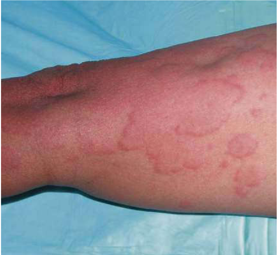
- Wheals: Edematous plaques, pale center + red border ("wheal and flare"), blanching on pressure
- Pruritus: Hallmark symptom
- Evanescent: Individual lesions typically last < 24 hours (this is a key diagnostic feature)
- New lesions continuously develop while old ones resolve
- May coexist with angioedema (deeper swelling - face, lips, tongue, throat)
- Angioedema without wheals occurs in 1-13% of CSU patients
Differential Diagnosis
- Drug eruption / viral exanthem
- Erythema multiforme
- Erythema marginatum
- Urticarial vasculitis (lesions persist > 24h, may bruise)
- Juvenile rheumatoid arthritis
- Bullous pemphigoid (urticarial stage)
- Neutrophilic urticaria
- Mastocytosis
- Fitzpatrick's Dermatology, p. 46
- Rosen's Emergency Medicine, p. 2413
Management
Step 1: Remove the trigger
Step 2: Antihistamines (First-line)
- H1 antihistamines (preferred non-sedating): Cetirizine, fexofenadine, loratadine
- Sedating H1: Hydroxyzine (useful for pruritus relief)
- H2 blocker addition: Can be added in chronic or refractory cases (e.g., ranitidine, famotidine)
- For chronic urticaria: long-term combination H1+H2 antihistamines
Step 3: Corticosteroids (Adjunctive)
- Moderate/severe urticaria: prednisone or dexamethasone
- Recurrent urticaria: 14-21 day course with taper
- Chronic steroid use is not recommended
Step 4: Advanced/Refractory Therapy
-
Omalizumab (anti-IgE monoclonal antibody): highly effective for chronic spontaneous urticaria - a 2025 meta-analysis confirms its efficacy and safety in pediatric CSU as well
-
Cyclosporine: second-line immunosuppressant for CSU
-
A 2024 systematic review on acute urticaria treatment found that antihistamines remain the cornerstone, with corticosteroids as adjuncts
-
Rosen's Emergency Medicine, p. 2414
-
Goldman-Cecil Medicine, p. 4302
Special Considerations
| Situation | Notes |
|---|---|
| Angioedema with airway compromise | Treat as anaphylaxis: epinephrine IM, airway management |
| Chronic spontaneous urticaria | Half of patients have disease for 5+ years; investigate for autoimmune cause |
| Dermatographism | Usually resolves within 5 years; reassurance + antihistamines |
| Cholinergic urticaria | Avoid triggers (heat, exercise); H1 antihistamines ± beta-blockers |
| Cold urticaria | Risk of anaphylaxis with cold water immersion; carry epinephrine |
| Pressure urticaria | Often accompanies CSU; responds poorly to antihistamines |
Key Points for Boards/Exams
- Urticaria lasting < 6 weeks = acute; ≥ 6 weeks = chronic
- Individual wheals resolve in < 24 hours - if they persist longer, consider urticarial vasculitis
- Most common drugs: penicillin and aspirin
- Most chronic urticaria is idiopathic/spontaneous
- First-line: non-sedating H1 antihistamines
- Refractory CSU: omalizumab (anti-IgE)
- Angioedema of the larynx/tongue = emergency - epinephrine
Types of Give me detailed notes about urticaria
urticaria types wheals angioedema dermatographism cholinergic
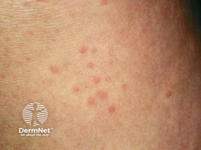
Clinical photography of a patient with cholinergic urticaria showing multiple small, erythematous, blanching wheals on the forearm. Imaging modality is clinical photography; technique involves a high-resolution, color-balanced image capturing superficial skin lesions in a macro-clinical view. The wheals are transient, pruritic, and vary in size from a few millimeters to up to several millimeters, with well-demarcated borders and pale centers. Triggered by heat, exercise, and emotional stress, the lesions can appear soon after sweating and typically resolve within minutes to hours. This presentation correlates with cholinergic urticaria, a physical urticaria subtype linked to cholinergic stimuli, requiring history of triggers and symptom timing. Differential diagnoses include other urticarias and heat-induced dermatoses; diagnostic significance lies in distinguishing exercise/sweat-induced wheals from dermatographism or drug reactions to guide management. Clinical utility includes educating patients on trigger mitigation, optimizing antihistamine therapy, and evaluating for underlying mast cell disorders if atypical features arise. Chronic management includes non-sedating H1 antihistamines at standard or increased dosing, with occasional H2 blockade. Patients are advised to avoid overheating, wear breathable clothing, and perform graded exercise; epinephrine is reserved for rare anaphylaxis. In ambiguous cases, exercise-provoked challenges or sweat tests may help; history differentiates from other physical urticarias.
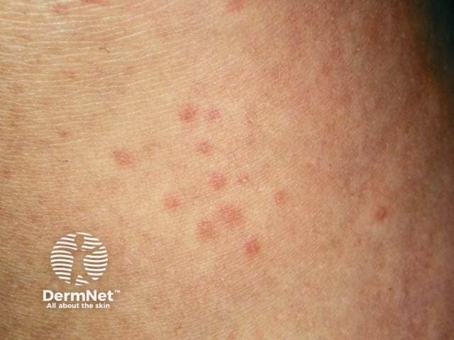
This is a dermatology clinical photograph of the skin surface demonstrating the typical small, transient wheals seen in cholinergic urticaria. The image shows numerous pinpoint to 3–4 mm erythematous, raised plaques scattered across the forearm in a crops-like distribution. Each wheal shows central blanching with a slightly edematous, ring-like border and a blanching halo consistent with superficial dermal edema. The lesions are pruritic and short-lived, usually resolving within 1–2 hours, often appearing after heat exposure, exercise, or emotional stress. In cholinergic urticaria, symptoms may be accompanied by warmth, sweating, or mild warmth-induced flushing. The focal photodermatographic appearance is not necessarily specific but the small size, rapid onset following provocative stimuli, and distribution pattern strongly support a physical urticaria rather than mast cell neoplasia or dermatitis. Clinically relevant features include the ability to trigger crops, readiness to respond to oral antihistamines, and potential improvement with pre-exercise cooling or preventive antihistamine therapy. The differential diagnosis includes other physical urticarias (dermographism, heat urticaria), ordinary urticaria, papular urticaria, and less commonly urticarial vasculitis. This image is most useful for educational illustration of cholinergic urticaria triggers: exercise, heat, stress; and for training in clinical recognition and patient counseling.
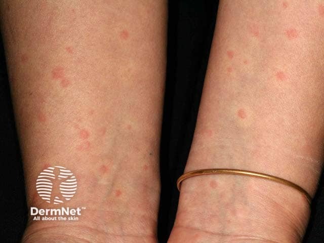
Clinical photograph of bilateral forearms showing multiple discrete 5 mm erythematous, blanchable wheals consistent with cholinergic urticaria precipitated by exercise. Lesions are superficial, well defined, round-to-oval, slightly raised, and distributed patchily across extensor surfaces of the forearms. The wheals appear shortly after exertion or warm ambient conditions and resolve within minutes to an hour, often preceding or accompanying mild pruritus. The image demonstrates a transient urticarial eruption without mucosal involvement or vesiculation. From a pathophysiologic perspective, cholinergic urticaria results from mast cell degranulation and histamine release triggered by increased core temperature and sweating; histologic findings (if biopsy were available) would show dermal edema with perivascular inflammatory infiltrate rich in eosinophils. Clinically, these findings aid differentiation from other physical urticarias such as heat urticaria, solar urticaria, or pressure urticaria, and from non-urticarial pruritic eruptions. This photograph supports diagnostic consideration in patients with exercise-induced pruritic wheals and provides material for education on recognition, documentation, and management planning. Relevance spans dermatology, allergology, pediatrics, and sports medicine; can guide antihistamine therapy, pre-exercise prophylaxis, and patient counseling on trigger avoidance and skin care during heat stress. Documentation of such patterns supports longitudinal monitoring and therapeutic response assessment.
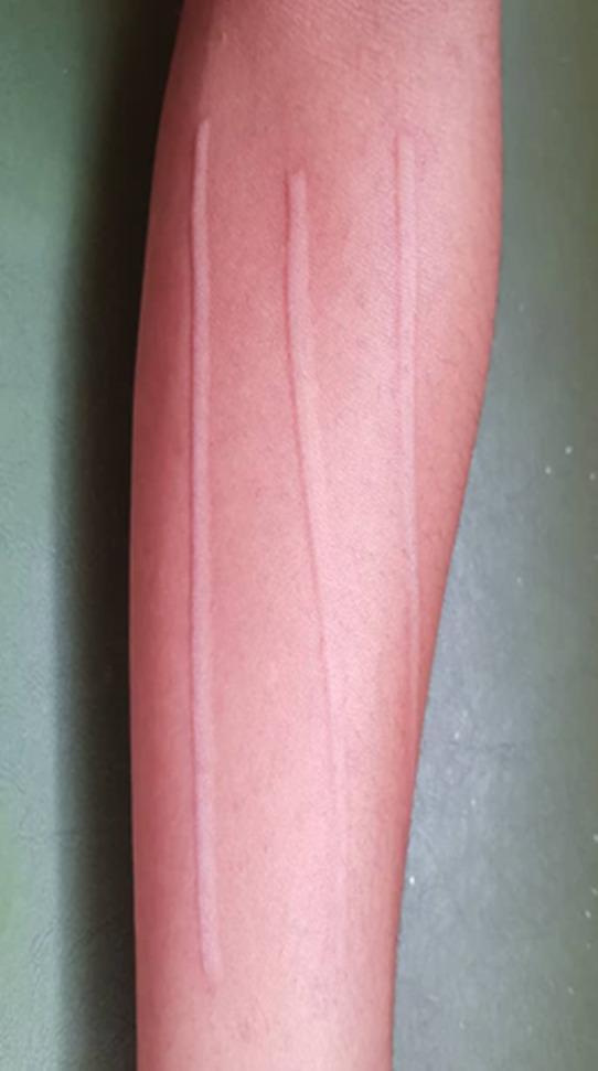
Clinical photograph of a left forearm demonstrating dermatographism (skin writing), a form of physical urticaria. The image shows three distinct, raised, linear wheals (urticarial lesions) running longitudinally along the forearm. These markings are characteristically pale or erythematous with well-defined borders against the surrounding skin. The appearance is a classic representation of the triple response of Lewis, specifically the localized edema (wheal) that occurs in response to firm stroking or mechanical pressure on the skin. This finding is often associated with mast cell degranulation and histamine release in sensitive individuals. The photograph serves as an educational example for diagnosing physical urticarias and understanding the cutaneous manifestations of hyperreactivity in the dermatology and allergy/immunology domains.
Detailed Notes on Types of Urticaria
Master Classification Framework
URTICARIA
├── SPONTANEOUS
│   ├── Acute Spontaneous (<6 weeks)
│   └── Chronic Spontaneous (≥6 weeks)
│       ├── Autoimmune
│       └── Idiopathic
└── INDUCIBLE (Physical)
    ├── Symptomatic Dermographism
    ├── Cholinergic Urticaria
    ├── Cold Urticaria
    ├── Heat Urticaria
    ├── Delayed Pressure Urticaria
    ├── Solar Urticaria
    ├── Aquagenic Urticaria
    ├── Vibratory Angioedema
    ├── Contact Urticaria
    └── Adrenergic Urticaria

SPECIAL ENTITIES
    ├── Urticarial Vasculitis
    ├── Angioedema (including HAE)
    └── Papular Urticaria
- Andrews' Diseases of the Skin, p. 176
- Fitzpatrick's Dermatology, Vol. 1-2
PART 1: SPONTANEOUS URTICARIA
1A. Acute Spontaneous Urticaria
| Feature | Details |
|---|---|
| Duration | < 6 weeks |
| Prevalence | Most new-onset urticaria (>2/3 of cases) |
| Common in | Children and young adults |
| Main causes | Viral URIs (most common in children), drugs (NSAIDs, antibiotics), foods, insect stings |
| Workup needed | Usually none; history + exam sufficient |
| Prognosis | Self-limited; resolves once trigger removed |
-
Infections: rhinovirus, rotavirus, EBV (mononucleosis), hepatitis B/C, Coxsackievirus, psittacosis
-
Drugs: penicillin, NSAIDs, opioids, radiocontrast dye, vancomycin
-
Foods: shellfish, peanuts, tree nuts, eggs, milk, strawberries, lobster, chocolate, tomatoes, spices
-
Food additives: azo dyes, benzoic acid, sulfites, tartrazine
-
Inhalants: grass pollen, house dust mites, animal dander, molds, formaldehyde
-
Andrews' Diseases of the Skin, p. 175
1B. Chronic Spontaneous Urticaria (CSU)
| Feature | Details |
|---|---|
| Duration | ≥ 6 weeks, occurring almost daily |
| Prevalence | ~0.5-1% of population; 66-93% of chronic urticaria cases |
| Demographics | Women > men (2:1); peak age 20-40 years |
| Cause identified | < 20% of cases; majority remain idiopathic |
| Duration of disease | 50% have symptoms ≥ 5 years |
- Wheals + angioedema: 33-67% of CSU patients
- Wheals only: 29-65%
- Angioedema only: 1-13%
- Flower-like or annular-shaped wheals are characteristic
- Stress and fatigue are common aggravating factors; depression/anxiety are comorbidities
-
Routine: CBC with differential, ESR or CRP
-
Extended (based on history): H. pylori testing, thyroid antibodies (anti-TPO, anti-Tg), autologous serum skin test (ASST), tryptase (rule out mastocytosis), allergy skin tests, skin biopsy
-
Fitzpatrick's Dermatology, p. 717-723
-
Harrison's 22E, p. 2852
1C. Autoimmune Urticaria (subset of CSU)
| Feature | Details |
|---|---|
| Prevalence | Up to 45% of chronic urticaria patients |
| Mechanism | IgG autoantibodies against IgE or against the alpha chain of the high-affinity IgE receptor (FcεRI) |
| Detection | Autologous serum skin test (ASST): inject patient's own serum intradermally → positive wheal-and-flare |
| Basophil testing | Autoantibodies induce CD63 or CD203c expression on basophils (activation markers) |
| Associated conditions | Hashimoto's thyroiditis (euthyroid or clinical), SLE |
| Response to Rx | Often requires omalizumab or cyclosporine; antihistamines alone frequently insufficient |
- Harrison's Principles of Internal Medicine 22E, p. 2851
PART 2: INDUCIBLE (PHYSICAL) URTICARIAS
2A. Symptomatic Dermographism (Urticaria Factitia)
| Feature | Details |
|---|---|
| Definition | Linear wheal + erythema at site of a brisk stroke with a firm object |
| Prevalence | 2-5% of population; most common inducible urticaria |
| Demographics | Peak in 2nd-3rd decades; not influenced by atopy |
| Mechanism | Mast cell degranulation (piecemeal/zonal pattern) with histamine release |
| Duration of disease | Generally < 5 years |
| Provocation test | Stroke skin firmly (e.g., tongue blade) - wheal appears within minutes |
| Treatment | H1 antihistamines (first-line); threshold testing to gauge response |
- Harrison's 22E, p. 2850; Andrews' Diseases of the Skin
2B. Cholinergic Urticaria
| Feature | Details |
|---|---|
| Trigger | Rise in core body temperature (exercise, hot bath/shower, fever, emotional stress) |
| Morphology | Minute 1-3 mm wheals surrounded by large erythematous flare - very distinctive |
| Location | Primarily trunk and face; spares palms and soles |
| Duration | Lesions persist 30-90 min; followed by refractory period up to 24 hours |
| Mechanism | Acetylcholine acts on mast cells; ~2/3 of patients have IgE antibodies to sweat antigens |
| Complications | Bronchospasm may occur; familial cases reported |
| Provocation test | Exercise or warm bath raising core temperature by 0.7-1.0°C |
| Abort an attack | Rapid cooling (cold shower) may abort an attack |
-
H1 antihistamines (first-line)
-
Add H2 antihistamine for additional benefit
-
Combine with montelukast and propranolol in refractory cases
-
Danazol (attenuated androgen) for refractory cases
-
Omalizumab for severe refractory cases
-
Andrews' Diseases of the Skin, p. 176; Harrison's 22E
2C. Cold Urticaria
| Feature | Details |
|---|---|
| Trigger | Exposure to cold air, cold water, cold objects |
| Key feature | Wheals develop on rewarming, NOT during cold exposure |
| Location | Usually localized to exposed areas (face, hands) |
| Life-threatening risk | Fatal anaphylaxis/shock from cold water immersion or cold shower - patients must carry epinephrine |
| Type | Features |
|---|---|
| Primary (Essential) | No underlying disease or cold-reactive proteins; begins in adulthood; localized to cold-exposed areas |
| Secondary | Associated with cryoglobulins, cold agglutinins (up to 5% of cases), infections (hepatitis, EBV), SLE |
| Familial | Rare; autosomal dominant; mutations in NLRP3 (inflammasome) → familial cold autoinflammatory syndrome |
- Andrews' Diseases of the Skin; Harrison's 22E, p. 2851
2D. Delayed Pressure Urticaria
| Feature | Details |
|---|---|
| Trigger | Sustained pressure after a delay of 4-6 hours |
| Examples | Shoulder strap, belt, running (feet), manual labor (hands), sitting |
| Morphology | Deep, painful edematous swellings at pressure sites |
| Association | Frequently accompanies chronic spontaneous urticaria |
| Delay | Characteristic 4-6 hour delay between pressure and wheal |
| Treatment | Responds poorly to antihistamines; NSAIDs, dapsone, or systemic steroids sometimes needed |
- Harrison's 22E; Andrews' Diseases of the Skin
2E. Solar Urticaria
| Feature | Details |
|---|---|
| Trigger | Ultraviolet (UV) or visible light exposure |
| Onset | Minutes after sun exposure |
| Resolution | Within hours of removing light source |
| Classification | 6 subtypes based on wavelength of light causing reaction |
| Complications | Severe cases: systemic reaction with exposed large body surface area |
| Treatment | Antihistamines; photoprotection; phototherapy (hardening/tolerance induction); omalizumab |
- Fitzpatrick's Dermatology, Table 41-1; Harrison's 22E
2F. Heat Urticaria
| Feature | Details |
|---|---|
| Trigger | Local heat application to skin |
| Distinction | Different from cholinergic urticaria (which is triggered by rise in core temperature; heat urticaria is local) |
| Provocation test | Apply warm object to skin; measure threshold temperature |
| Treatment | Antihistamines |
2G. Aquagenic Urticaria
| Feature | Details |
|---|---|
| Trigger | Contact with water at any temperature |
| Rare condition | Very uncommon |
| Association | Sometimes associated with polycythemia vera |
| Distinction | Different from cold urticaria (which is temperature-dependent) |
| Treatment | Antihistamines; barrier creams before water contact |
2H. Vibratory Angioedema/Urticaria
| Feature | Details |
|---|---|
| Trigger | Vibration (using vibrating machinery, running, towel drying) |
| Onset | Within minutes of vibration exposure |
| Hereditary form | Autosomal dominant; mutation in ADGRE2 gene (mast cell mechanoreceptor) - identified recently |
| Acquired form | After years of occupational exposure (e.g., jackhammer operators) |
| Association | May accompany cholinergic urticaria |
| Diagnostic test | Vortex mixer or vibration device applied to forearm |
| Treatment | Avoidance; antihistamines |
- Harrison's 22E, p. 2851; Fitzpatrick's Dermatology
2I. Contact Urticaria
| Feature | Details |
|---|---|
| Trigger | Direct skin/mucosal contact with chemical or protein allergen |
| Mechanism | Immunologic (IgE-mediated) or non-immunologic |
| Common allergens | Latex (with cross-reactivity to chestnuts, bananas, avocado, kiwi, passion fruit), foods, plants, medications, cosmetics |
| Latex cross-reaction | Important to recognize - latex allergy patients may react to these foods |
| Treatment | Avoidance; antihistamines |
2J. Adrenergic Urticaria
| Feature | Details |
|---|---|
| Trigger | Emotional upset, coffee, chocolate |
| Morphology | Small (1-5 mm) red macules/papules with pale halo (distinguishes from cholinergic) |
| Onset | Within 10-15 minutes of trigger |
| Mechanism | Mediated by norepinephrine (not acetylcholine) - serum catecholamines elevated during attacks |
| Key distinction | Histamine and serotonin levels are NORMAL (unlike cholinergic) |
| May coexist | Can occur with cholinergic urticaria |
| Provocation test | Intradermal injection of 3-10 ng of norepinephrine |
| Treatment | Propranolol 10 mg QID (effective); atenolol is ineffective; anxiolytic benzodiazepines for refractory cases |
- Andrews' Diseases of the Skin, p. 176
PART 3: SPECIAL ENTITIES
3A. Urticarial Vasculitis
| Feature | Details |
|---|---|
| Prevalence | 5-10% of patients with "fixed" urticarial lesions |
| Key distinguishing features | See table below |
| Feature | True Urticaria | Urticarial Vasculitis |
|---|---|---|
| Symptom | Pruritic | Burning or painful (not pruritic) |
| Duration | < 24 hours, transient | > 24 hours, fixed |
| Resolution | Clean | Postinflammatory purpura/hyperpigmentation |
| Histology | Dermal edema | Leukocytoclastic vasculitis |
| Antihistamine response | Good | Poor |
| Type | Features |
|---|---|
| Normocomplementemic | Limited to skin; idiopathic; self-resolving; subset of cutaneous small-vessel vasculitis |
| Hypocomplementemic | Almost always in women; ↓C1q, C3, C4; anti-C1q antibodies; arthritis (50-82%), angioedema, eye symptoms (56%), asthma (20%), GI symptoms (20%), glomerulonephritis; >50% eventually meet SLE criteria |
-
Antihistamines (often ineffective for hypocomplementemic type)
-
Indomethacin (particularly effective)
-
Dapsone, antimalarials (hydroxychloroquine), colchicine
-
Pentoxifylline as add-on
-
Severe cases: corticosteroids, immunosuppressives, omalizumab
-
Andrews' Diseases of the Skin, p. 4254-4282
3B. Angioedema
| Type | Features |
|---|---|
| Histaminergic (deep urticaria) | Solitary or multiple sites; associated with hives; pruritic; vasomotor lability; responds to antihistamines/epinephrine |
| Bradykinin-mediated (HAE/ACE-inhibitor) | No hives, no pruritus; pain predominates; GI involvement; lasts >24h; does NOT respond to antihistamines/epinephrine |
- Also called Quincke edema; originally described by Osler (1888)
- Autosomal dominant; SERPING1 gene mutation; 1 in 30,000-80,000 population
- Onset before age 20; attacks every 2 weeks lasting 2-5 days
- Asymmetric swelling; no urticaria, no pruritus
- Life-threatening: laryngeal edema (fatal if untreated)
- GI attacks mimic appendicitis (can lead to unnecessary surgery)
| HAE Type | C1INH Antigen | C1INH Function | C4 | C1q |
|---|---|---|---|---|
| Type I (85%) | Low | Low | Low | Normal |
| Type II | Normal/High | Low | Low | Normal |
| Type III | Normal | Normal | Normal | Normal |
-
Acute attack: Plasma-derived or recombinant C1 inhibitor; ecallantide (kallikrein inhibitor); icatibant (bradykinin B2 receptor antagonist)
-
Short-term prophylaxis (pre-surgery/dental): C1 inhibitor replacement or danazol
-
Long-term prophylaxis: Attenuated androgens (danazol), antifibrinolytics (tranexamic acid), C1 inhibitor replacement, lanadelumab (anti-kallikrein monoclonal antibody)
-
Estrogens/OCPs precipitate attacks - avoid!
-
Antihistamines, epinephrine, corticosteroids have minimal/no effect on HAE
-
Andrews' Diseases of the Skin, p. 3300-3311; Harrison's 22E, p. 2852
3C. Papular Urticaria
| Feature | Details |
|---|---|
| Cause | Hypersensitivity reaction to insect bites (fleas, mosquitoes, bed bugs, mites) |
| Common in | Children |
| Morphology | Pruritic papules/papulovesicles at bite sites; may be grouped |
| Distribution | Exposed areas (legs, arms) |
| Treatment | Insect avoidance, antihistamines, low-potency topical steroids |
PART 4: DIAGNOSTIC TESTING SUMMARY (EAACI Guidelines)
| Urticaria Subtype | Routine Test | Extended Test |
|---|---|---|
| Acute spontaneous | None | None (unless allergy suspected) |
| CSU | CBC, ESR/CRP | H. pylori, thyroid antibodies, ASST, tryptase, biopsy |
| Cold urticaria | Cold provocation + threshold test | Differential CBC, ESR/CRP, rule out infections |
| Delayed pressure | Pressure test + threshold | None |
| Heat urticaria | Heat provocation + threshold | None |
| Solar urticaria | Multi-wavelength UV/visible light testing | Rule out other photodermatoses |
| Dermographism | Dermographometer + threshold | CBC, ESR/CRP |
| Cholinergic | Exercise/heat provocation + threshold | None |
| Vibratory | Vortex or mixer vibration test | None |
| Aquagenic | Water provocation | None |
| Contact urticaria | Provocation at contact site | None |
PART 5: TREATMENT ALGORITHM (Step-Up Approach)
STEP 1 ─── Identify and remove trigger
            Non-sedating H1 antihistamine (cetirizine, loratadine, fexofenadine)
            [Standard dose]

STEP 2 ─── Increase H1 antihistamine up to 4x daily dosing
            Add H2 antihistamine (ranitidine/famotidine)
            Add leukotriene receptor antagonist (montelukast 10 mg/day)

STEP 3 ─── OMALIZUMAB (anti-IgE monoclonal antibody)
            [For CSU failing Steps 1-2]
            Evidence: very effective, including pediatric CSU

STEP 4 ─── CYCLOSPORINE (immunosuppressant)
            [For CSU with autoimmune component, omalizumab failure]
            Second-line immunosuppression

ADJUNCTS
  • Short course corticosteroids (prednisone 14-21 days with taper)
  • Not for chronic use
  • For HAE: C1 inhibitor / icatibant / ecallantide (NOT antihistamines)
  • For urticarial vasculitis: indomethacin, dapsone, antimalarials
QUICK COMPARISON TABLE: Key Inducible Urticarias
| Type | Trigger | Wheal Size | Key Feature | Treatment |
|---|---|---|---|---|
| Dermographism | Scratching/stroking | Linear | "Skin writing"; 2-5% prevalence | H1 antihistamines |
| Cholinergic | Heat, exercise, fever | Tiny 1-3 mm + large flare | Spares palms/soles; refractory period | H1 ± H2, propranolol |
| Adrenergic | Stress, coffee, chocolate | Small 1-5 mm, pale halo | Normal histamine; high catecholamines | Propranolol |
| Cold | Cold exposure | Varies | Wheals on rewarming; anaphylaxis risk | Antihistamines; epinephrine |
| Pressure (delayed) | Sustained pressure | Deep, painful | 4-6 hour delay | Steroids; dapsone |
| Solar | UV/visible light | Varies | 6 subtypes by wavelength | Antihistamines; photoprotection |
| Aquagenic | Water (any temp) | Tiny punctate | Associated with polycythemia vera | Antihistamines; barrier cream |
| Vibratory | Vibration | Localized | ADGRE2 mutation (hereditary form) | Avoidance |
Pathology of urticaria
urticaria histology mast cell degranulation dermal edema microscopy
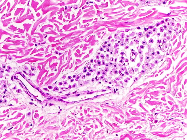
Imaging modality: Light microscopy of a hematoxylin and eosin stained skin biopsy (dermis). The section demonstrates a sparse dermal infiltrate of mast cells in a perivascular distribution within the superficial to mid dermis, adjacent to collagen and small vessels. Mast cells appear as round to oval basophilic nuclei with granulated cytoplasm; the background stroma shows mild edema and mild perivascular lymphocytic inflammation. The infiltrate is not dense, supporting a clinical diagnosis of urticaria pigmentosa (cutaneous mastocytosis) rather than a mast cell proliferation with extensive infiltration. The epidermis is relatively preserved; no pseudoepitheliomatous hyperplasia; no significant fibrosis. Special stains or immunohistochemistry with CD117 (c-kit) or tryptase would enhance confirmation, as normal skin contains few dermal mast cells. Clinically, this histology correlates with pruritus, flush, Darier sign, and pigmentary changes. Diagnostic significance: Supports cutaneous mastocytosis; helps differentiate from pigmented nevi or dermatitis without mast cell infiltration. Differential considerations include other forms of cutaneous mastocytosis, pigmented lesions, and inflammatory dermatoses with inflammatory infiltrates. This image is educational for dermatopathology and histology of urticaria pigmentosa, and can guide workup for systemic involvement if clinically indicated. Terminology: cutaneous mastocytosis; mast cell tryptase testing; KIT mutation assessment may follow.
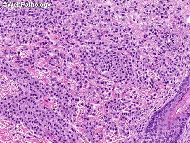
This histopathology image demonstrates a dermatologic mast cell infiltrate associated with urticaria pigmentosa (cutaneous mastocytosis). Acquired lesion is evaluated on a formalin-fixed skin biopsy processed for light microscopy. The section shows dense sheets of round to oval, sometimes spindle-shaped mast cells occupying the superficial to mid-dermis. Individual cells exhibit uniform nuclei with clumped chromatin and moderate eosinophilic to amphophilic cytoplasm; cytoplasmic granules are faintly visible. The overall pattern lacks conspicuous epithelial involvement and displays a perivascular, often diffuse dermal distribution rather than compact clusters. Immunophenotypic context typically includes CD117 (c-KIT) positivity of neoplastic mast cells; aberrant CD25/CD2 expression may be present in some cases of systemic mastocytosis but is not consistently seen in reactive mast cell hyperplasia. In reactive conditions, mast cells are more loosely scattered with a perivascular tendency and are usually CD25/CD2 negative. Clinically, this histology supports a diagnosis of cutaneous mastocytosis when correlated with clinical findings such as pruritus, Darier sign, or urticarial plaques. The image is educational for dermatopathology, pathology residents, and clinicians, illustrating differential considerations with reactive mast cell hyperplasia and mastocytosis. It may guide immunohistochemical workup and subsequent systemic evaluation if warranted. for education.
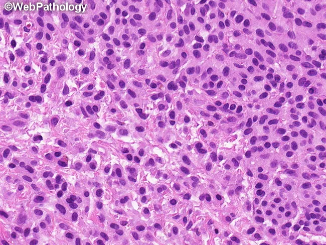
This is a histopathology image of a skin biopsy stained with hematoxylin and eosin, showing a dense dermal mast cell infiltrate consistent with urticaria pigmentosa (cutaneous mastocytosis). The neoplastic mast cells are uniform, arranged in sheets within the reticular dermis, with oval to spindle-shaped morphology and ample eosinophilic or amphophilic cytoplasm containing visible granules. Nuclei are round to oval with clumped chromatin and inconspicuous nucleoli; binucleated cells may be present. Cytologic atypia is not a prominent feature, supporting a benign or indolent mast cell proliferation rather than high-grade disease. The surrounding stroma shows mild edema and occasional inflammatory cells; blood vessels are unaffected. Immunohistochemistry for CD117 (c-KIT) would typically be positive, and toluidine blue or other mast cell stains would highlight granules, confirming mast cell lineage. Clinically, this histology correlates with pigmented, pruritic lesions that characteristically degranulate on mechanical stimulation (Darier's sign). The image captures the key diagnostic features: sheets of uniform mast cells in the dermis, minimal pleomorphism, and absence of marked mitotic activity. This pattern helps distinguish cutaneous mastocytosis from reactive mast cell hyperplasia; however, assessment for systemic involvement (serum tryptase, KIT mutational status, organomegaly) may be warranted in broader clinical contexts.
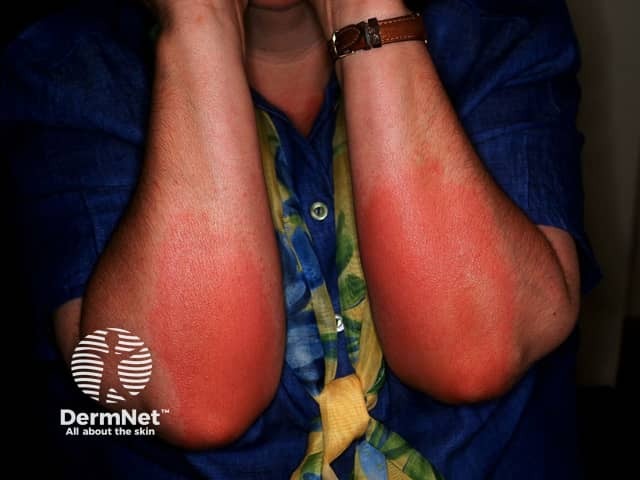
Clinical photograph documenting pressure urticaria on the forearms. This in vivo, noninvasive Digital photography captures bilateral erythematous plaques and diffuse surface redness following sustained mechanical pressure. The forearms (bilateral) show symmetric involvement of the ventral/volar aspects near the antecubital region with a uniform, diffuse erythema and subtle edema without purpura or necrosis. The appearance is consistent with urticarial wheals produced by mast cell degranulation and superficial dermal edema in response to physical stimulation. The photo exemplifies a transient, pressure-induced skin reaction that typically waxes and wanes with time and relief of pressure. The imaging modality is clinical photography using a standard digital camera under ambient lighting, not contrast-enhanced, not magnified. Staining or histology are Not applicable. This image is valuable for clinical documentation, patient education, and research on physical urticarias, including pressure urticaria and dermographism. Differential considerations include allergic contact dermatitis, erythema multiforme, cellulitis, and vasculitic eruptions, all of which can mimic redness but differ in onset, warmth, and distribution. Clinical correlation with history of pressure exposure, timing, pruritus, and response to antihistamines is essential for accurate diagnosis and management planning. Images of such reactions support education, triage, and monitoring treatment outcomes in dermatology and allergy clinics for broader research.
Pathology of Urticaria - Detailed Notes
1. GROSS / MACROSCOPIC PATHOLOGY
| Feature | Description |
|---|---|
| Morphology | Raised, edematous plaques with central pallor (pale/white center) and peripheral erythematous flare |
| Size | A few mm to as large as a hand; may coalesce into larger plaques |
| Color | Pink to pale (whiter when edema is more prominent) |
| Surface | Smooth; no scaling, no vesicles, no necrosis |
| Hallmark | Each individual lesion comes and goes within < 24 hours |
| Pruritus | Hallmark symptom |
| Angioedema | When edema extends into deep dermis/subcutaneous tissue: faint pink, painful, less defined, lasts 2-3 days |
- Dermatology 5e (Grattan, Kocatürk, Saini), p. 372
- Robbins & Kumar Basic Pathology, p. 879
2. HISTOPATHOLOGY (Microscopic Pathology)
Classic Histologic Findings
| Layer | Findings |
|---|---|
| Epidermis | Normal; no spongiosis, no acanthosis |
| Superficial dermis | Edema - collagen bundles are widely spaced ("splaying") due to accumulated fluid |
| Blood vessels | Dilated postcapillary venules; endothelium normal (in ordinary urticaria) |
| Inflammatory infiltrate | Sparse mixed perivascular infiltrate around superficial and mid-dermal venules |
| Cell types | Lymphocytes (predominant), eosinophils, basophils, some neutrophils |
| Mast cells | Degranulated mast cells around superficial dermal venules; appear pale/empty |
- Robbins & Kumar Basic Pathology, p. 879
- Dermatology 5e, p. 382
Histologic Variants
| Variant | Microscopic Pattern | Clinical Significance |
|---|---|---|
| Classic/Lymphocytic urticaria | Sparse perivascular lymphocytes + mild edema | Most common; typical CSU |
| Neutrophilic urticaria | Predominantly neutrophilic dermal infiltrate (no vasculitis features) | May indicate autoinflammatory syndrome (CAPS, Schnitzler, Still disease, SLE) |
| Urticarial vasculitis | Leukocytoclastic vasculitis: endothelial damage, perivenular neutrophil-rich infiltrate, karyorrhexis (nuclear dust), fibrinoid deposits in vessel walls, erythrocyte extravasation | Hypocomplementemic form = systemic disease (SLE overlap) |
| Urticarial bullous pemphigoid | Eosinophil-rich infiltrate; subepidermal blister | Pre-bullous pemphigoid; lesions last >24h |
Rule: If urticarial lesions consistently persist >24-36 hours, are painful rather than pruritic, and leave bruising/pigmentation on resolution → biopsy is mandatory to rule out urticarial vasculitis.
- Dermatology 5e, p. 382
3. PATHOGENESIS - CELLULAR AND MOLECULAR
The Central Event: Mast Cell Degranulation
| Type | Location | Contents | IgE receptor |
|---|---|---|---|
| MCTC (tryptase + chymase) | Skin, intestinal submucosa | Tryptase + chymase | Yes (FcεRI) |
| MCT (tryptase only) | Bowel mucosa, alveolar wall, nasal mucosa | Tryptase only | Yes (FcεRI) |
Step-by-Step Degranulation Mechanism
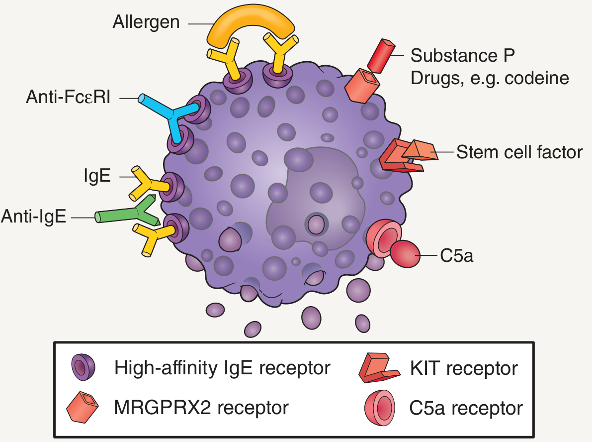
Mast Cell Degranulating Stimuli
| Pathway | Stimulus | Receptor |
|---|---|---|
| Immunologic (IgE-dependent) | Allergen cross-links IgE-bound FcεRI | FcεRI |
| Immunologic (autoantibody) | Anti-IgE IgG, anti-FcεRI IgG | FcεRI |
| Complement-mediated | C5a anaphylatoxin | C5a receptor |
| Non-immunologic | Opioids, codeine, substance P, polymyxin B | MRGPRX2 receptor |
| Growth factor | Stem cell factor | KIT receptor |
| Physical stimuli | Cold, pressure, vibration, UV, heat | Various / indirect |
4. INFLAMMATORY MEDIATORS RELEASED
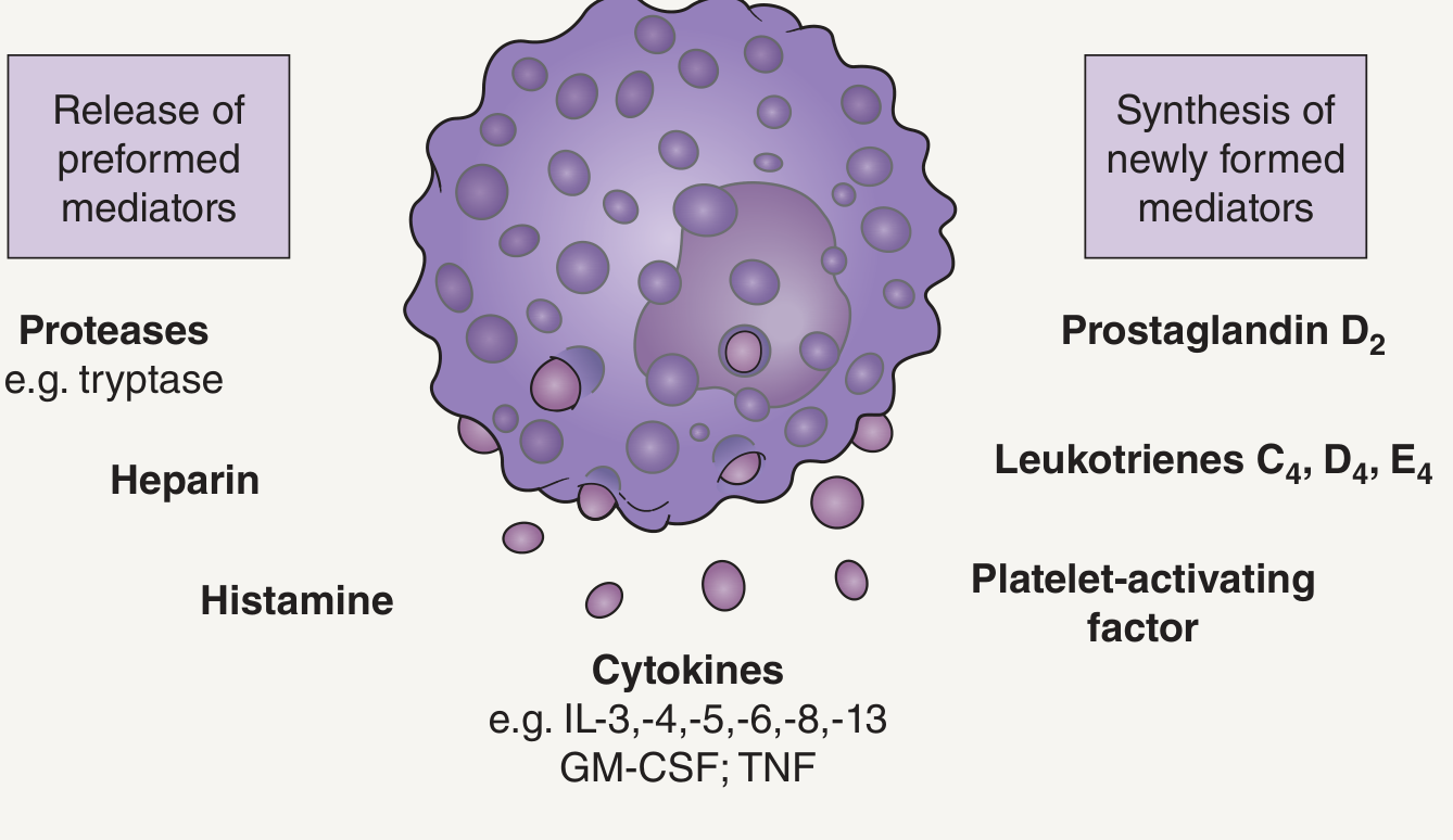
Preformed Mediators (stored in granules, released immediately)
| Mediator | Role in Urticaria |
|---|---|
| Histamine | Primary mediator; binds H1 receptors on postcapillary venules → vasodilation + increased permeability; acts on sensory nerves → itch |
| Tryptase | Serine protease; activates bradykinin; used as marker of mast cell activation (serum tryptase) |
| Chymase | Serine protease; cleaves angiotensin I → angiotensin II; modulates matrix |
| Heparin | Anticoagulant; binds growth factors; contributes to wheal formation |
| Chondroitin sulfate | Structural proteoglycan component of granules |
Newly Synthesized Mediators (formed after activation from membrane phospholipids)
| Mediator | Role |
|---|---|
| Prostaglandin D2 (PGD2) | Most important eicosanoid; vasodilation + bronchoconstriction |
| PGE2 | Inhibitory - has protective role by inhibiting immunologic mast cell degranulation |
| Leukotriene C4, D4, E4 | = "Slow-reacting substance of anaphylaxis (SRS-A)"; prolonged vasodilation + wheal formation |
| Platelet-activating factor (PAF) | Platelet aggregation; amplifies wheal formation; especially in cold urticaria |
| Cytokines: TNF, IL-1β, IL-3, IL-5, IL-6, IL-8, IL-10, IL-12p70, IL-13 | Upregulate endothelial adhesion molecules (ICAM-1, E-selectin); recruit eosinophils, basophils, neutrophils, Th0 cells into lesion |
| GM-CSF | Promotes eosinophil/basophil survival |
| BAFF (B-cell activating factor) | Elevated in CSU sera; may sustain autoantibody production |
- Dermatology 5e, p. 374
- Fitzpatrick's Dermatology, p. 720-721
5. IMMUNOLOGIC MECHANISMS (DETAILED)
5A. Type I Hypersensitivity (IgE-Mediated)
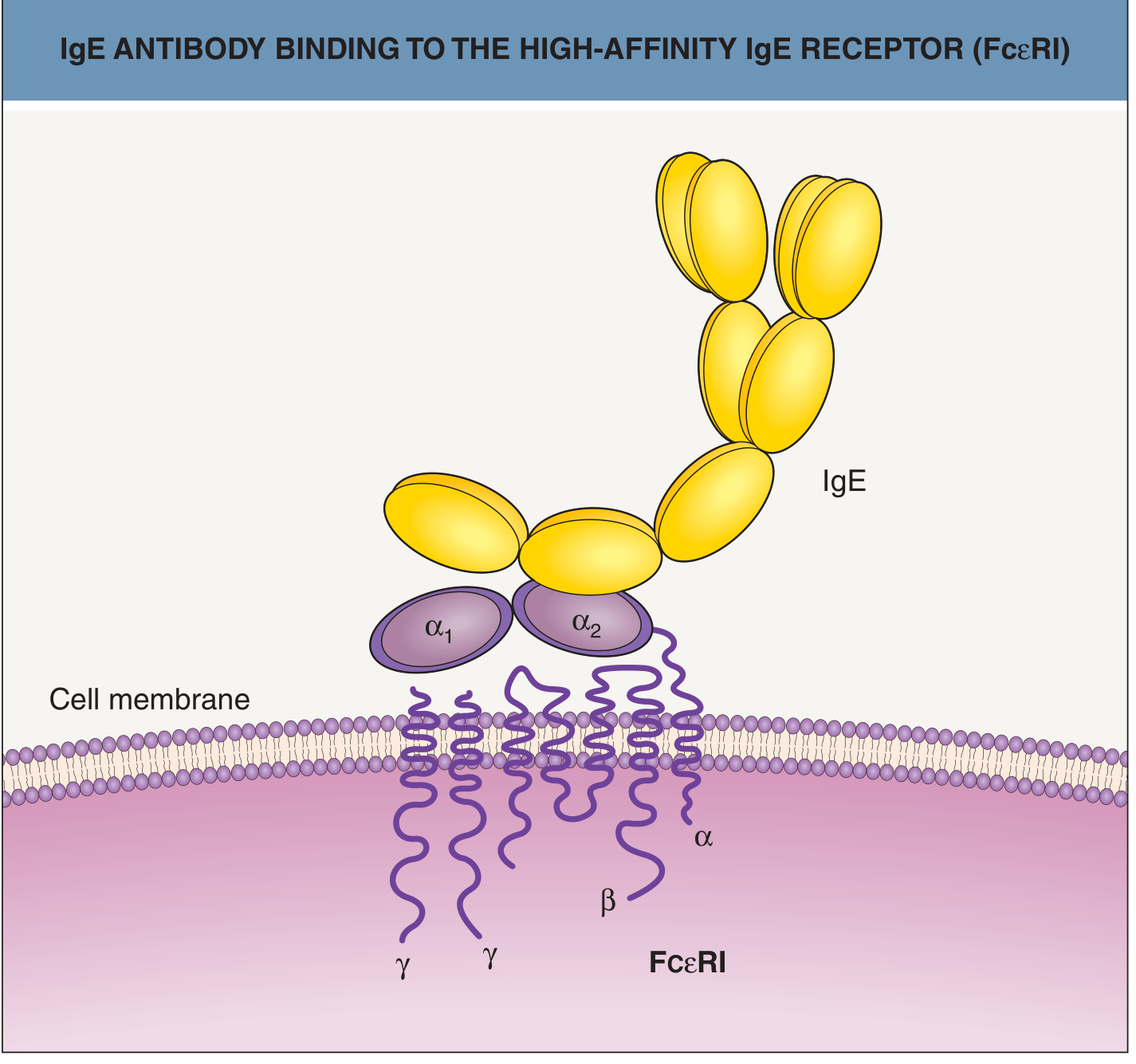
- Antigen-specific IgE coats mast cells via FcεRI
- Re-exposure to antigen cross-links adjacent FcεRI → degranulation
- Symptoms begin 15 min to 1 hour after allergen exposure
- Probably accounts for < 10% of all urticaria (mainly acute/episodic forms)
- Classic examples: latex contact urticaria, food allergy urticaria (nuts, fish, fruit)
5B. Autoimmune Mechanism (IgG Autoantibodies)
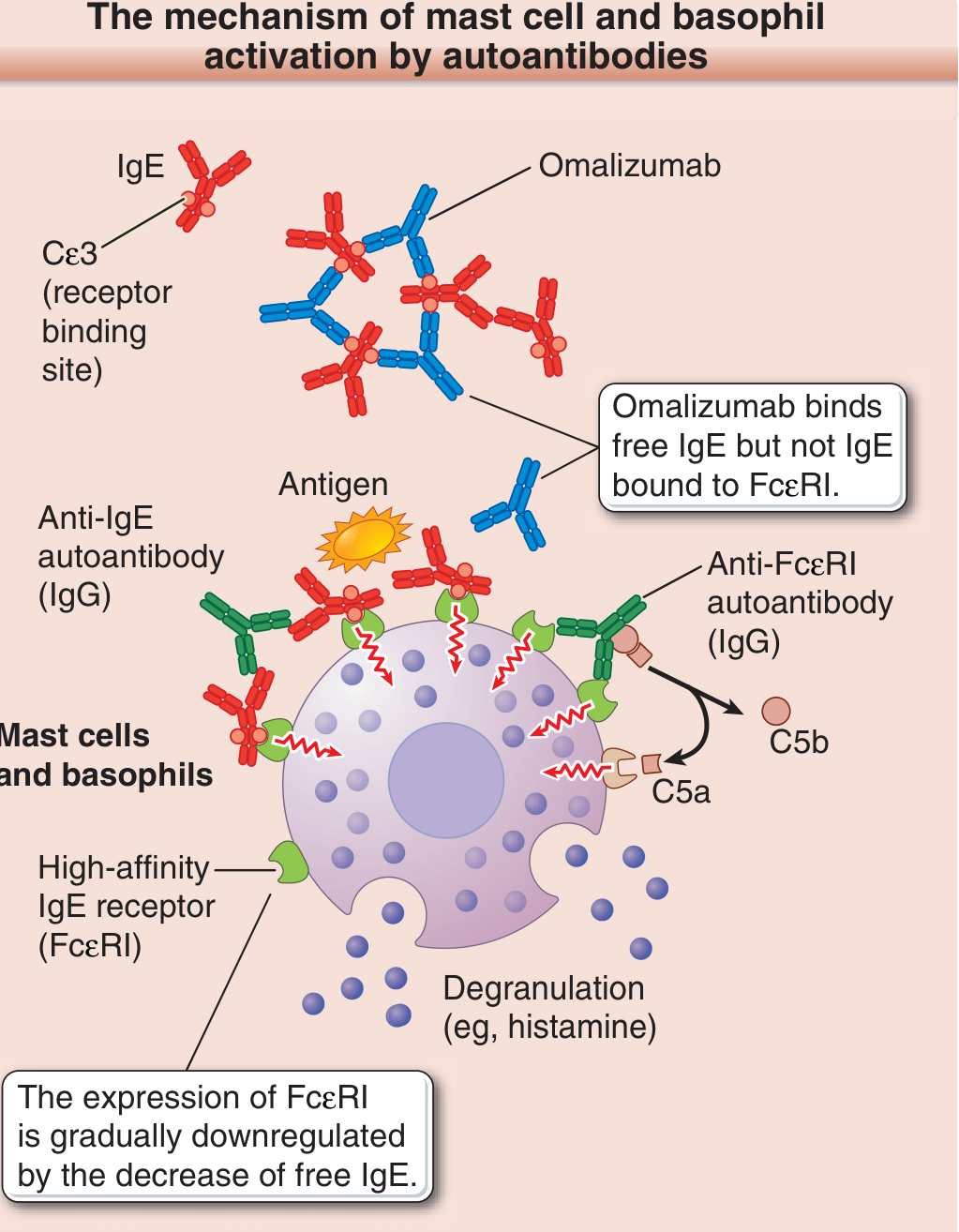
- Affects 30-50% of CSU patients (up to 45% by some estimates)
- Two types of pathogenic autoantibodies:
| Autoantibody | Target | Mechanism |
|---|---|---|
| IgG anti-FcεRI (majority) | α subunit of high-affinity IgE receptor | Cross-links FcεRI → mast cell/basophil degranulation |
| IgG anti-IgE (~10%) | Fc portion of IgE itself | Cross-links IgE bound to FcεRI → degranulation |
-
Anti-FcεRI subtypes:
- Anti-α2 domain: competitive with IgE (blocks binding)
- Anti-α1 domain: non-competitive (binds even in presence of IgE)
-
Complement amplification: Autoantibody binding → complement activation → C5a anaphylatoxin generated → further augments mast cell degranulation
-
Detection methods: Autologous serum skin test (ASST), basophil CD63/CD203c activation test, in vitro histamine release assay
-
IgE autoantibodies also found in CSU against: thyroid peroxidase (TPO), thyroglobulin, tissue factor, IL-24 → these IgE anti-TPO antibodies can produce a local wheal-and-flare, explaining the thyroid disease-urticaria association
-
Basophils in CSU: Blood basophils are reduced in number and less responsive to anti-IgE stimulus in active disease; responsiveness returns during remission
-
Omalizumab mechanism: Binds free IgE at its Cε3 domain (receptor binding site) → prevents IgE binding to FcεRI → gradual downregulation of FcεRI expression on mast cells → reduced mast cell "readiness" to degranulate
-
Dermatology 5e, p. 373-374; Fitzpatrick's Dermatology, p. 720-721; Harrison's 22E
5C. Immune Complex / Complement-Mediated
- Urticarial vasculitis and serum-sickness type urticaria
- Antigen-antibody complexes deposit in vessel walls → activate complement → C3a + C5a anaphylatoxins → mast cell degranulation AND direct vascular damage
- Leads to leukocytoclastic vasculitis histologically
5D. Non-Immunologic (Direct Mast Cell Activation)
| Agent | Receptor/Mechanism |
|---|---|
| Opioids (morphine, codeine) | MRGPRX2 receptor |
| Substance P | MRGPRX2 receptor |
| Vancomycin ("red man syndrome") | Direct MRGPRX2 |
| Radiocontrast media | Direct mast cell degranulation |
| Aspirin / NSAIDs | Shift arachidonic acid to leukotriene pathway (LOX > COX); non-immunologic |
| Strawberries, lobster | Directly release histamine from mast cells |
6. PATHOGENESIS OF SPECIFIC URTICARIA TYPES
| Type | Specific Pathological Mechanism |
|---|---|
| Cholinergic urticaria | Acetylcholine released from cholinergic sympathetic nerves → mast cell activation; ~2/3 have IgE antibodies to MGL_1304 (protein from Malassezia globosa in sweat) |
| Cold urticaria | Proposed IgE-mediated neoantigen (cold-induced); cryoglobulins (5%); platelet clumping → PAF/platelet factor 4 release; familial form: F12 mutation → bradykinin pathway; FCAS: NLRP3 mutation → IL-1β |
| Dermatographism | Zonal (piecemeal) mast cell degranulation pattern on electron microscopy; elevated histamine in suction blisters at dermographic sites |
| Pressure urticaria | Possibly late-phase IgE reaction; no antigen identified |
| Vibratory urticaria (hereditary) | ADGRE2 missense mutation → destabilizes inhibitory α/β subunit interaction → sensitizes mast cells to vibration-induced degranulation (IgE-independent) |
| Solar urticaria | Physical stimulus creates neoantigen → reacts with IgE on mast cells; 6 subtypes by action spectrum |
| Aquagenic urticaria | Water dilutes inhibitory substance in skin → IgE mechanism? Pathomechanism unclear |
| HAE (not true urticaria) | C1INH deficiency → failure to inhibit factor XII (Hageman factor) and kallikrein → bradykinin excess → vascular permeability (NOT histamine-dependent) |
7. VASCULAR PATHOLOGY: HOW THE WHEAL FORMS
Mast cell degranulation
         ↓
Histamine released → binds H1 receptors on postcapillary venules
         ↓
┌─────────────────────────────────────┐
│ Vasodilation of arterioles          │  → ERYTHEMA (red flare)
│ Increased permeability of venules   │  → PLASMA EXTRAVASATION → EDEMA (wheal center pale)
│ Sensory nerve activation (itch)     │  → PRURITUS
└─────────────────────────────────────┘
         ↓
TNF + IL-8 upregulate endothelial adhesion molecules
(ICAM-1, E-selectin, VCAM-1)
         ↓
Eosinophils, basophils, neutrophils, Th0 cells migrate into dermis
         ↓
Late-phase reaction: secondary cytokine release → prolongs/amplifies inflammation
- Local red reaction (direct vasodilation)
- Surrounding flare (axon reflex via sensory nerves)
- Wheal (local edema from vascular leakage)
8. ROLE OF COAGULATION IN URTICARIA
- Tissue factor is upregulated on eosinophils and monocytes in CSU
- Leads to activation of the extrinsic coagulation cascade
- Generates thrombin, which can directly activate PAR-1 receptors on mast cells → degranulation
- D-dimer levels are elevated in active CSU and correlate with disease severity
- This explains why anticoagulants (heparin) may reduce urticaria symptoms
9. SUMMARY TABLE: PATHOLOGY AT A GLANCE
| Level | Finding |
|---|---|
| Gross | Edematous wheal; pale center + red flare; transient (<24h); no scaling |
| Histology | Superficial dermal edema; splayed collagen bundles; sparse perivascular mixed infiltrate (lymphocytes, eos, basophils, neutrophils); degranulated mast cells |
| Special stains | Mast cells: Giemsa/toluidine blue (metachromatic granules); tryptase immunostain |
| Key effector cell | Mast cell (cutaneous MCTC phenotype) |
| Primary mediator | Histamine (preformed, immediate) |
| Newly synthesized | PGD2, LTC4/D4/E4, PAF, cytokines (TNF, IL-6, IL-8) |
| Main receptor | FcεRI (high-affinity IgE receptor) - α1β(γγ) subunit structure |
| Immunologic trigger | IgE cross-linking (type I HSR) or IgG autoantibodies (anti-FcεRI / anti-IgE) |
| Non-immunologic trigger | MRGPRX2 agonists (opioids, substance P, codeine) |
| Autoimmune mechanism | IgG anti-FcεRI or anti-IgE in ~30-50% of CSU; C5a amplifies |
| Vasculitis variant | Leukocytoclastic vasculitis; fibrinoid necrosis; erythrocyte extravasation |